Opponens digiti minimi muscle of hand

Last updated
Opponens digiti minimi muscle
1121 Intrinsic Muscles of the Hand Deep LD.png
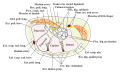
Deep muscles of the right hand, palmar view.
Details
Origin Hook of hamate and flexor retinaculum
Insertion Medial border of 5th metacarpal (in hand, 3rd digit is orientation of mid-line)
Artery Ulnar artery
Nerve Deep branch of ulnar nerve (C8 and T1)
Actions Draws 5th metacarpal anteriorly and rotates it, bringing little finger (5th digit) into opposition with thumb
Identifiers
Latin musculus opponens digiti minimi
(Old: opponens quinti digiti)
TA98 A04.6.02.064
TA2 2531
FMA 37384
Anatomical terms of muscle

The opponens digiti minimi (opponens digiti quinti in older texts) is a muscle in the hand. It is of a triangular form, and placed immediately beneath the palmaris brevis, abductor digiti minimi and flexor digiti minimi brevis. It is one of the three hypothenar muscles that control the little finger. [1]

Contents

It arises from the convexity of the hamulus of the hamate bone and the contiguous portion of the transverse carpal ligament; it is inserted into the whole length of the metacarpal bone of the little finger, along its ulnar margin.

The opponens digiti minimi muscle serves to flex and laterally rotate the 5th metacarpal about the 5th carpometacarpal joint, as when bringing the little finger and thumb into opposition. It is innervated by the deep branch of the ulnar nerve.

See also

Additional images

Related Research Articles

<span class="mw-page-title-main">Thenar eminence</span> Mound at the base of the thumb formed by the intrinsic group of muscles

The thenar eminence is the mound formed at the base of the thumb on the palm of the hand by the intrinsic group of muscles of the thumb. The skin overlying this region is the area stimulated when trying to elicit a palmomental reflex. The word thenar comes from Ancient Greek θέναρ (thenar) 'palm of the hand'.

<span class="mw-page-title-main">Ulnar nerve</span> Nerve which runs near the ulna bone

The ulnar nerve is a nerve that runs near the ulna, one of the two long bones in the forearm. The ulnar collateral ligament of elbow joint is in relation with the ulnar nerve. The nerve is the largest in the human body unprotected by muscle or bone, so injury is common. This nerve is directly connected to the little finger, and the adjacent half of the ring finger, innervating the palmar aspect of these fingers, including both front and back of the tips, perhaps as far back as the fingernail beds.

<span class="mw-page-title-main">Trapezium (bone)</span> Bone of the wrist

The trapezium bone is a carpal bone in the hand. It forms the radial border of the carpal tunnel.

<span class="mw-page-title-main">Metacarpal bones</span> Bones of hand

In human anatomy, the metacarpal bones or metacarpus, also known as the "palm bones", are the appendicular bones that form the intermediate part of the hand between the phalanges (fingers) and the carpal bones, which articulate with the forearm. The metacarpal bones are homologous to the metatarsal bones in the foot.

<span class="mw-page-title-main">Hamate bone</span> Carpal bone in the wrist

The hamate bone, or unciform bone, Latin os hamatum and occasionally abbreviated as just hamatum, is a bone in the human wrist readily distinguishable by its wedge shape and a hook-like process ("hamulus") projecting from its palmar surface.

<span class="mw-page-title-main">Upper limb</span> Consists of the arm, forearm, and hand

The upper limbs or upper extremities are the forelimbs of an upright-postured tetrapod vertebrate, extending from the scapulae and clavicles down to and including the digits, including all the musculatures and ligaments involved with the shoulder, elbow, wrist and knuckle joints. In humans, each upper limb is divided into the shoulder, arm, elbow, forearm, wrist and hand, and is primarily used for climbing, lifting and manipulating objects. In anatomy, just as arm refers to the upper arm, leg refers to the lower leg.

<span class="mw-page-title-main">Little finger</span> Smallest finger of the human hand

The little finger or pinkie, also known as the baby finger, fifth digit, or pinky finger, is the most ulnar and smallest digit of the human hand, and next to the ring finger.

<span class="mw-page-title-main">Adductor pollicis muscle</span> Muscle in the thenar compartment

In human anatomy, the adductor pollicis muscle is a muscle in the hand that functions to adduct the thumb. It has two heads: transverse and oblique.

<span class="mw-page-title-main">Opponens pollicis muscle</span> Muscle in the thenar compartment

The opponens pollicis is a small, triangular muscle in the hand, which functions to oppose the thumb. It is one of the three thenar muscles. It lies deep to the abductor pollicis brevis and lateral to the flexor pollicis brevis.

<span class="mw-page-title-main">Hypothenar eminence</span> Group of three muscles of the palm

The hypothenar muscles are a group of three muscles of the palm that control the motion of the little finger.

<span class="mw-page-title-main">Carpometacarpal joint</span> Joint between the wrist and hand

The carpometacarpal (CMC) joints are five joints in the wrist that articulate the distal row of carpal bones and the proximal bases of the five metacarpal bones.

<span class="mw-page-title-main">Flexor digiti minimi brevis muscle of foot</span> Muscles of the sole of the foot in the third layer

The flexor digiti minimi brevis lies under the metatarsal bone on the little toe, and resembles one of the interossei.

<span class="mw-page-title-main">Dorsal interossei of the hand</span> Muscles between the metacarpals

In human anatomy, the dorsal interossei (DI) are four muscles in the back of the hand that act to abduct (spread) the index, middle, and ring fingers away from the hand's midline and assist in flexion at the metacarpophalangeal joints and extension at the interphalangeal joints of the index, middle and ring fingers.

<span class="mw-page-title-main">Abductor digiti minimi muscle of the hand</span> Muscle in the hypothenar compartment

In human anatomy, the abductor digiti minimi is a skeletal muscle situated on the ulnar border of the palm of the hand. It forms the ulnar border of the palm and its spindle-like shape defines the hypothenar eminence of the palm together with the skin, connective tissue, and fat surrounding it. Its main function is to pull the little finger away from the other fingers.

<span class="mw-page-title-main">Flexor digiti minimi brevis muscle of hand</span> Muscle in the hypothenar compartment

The flexor digiti minimi brevis is a hypothenar muscle in the hand that flexes the little finger at the metacarpophalangeal joint. It lies lateral to the abductor digiti minimi when the hand is in anatomical position.

<span class="mw-page-title-main">Deep branch of ulnar nerve</span>

The deep branch of the ulnar nerve is a terminal, primarily motor branch of the ulnar nerve. It is accompanied by the deep palmar branch of ulnar artery.

<span class="mw-page-title-main">Cervical spinal nerve 8</span> Spinal nerve of the cervical segment

The cervical spinal nerve 8 (C8) is a spinal nerve of the cervical segment.

<span class="mw-page-title-main">Muscles of the hand</span> Muscles of the human hand

The muscles of the hand are the skeletal muscles responsible for the movement of the hand and fingers. The muscles of the hand can be subdivided into two groups: the extrinsic and intrinsic muscle groups. The extrinsic muscle groups are the long flexors and extensors. They are called extrinsic because the muscle belly is located on the forearm. The intrinsic group are the smaller muscles located within the hand itself. The muscles of the hand are innervated by the radial, median, and ulnar nerves from the brachial plexus.

<span class="mw-page-title-main">Extrinsic extensor muscles of the hand</span>

The extrinsic extensor muscles of the hand are located in the back of the forearm and have long tendons connecting them to bones in the hand, where they exert their action. Extrinsic denotes their location outside the hand. Extensor denotes their action which is to extend, or open flat, joints in the hand. They include the extensor carpi radialis longus (ECRL), extensor carpi radialis brevis (ECRB), extensor digitorum (ED), extensor digiti minimi (EDM), extensor carpi ulnaris (ECU), abductor pollicis longus (APL), extensor pollicis brevis (EPB), extensor pollicis longus (EPL), and extensor indicis (EI).

<span class="mw-page-title-main">Muscles of the thumb</span> Set of nine human skeletal muscles

The muscles of the thumb are nine skeletal muscles located in the hand and forearm. The muscles allow for flexion, extension, adduction, abduction and opposition of the thumb. The muscles acting on the thumb can be divided into two groups: The extrinsic hand muscles, with their muscle bellies located in the forearm, and the intrinsic hand muscles, with their muscles bellies located in the hand proper.

References

PD-icon.svgThis article incorporates text in the public domain from page 464 of the 20th edition of Gray's Anatomy (1918)

  1. Uysal, Ahmet Çaǧri; Alagöz, Murat Şahin; Tüccar, Eray; Şensöz, Ömer; Tekdemir, Ibrahim (2005-01-01). "The vascular anatomy of the abductor digiti minimi and the flexor digiti minimi brevis muscles". The Journal of Hand Surgery. 30 (1): 172–176. doi:10.1016/j.jhsa.2004.06.001. ISSN   0363-5023. PMID   15680577.