PHEX

Last updated
PHEX
Identifiers
Aliases PHEX , HPDR, HPDR1, HYP, HYP1, LXHR, PEX, XLH, phosphate regulating endopeptidase homolog, X-linked, phosphate regulating endopeptidase homolog X-linked
External IDs OMIM: 300550 MGI: 107489 HomoloGene: 37310 GeneCards: PHEX
Orthologs
SpeciesHumanMouse
Entrez
Ensembl
UniProt
RefSeq (mRNA)

NM_000444
NM_001282754

NM_011077

RefSeq (protein)

NP_000435
NP_001269683

NP_035207

Location (UCSC) Chr X: 22.03 – 22.49 Mb Chr X: 155.95 – 156.2 Mb
PubMed search [3] [4]
Wikidata
View/Edit Human View/Edit Mouse

Phosphate-regulating endopeptidase homolog X-linked also known as phosphate-regulating gene with homologies to endopeptidases on the X chromosome or metalloendopeptidase homolog PEX is an enzyme that in humans is encoded by the PHEX gene. [5] [6] This gene contains 18 exons and is located on the X chromosome.

Contents

Function

The protein encoded by this gene is a transmembrane endopeptidase that belongs to the type II integral membrane zinc-dependent endopeptidase family. The protein is thought to be involved in bone and dentin mineralization and renal phosphate reabsorption. [7] The bone and dentin protein osteopontin (OPN) which inhibits mineralization in the skeleton and in teeth is a substrate for PHEX. [8] In the absence of functional PHEX in the mouse model (Hyp) of X-linked hypophosphatemia (XLH), and in human XLH where PHEX activity is decreased or absent, increased circulating FGF23 hormone results in low serum phosphate (caused by renal phosphate wasting) such that there is an insufficient level of this mineral ion in the blood in transit to mineralized tissues compared to the normal amount that is required for proper bone and tooth mineralization; this leads to soft bones and teeth. In addition to renal phosphate wasting, the mineralization-inhibiting phosphoprotein osteopontin and osteopontin fragments accumulate in the extracellular matrix of bones and teeth to contribute locally to the reduction in mineralization, which together with the systemic lower level of circulating serum phosphate, both lead to the decreased mineralization (hypomineralization) characteristic of the osteomalacia and odontomalacia typically seen in XLH/Hyp. [9] [10] [11] [12] [13] XLH patients have soft and deformed skeletons, and soft teeth that easily become infected. Osteopontin (OPN) is a substrate protein for the enzyme PHEX whose enzymatic activity degrades/removes the mineralization-inhibiting function of OPN in normal mineralized tissue physiology, [14] In disease, when the PHEX gene is mutated causing reduced or absent PHEX enzymatic activity, OPN that would normally be degraded and cleared remains behind in the extracellular matrix of bones and teeth, accumulating locally in the tissue to contribute to the osteomalacia and odontomalacia. [15] [16] A relationship describing local, physiologic double-negative (inhibiting inhibitors) regulation of mineralization involving OPN has been termed the Stenciling Principle of mineralization, whereby enzyme-substrate pairs imprint mineralization patterns into the extracellular matrix (most notably for bone) by degrading mineralization inhibitors (e.g. TNAP/TNSALP/ALPL enzyme degrading the pyrophosphate inhibition, and PHEX enzyme degrading the osteopontin inhibition). [17] [18] [19] The Stenciling Principle for mineralization is particularly relevant to the osteomalacia and odontomalacia observed in hypophosphatasia and X-linked hypophosphatemia. [20]

Clinical significance

Mutation of PHEX leads to X-linked hypophosphatemia. [5]

Related Research Articles

Osteoblast Cells secreting extracellular matrix

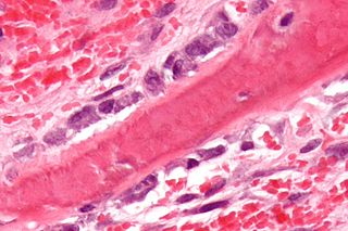
Osteoblasts are cells with a single nucleus that synthesize bone. However, in the process of bone formation, osteoblasts function in groups of connected cells. Individual cells cannot make bone. A group of organized osteoblasts together with the bone made by a unit of cells is usually called the osteon.

Osteomalacia Medical condition

Osteomalacia is a disease characterized by the softening of the bones caused by impaired bone metabolism primarily due to inadequate levels of available phosphate, calcium, and vitamin D, or because of resorption of calcium. The impairment of bone metabolism causes inadequate bone mineralization. Osteomalacia in children is known as rickets, and because of this, use of the term "osteomalacia" is often restricted to the milder, adult form of the disease. Signs and symptoms can include diffuse body pains, muscle weakness, and fragility of the bones. In addition to low systemic levels of circulating mineral ions that result in decreased bone and tooth mineralization, accumulation of mineralization-inhibiting proteins and peptides, and small inhibitory molecules, can occur in the extracellular matrix of bones and teeth, contributing locally to cause matrix hypomineralization (osteomalacia/odontomalacia). A relationship describing local, physiologic double-negative regulation of mineralization has been termed the Stenciling Principle of mineralization, whereby enzyme-substrate pairs imprint mineralization patterns into the extracellular matrix by degrading mineralization inhibitors. The Stenciling Principle for mineralization is particularly relevant to the osteomalacia and odontomalacia observed in hypophosphatasia (HPP) and X-linked hypophosphatemia (XLH).

Hypophosphatemia Medical condition

Hypophosphatemia is an electrolyte disorder in which there is a low level of phosphate in the blood. Symptoms may include weakness, trouble breathing, and loss of appetite. Complications may include seizures, coma, rhabdomyolysis, or softening of the bones.

Osteocyte Mature osteoblasts which helps in communication between cells and also in molecular synthesis

An osteocyte, an oblate shaped type of bone cell with dendritic processes, is the most commonly found cell in mature bone tissue, and can live as long as the organism itself. The adult human body has about 42 billion of them. Osteocytes do not divide and have an average half life of 25 years. They are derived from osteoprogenitor cells, some of which differentiate into active osteoblasts. Osteoblasts/osteocytes develop in mesenchyme.

Hypophosphatasia Medical condition

Hypophosphatasia (; also called deficiency of alkaline phosphatase, phosphoethanolaminuria, or Rathbun's syndrome; sometimes abbreviated HPP) is a rare, and sometimes fatal, inherited metabolic bone disease. Clinical symptoms are heterogeneous, ranging from the rapidly fatal, perinatal variant, with profound skeletal hypomineralization, respiratory compromise or vitamin B6 dependent seizures to a milder, progressive osteomalacia later in life. Tissue non-specific alkaline phosphatase (TNSALP) deficiency in osteoblasts and chondrocytes impairs bone mineralization, leading to rickets or osteomalacia. The pathognomonic finding is subnormal serum activity of the TNSALP enzyme, which is caused by one of 388 genetic mutations identified to date, in the gene encoding TNSALP. Genetic inheritance is autosomal recessive for the perinatal and infantile forms but either autosomal recessive or autosomal dominant in the milder forms.

Osteopontin

Osteopontin (OPN), also known as bone /sialoprotein I, early T-lymphocyte activation (ETA-1), secreted phosphoprotein 1 (SPP1), 2ar and Rickettsia resistance (Ric), is a protein that in humans is encoded by the SPP1 gene. The murine ortholog is Spp1. Osteopontin is a SIBLING (glycoprotein) that was first identified in 1986 in osteoblasts.

Fibroblast growth factor 23

Fibroblast growth factor 23 (FGF23) is a protein that in humans is encoded by the FGF23 gene. FGF23 is a member of the fibroblast growth factor (FGF) family which participates in phosphate and vitamin D metabolism and regulation.

X-linked hypophosphatemia X-linked dominant disorder that causes rickets

X-linked hypophosphatemia (XLH) is an X-linked dominant form of rickets that differs from most cases of dietary deficiency rickets in that vitamin D supplementation does not cure it. It can cause bone deformity including short stature and genu varum (bow-leggedness). It is associated with a mutation in the PHEX gene sequence (Xp.22) and subsequent inactivity of the PHEX protein. PHEX mutations lead to an elevated circulating (systemic) level of the hormone FGF23 which results in renal phosphate wasting, and locally in the extracellular matrix of bones and teeth an elevated level of the mineralization/calcification-inhibiting protein osteopontin. An inactivating mutation in the PHEX gene results in an increase in systemic circulating FGF23, and a decrease in the enzymatic activity of the PHEX enzyme which normally removes (degrades) mineralization-inhibiting osteopontin protein; in XLH, the decreased PHEX enzyme activity leads to an accumulation of inhibitory osteopontin locally in bones and teeth to block mineralization which, along with renal phosphate wasting, both cause osteomalacia and odontomalacia. For both XLH and hypophosphatasia, inhibitor-enzyme pair relationships function to regulate mineralization in the extracellular matrix through a double-negative activation effect in a manner described as the Stenciling Principle. Both these underlying mechanisms contribute to the pathophysiology of XLH that leads to soft bones and teeth. The prevalence of the disease is 1 in 20,000.

Osteonectin

Osteonectin (ON) also known as secreted protein acidic and rich in cysteine (SPARC) or basement-membrane protein 40 (BM-40) is a protein that in humans is encoded by the SPARC gene.

Bone sialoprotein Protein-coding gene in the species Homo sapiens

Bone sialoprotein (BSP) is a component of mineralized tissues such as bone, dentin, cementum and calcified cartilage. BSP is a significant component of the bone extracellular matrix and has been suggested to constitute approximately 8% of all non-collagenous proteins found in bone and cementum. BSP, a SIBLING protein, was originally isolated from bovine cortical bone as a 23-kDa glycopeptide with high sialic acid content.
Tartrate-resistant acid phosphatase Protein-coding gene in the species Homo sapiens

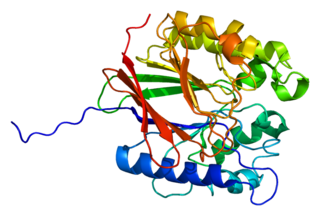
Tartrate-resistant acid phosphatase, also called acid phosphatase 5, tartrate resistant (ACP5), is a glycosylated monomeric metalloprotein enzyme expressed in mammals. It has a molecular weight of approximately 35kDa, a basic isoelectric point (7.6–9.5), and optimal activity in acidic conditions. TRAP is synthesized as latent proenzyme and activated by proteolytic cleavage and reduction. It is differentiated from other mammalian acid phosphatases by its resistance to inhibition by tartrate and by its molecular weight.

<span class="mw-page-title-main">GDF6</span> Protein-coding gene in the species Homo sapiens

Growth differentiation factor 6 (GDF6) is a protein that in humans is encoded by the GDF6 gene.

LRP5

Low-density lipoprotein receptor-related protein 5 is a protein that in humans is encoded by the LRP5 gene. LRP5 is a key component of the LRP5/LRP6/Frizzled co-receptor group that is involved in canonical Wnt pathway. Mutations in LRP5 can lead to considerable changes in bone mass. A loss-of-function mutation causes osteoporosis-pseudoglioma, while a gain-of-function mutation causes drastic increases in bone mass.

alpha-2-HS-glycoprotein Protein-coding gene in the species Homo sapiens

alpha-2-HS-glycoprotein also known as fetuin-A is a protein that in humans is encoded by the AHSG gene. Fetuin-A belongs to the fetuin class of plasma binding proteins and is more abundant in fetal than adult blood.

DMP1

Dentin matrix acidic phosphoprotein 1 is a protein that in humans is encoded by the DMP1 gene.

MEPE

Matrix extracellular phosphoglycoprotein is a protein that in humans is encoded by the MEPE gene. A conserved RGD motif is found in this protein, and this is potentially involved in integrin recognition.

Oncogenic osteomalacia, also known as oncogenic hypophosphatemic osteomalacia, is an uncommon disorder resulting in increased renal phosphate excretion, hypophosphatemia and osteomalacia. It may be caused by a phosphaturic mesenchymal tumor.

<span class="mw-page-title-main">FAM20C</span> Protein-coding gene in the species Homo sapiens

Family with sequence similarity 20, member C also known as FAM20C or DMP4 is a protein which in humans is encoded by the FAM20C gene. Fam20C, a Golgi localized protein kinase, is a serine kinase that phosphorylates both casein and other highly acidic proteins and members of the small integrin-binding ligand, the N-linked glycoproteins (SIBLING) family at the target motif SerXGlu.

The family of non-collagenous proteins known as SIBLING proteins, standing for small integrin-binding ligand, N-linked glycoprotein, are components of the extracellular matrix of bone and dentin. Evidence shows that these proteins play key roles in the mineralization of these tissues.

Carboxypeptidase A6 Protein-coding gene in the species Homo sapiens

Carboxypeptidase A6 (CPA6) is a metallocarboxypeptidase enzyme that in humans is encoded by the CPA6 gene. It is highly expressed in the adult mouse olfactory bulb and is broadly expressed in the embryonic brain and other tissues.

References

  1. 1 2 3 GRCh38: Ensembl release 89: ENSG00000102174 - Ensembl, May 2017
  2. 1 2 3 GRCm38: Ensembl release 89: ENSMUSG00000057457 - Ensembl, May 2017
  3. "Human PubMed Reference:". National Center for Biotechnology Information, U.S. National Library of Medicine.
  4. "Mouse PubMed Reference:". National Center for Biotechnology Information, U.S. National Library of Medicine.
  5. 1 2 Francis F, Hennig S, Korn B, Reinhardt R, De Jong P, Poustka A, et al. (October 1995). "A gene (PEX) with homologies to endopeptidases is mutated in patients with X-linked hypophosphatemic rickets. The HYP Consortium". Nature Genetics. 11 (2): 130–136. doi:10.1038/ng1095-130. PMID   7550339. S2CID   6424732.
  6. Grieff M, Mumm S, Waeltz P, Mazzarella R, Whyte MP, Thakker RV, Schlessinger D (February 1997). "Expression and cloning of the human X-linked hypophosphatemia gene cDNA". Biochemical and Biophysical Research Communications. 231 (3): 635–639. doi:10.1006/bbrc.1997.6153. PMID   9070861.
  7. "Entrez Gene: phosphate regulating endopeptidase homolog".
  8. Barros NM, Hoac B, Neves RL, Addison WN, Assis DM, Murshed M, et al. (March 2013). "Proteolytic processing of osteopontin by PHEX and accumulation of osteopontin fragments in Hyp mouse bone, the murine model of X-linked hypophosphatemia". Journal of Bone and Mineral Research. 28 (3): 688–699. doi: 10.1002/jbmr.1766 . PMID   22991293.
  9. Boukpessi T, Gaucher C, Léger T, Salmon B, Le Faouder J, Willig C, et al. (August 2010). "Abnormal presence of the matrix extracellular phosphoglycoprotein-derived acidic serine- and aspartate-rich motif peptide in human hypophosphatemic dentin". The American Journal of Pathology. 177 (2): 803–812. doi:10.2353/ajpath.2010.091231. PMC   2913338 . PMID   20581062.
  10. Boukpessi T, Hoac B, Coyac BR, Leger T, Garcia C, Wicart P, et al. (February 2017). "Osteopontin and the dento-osseous pathobiology of X-linked hypophosphatemia". Bone. 95: 151–161. doi:10.1016/j.bone.2016.11.019. PMID   27884786.
  11. Barros NM, Hoac B, Neves RL, Addison WN, Assis DM, Murshed M, et al. (March 2013). "Proteolytic processing of osteopontin by PHEX and accumulation of osteopontin fragments in Hyp mouse bone, the murine model of X-linked hypophosphatemia". Journal of Bone and Mineral Research. 28 (3): 688–699. doi: 10.1002/jbmr.1766 . PMID   22991293.
  12. McKee MD, Hoac B, Addison WN, Barros NM, Millán JL, Chaussain C (October 2013). "Extracellular matrix mineralization in periodontal tissues: Noncollagenous matrix proteins, enzymes, and relationship to hypophosphatasia and X-linked hypophosphatemia". Periodontology 2000. 63 (1): 102–122. doi:10.1111/prd.12029. PMC   3766584 . PMID   23931057.
  13. Salmon B, Bardet C, Coyac BR, Baroukh B, Naji J, Rowe PS, et al. (August 2014). "Abnormal osteopontin and matrix extracellular phosphoglycoprotein localization, and odontoblast differentiation, in X-linked hypophosphatemic teeth". Connective Tissue Research. 55 (Suppl 1): 79–82. doi:10.3109/03008207.2014.923864. PMID   25158186. S2CID   19702315.
  14. Barros NM, Hoac B, Neves RL, Addison WN, Assis DM, Murshed M, et al. (March 2013). "Proteolytic processing of osteopontin by PHEX and accumulation of osteopontin fragments in Hyp mouse bone, the murine model of X-linked hypophosphatemia". Journal of Bone and Mineral Research. 28 (3): 688–699. doi:10.1002/jbmr.1766. PMID   22991293. S2CID   20840491.
  15. Boukpessi T, Hoac B, Coyac BR, Leger T, Garcia C, Wicart P, et al. (February 2017). "Osteopontin and the dento-osseous pathobiology of X-linked hypophosphatemia". Bone. 95: 151–161. doi:10.1016/j.bone.2016.11.019. PMID   27884786.
  16. Barros NM, Hoac B, Neves RL, Addison WN, Assis DM, Murshed M, et al. (March 2013). "Proteolytic processing of osteopontin by PHEX and accumulation of osteopontin fragments in Hyp mouse bone, the murine model of X-linked hypophosphatemia". Journal of Bone and Mineral Research. 28 (3): 688–699. doi:10.1002/jbmr.1766. PMID   22991293. S2CID   20840491.
  17. McKee MD, Buss DJ, Reznikov N (December 2021). "Mineral tessellation in bone and the stenciling principle for extracellular matrix mineralization". Journal of Structural Biology. 214 (1): 107823. doi:10.1016/j.jsb.2021.107823. PMID   34915130. S2CID   245187449.
  18. Buss DJ, Reznikov N, McKee MD (November 2020). "Crossfibrillar mineral tessellation in normal and Hyp mouse bone as revealed by 3D FIB-SEM microscopy". Journal of Structural Biology. 212 (2): 107603. doi:10.1016/j.jsb.2020.107603. PMID   32805412. S2CID   221164596.
  19. Reznikov N, Hoac B, Buss DJ, Addison WN, Barros NM, McKee MD (September 2020). "Biological stenciling of mineralization in the skeleton: Local enzymatic removal of inhibitors in the extracellular matrix". Bone. 138: 115447. doi:10.1016/j.bone.2020.115447. PMID   32454257. S2CID   218909350.
  20. McKee MD, Buss DJ, Reznikov N (December 2021). "Mineral tessellation in bone and the stenciling principle for extracellular matrix mineralization". Journal of Structural Biology. 214 (1): 107823. doi:10.1016/j.jsb.2021.107823. PMID   34915130. S2CID   245187449.

This article incorporates text from the United States National Library of Medicine, which is in the public domain.