Pterygium (conjunctiva)

Last updated

Pterygium (conjunctiva)
Other namesSurfer's eye [1]
Pterygium (from Michigan Uni site, CC-BY).jpg
Pterygium growing onto the cornea
Specialty Ophthalmology
Symptoms Pinkish, triangular tissue growth on the cornea [2]
Complications Vision loss [2]
Usual onsetGradual [2]
CausesUnknown [2]
Risk factors UV light, dust, genetics [2] [3] [4]
Differential diagnosis Pinguecula, tumor, Terrien's marginal degeneration [5]
PreventionSunglasses, hat [2]
TreatmentNone, eye lubricant, surgery [2]
Prognosis Benign [6]
Frequency1% to 33% [7]

A pterygium is a pinkish, triangular tissue growth on the cornea of the eye. [2] It typically starts on the cornea near the nose. [3] It may slowly grow but rarely grows so large that it covers the pupil and impairs vision. [2] Often both eyes are involved. [5]

Contents

The cause is unclear. [2] It appears to be partly related to long term exposure to UV light and dust. [2] [3] Genetic factors also appear to be involved. [4] It is a benign growth. [6] Other conditions that can look similar include a pinguecula, tumor, or Terrien's marginal corneal degeneration. [5]

Prevention may include wearing sunglasses and a hat if in an area with strong sunlight. [2] Among those with the condition, an eye lubricant can help with symptoms. [2] Surgical removal is typically only recommended if the ability to see is affected. [2] Following surgery a pterygium may recur in around half of cases. [2] [6]

The frequency of the condition varies from 1% to 33% in various regions of the world. [7] It occurs more commonly among males than females and in people who live closer to the equator. [7] The condition becomes more common with age. [7] The condition has been described since at least 1000 BC. [8]

Signs and symptoms

Small pterygium Pterygium.png
Small pterygium
Large primary pterygium invading the pupil (visual axis) Large Pterygium.jpg
Large primary pterygium invading the pupil (visual axis)

Symptoms of pterygium include persistent redness, [9] inflammation, [10] foreign body sensation, tearing, dry and itchy eyes. In advanced cases the pterygium can affect vision [10] as it invades the cornea with the potential of obscuring the optical center of the cornea and inducing astigmatism and corneal scarring. [11] Many patients do complain of the cosmetic appearance of the eye either with some of the symptoms above or as their major complaint. The use of standard contact lenses can become uncomfortable or even impossible although custom shaping may improve the fit to some extent.

Cause

The exact cause is unknown, but it is associated with excessive exposure to wind, sunlight, or sand. Therefore, it is more likely to occur in populations that inhabit the areas near the equator, as well as windy locations. In addition, pterygia are twice as likely to occur in men than women.

Pathology

Micrograph of a conjunctiva associated with prominent blood vessels (left-bottom of image) and elastotic collagen (centre of image), as may be seen in a pterygium. H&E stain. Pterygium - intermed mag.jpg
Micrograph of a conjunctiva associated with prominent blood vessels (left-bottom of image) and elastotic collagen (centre of image), as may be seen in a pterygium. H&E stain.

Pterygium in the conjunctiva is characterized by elastotic degeneration of collagen (actinic elastosis [12] ) and fibrovascular proliferation. It has an advancing portion called the head of the pterygium, which is connected to the main body of the pterygium by the neck. Sometimes a line of iron deposition can be seen adjacent to the head of the pterygium called Stocker's line. The location of the line can give an indication of the pattern of growth.

The predominance of pterygia on the nasal side is possibly a result of peripheral light focusing, where the sun's rays passing laterally through the cornea, where they undergo refraction and become focused on the limbic area. Sunlight passes unobstructed from the lateral side of the eye, focusing on the medial limbus after passing through the cornea. On the contralateral (medial) side, however, the shadow of the nose medially reduces the intensity of sunlight focused on the lateral/temporal limbus. [9]

Some research also suggests a genetic predisposition due to an expression of vimentin, which indicates cellular migration by the keratoblasts embryological development, which are the cells that give rise to the layers of the cornea. Supporting this fact is the congenital pterygium, in which pterygium is seen in infants. [13] These cells also exhibit an increased P53 expression likely due to a deficit in the tumor suppressor gene. These indications give the impression of a migrating limbus because the cellular origin of the pterygium is actually initiated by the limbal epithelium. [14]

The pterygium is composed of several segments:

Diagnosis

Pterygium (conjunctiva) can be diagnosed without need for a specific exam, however corneal topography is a practical test (technique) as the condition worsens. [15] [16]

Differential Diagnosis

Pterygium should be differentiated from pinguecula, which is histologically and etiologically similar to pterygium. [17] [18] Unlike pterygium, pinguecula is seen only on the conjunctiva, it will not progress to limbus or cornea.

Another condition which is similar to pterygium is inflammatory adhesion of conjunctiva to cornea known as pseudopterygium. Unlike pterygium, it may occur anywhere around cornea and the adhesion is usually limited to its apex. [18]

Prevention

As it is associated with excessive sun [19] or wind exposure, wearing protective sunglasses with side shields and/or wide brimmed hats and using artificial tears throughout the day may help prevent their formation or stop further growth. Surfers and other water-sport athletes should wear eye protection that blocks 100% of the UV rays from the water, as is often used by snow-sport athletes. Many of those who are at greatest risk of pterygium from work or play sun exposure do not understand the importance of protection. [20] [21]

Treatment

Pterygium typically do not require surgery unless it grows to such an extent that it causes visual problems. [2] Some of the symptoms such as irritation can be addressed with artificial tears. [2] Surgery may also be considered for unmanageable symptoms. [22]

Surgery

Pterygium removal surgery Pterygium.jpg
Pterygium removal surgery

A Cochrane review found conjunctival autograft surgery was less likely to have reoccurrence of the pterygium at 6 months compared to amniotic membrane transplant. [23] More research is needed to determine which type of surgery resulted in better vision or quality of life. [23] The additional use of mitomycin C is of unclear effect. [23] Radiotherapy has also been used in an attempt to reduce the risk of recurrence. [24]

Auto-grafting

Conjunctival auto-grafting is a surgical technique that is an effective and safe procedure for pterygium removal. [25] When the pterygium is removed, the tissue that covers the sclera known as the Tenons layer is also removed. Auto-grafting covers the bare sclera with conjunctival tissue that is surgically removed from an area of healthy conjunctiva. That "self-tissue" is then transplanted to the bare sclera and is fixated using sutures or tissue adhesive.

Amniotic membrane transplantation

Amniotic membrane transplantation is an effective and safe procedure for pterygium removal. Amniotic membrane transplantation offers practical alternative to conjunctival auto graft transplantation for extensive pterygium removal. Amniotic membrane transplantation is tissue that is acquired from the innermost layer of the human placenta and has been used to replace and heal damaged mucosal surfaces including successful reconstruction of the ocular surface. It has been used as a surgical material since the 1940s, and has been shown to have a strong anti-adhesive effect.

Using an amniotic graft facilitates epithelialization, and has anti-inflammatory as well as surface rejuvenation properties. Amniotic membrane transplantation can also be fixated to the sclera using sutures, or glue adhesive. [26] [27] [28] Amniotic membrane by itself does not provide an acceptable recurrence rate. [29]

Related Research Articles

Keratoconus Medical condition

Keratoconus (KC) is a disorder of the eye that results in progressive thinning of the cornea. This may result in blurry vision, double vision, nearsightedness, irregular astigmatism, and light sensitivity leading to poor quality-of-life. Usually both eyes are affected. In more severe cases a scarring or a circle may be seen within the cornea.

Cornea Structure of the eye

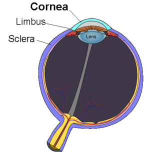
The cornea is the transparent front part of the eye that covers the iris, pupil, and anterior chamber. The cornea, with the anterior chamber and lens, refracts light, with the cornea accounting for approximately two-thirds of the eye's total optical power. In humans, the refractive power of the cornea is approximately 43 dioptres. The cornea can be reshaped by surgical procedures such as LASIK.

Eye surgery Surgery performed on the eye or its adnexa

Eye surgery, also known as ocular surgery, is surgery performed on the eye or its adnexa, typically by an ophthalmologist. The eye is a very fragile organ, and requires extreme care before, during, and after a surgical procedure to minimise or prevent further damage. An expert eye surgeon is responsible for selecting the appropriate surgical procedure for the patient, and for taking the necessary safety precautions. Mentions of eye surgery can be found in several ancient texts dating back as early as 1800 BC, with cataract treatment starting in the fifth century BC. Today it continues to be a widely practiced type of surgery, with various techniques having been developed for treating eye problems.

Corneal endothelium

The corneal endothelium is a single layer of endothelial cells on the inner surface of the cornea. It faces the chamber formed between the cornea and the iris.

Red eye (medicine) Eye that appears red due to illness or injury

A red eye is an eye that appears red due to illness or injury. It is usually injection and prominence of the superficial blood vessels of the conjunctiva, which may be caused by disorders of these or adjacent structures. Conjunctivitis and subconjunctival hemorrhage are two of the less serious but more common causes.

Corneal transplantation Surgical procedure

Corneal transplantation, also known as corneal grafting, is a surgical procedure where a damaged or diseased cornea is replaced by donated corneal tissue. When the entire cornea is replaced it is known as penetrating keratoplasty and when only part of the cornea is replaced it is known as lamellar keratoplasty. Keratoplasty simply means surgery to the cornea. The graft is taken from a recently deceased individual with no known diseases or other factors that may affect the chance of survival of the donated tissue or the health of the recipient.

Eye banks recover, prepare and deliver donated eyes for cornea transplants and research. The first successful cornea transplant was performed in 1905 and the first eye bank was founded in 1944. Currently, in the United States, eye banks provide tissue for over 80,000 cornea transplants each year to treat conditions such as keratoconus and corneal scarring. In some cases, the white of the eye (sclera) is used to surgically repair recipient eyes. Unlike other organs and tissues, there is an adequate supply of corneas for transplants in the United States, and excess tissue is exported internationally, where there are shortages in many countries, due to greater demand and a less-developed eye banking infrastructure.

Corneal ulcers in animals

A corneal ulcer, or ulcerative keratitis, is an inflammatory condition of the cornea involving loss of its outer layer. It is very common in dogs and is sometimes seen in cats. In veterinary medicine, the term corneal ulcer is a generic name for any condition involving the loss of the outer layer of the cornea, and as such is used to describe conditions with both inflammatory and traumatic causes.

Vernal keratoconjunctivitis Medical condition

Vernal keratoconjunctivitis (VKC) is a recurrent, bilateral, and self-limiting inflammation of conjunctiva, having a periodic seasonal incidence.

Pinguecula Medical condition

A pinguecula is a common type of conjunctival stromal degeneration in the eye. It appear as an elevated yellow-white plaque in the bulbar conjunctiva near to limbus. Calcification may also seen occasionally.

Corneal neovascularization Medical condition

Corneal neovascularization (CNV) is the in-growth of new blood vessels from the pericorneal plexus into avascular corneal tissue as a result of oxygen deprivation. Maintaining avascularity of the corneal stroma is an important aspect of corneal pathophysiology as it is required for corneal transparency and optimal vision. A decrease in corneal transparency causes visual acuity deterioration. Corneal tissue is avascular in nature and the presence of vascularization, which can be deep or superficial, is always pathologically related.

Corneal ulcer Medical condition

Corneal ulcer is an inflammatory or, more seriously, infective condition of the cornea involving disruption of its epithelial layer with involvement of the corneal stroma. It is a common condition in humans particularly in the tropics and the agrarian societies. In developing countries, children afflicted by Vitamin A deficiency are at high risk for corneal ulcer and may become blind in both eyes, which may persist lifelong. In ophthalmology, a corneal ulcer usually refers to having an infectious cause while the term corneal abrasion refers more to physical abrasions.

Pellucid marginal degeneration Degenerative corneal condition

Pellucid marginal degeneration (PMD), is a degenerative corneal condition, often confused with keratoconus. It typically presents with painless vision loss affecting both eyes. Rarely, it may cause acute vision loss with severe pain due to perforation of the cornea. It is typically characterized by a clear, bilateral thinning (ectasia) in the inferior and peripheral region of the cornea, although some cases affect only one eye. The cause of the disease remains unclear.

Pterygium

Pterygium refers to any winglike triangular membrane occurring in the neck, eyes, knees, elbows, ankles or digits.

Conjunctivochalasis Medical condition

Conjunctivochalasis is a common eye surface condition characterized by the presence of excess folds of the conjunctiva located between the globe of the eye and the eyelid margin.

Limbal stem cell

Limbal stem cells, also known as corneal epithelial stem cells, are stem cells located in the basal epithelial layer of the corneal limbus. They form the border between the cornea and the sclera. Characteristics of limbal stem cells include a slow turnover rate, high proliferative potential, clonogenicity, expression of stem cell markers, as well as the ability to regenerate the entire corneal epithelium. Limbal stem cell proliferation has the role of maintaining the cornea; for example, by replacing cells that are lost via tears. Additionally, these cells also prevent the conjunctival epithelial cells from migrating onto the surface of the cornea.

Pre Descemet's endothelial keratoplasty (PDEK) is a kind of endothelial keratoplasty, where the pre descemet's layer (PDL) along with descemet's membrane (DM) and endothelium is transplanted. Conventionally in a corneal transplantation, doctors use a whole cornea or parts of the five layers of the cornea to perform correction surgeries. In May 2013, Dr Harminder Dua discovered a sixth layer between the stroma and the descemet membrane which was named after him as the Dua's layer. In the PDEK technique, doctors take the innermost two layers of the cornea, along with the Dua's layer and graft it in the patient's eye.

Corneal opacity Medical condition

The human cornea is a transparent membrane which allows light to pass through it. The word corneal opacification literally means loss of normal transparency of cornea. The term corneal opacity is used particularly for the loss of transparency of cornea due to scarring. Transparency of the cornea is dependent on the uniform diameter and the regular spacing and arrangement of the collagen fibrils within the stroma. Alterations in the spacing of collagen fibrils in a variety of conditions including corneal edema, scars, and macular corneal dystrophy is clinically manifested as corneal opacity. The term "corneal blindness" is commonly used to describe blindness due to corneal opacity.

Pseudopterygium is the conjunctival adhesion to cornea caused by limbal or corneal inflammation or trauma. The pseudopterygium can be easily distinguished from pterygium by bowman's probe test. Because of the lack of adherence of pseudopterygium at every point, the probe can be passed beneath it at some point.

Exposure keratopathy is medical condition affecting the cornea of eyes. It can lead to corneal ulceration and permanent loss of vision due to corneal opacity.

References

  1. Tollefsbol, Trygve (2016). Medical Epigenetics. Academic Press. p. 395. ISBN   9780128032404. Archived from the original on 22 October 2016.
  2. 1 2 3 4 5 6 7 8 9 10 11 12 13 14 15 16 17 "Facts About the Cornea and Corneal Disease | National Eye Institute". The National Eye Institute (NEI). May 2016. Archived from the original on 16 April 2017. Retrieved 16 April 2017.
  3. 1 2 3 Yanoff, Myron; Duker, Jay S. (2009). Ophthalmology. Elsevier Health Sciences. p. 364. ISBN   978-0323043328. Archived from the original on 16 April 2017.
  4. 1 2 Anguria, P; Kitinya, J; Ntuli, S; Carmichael, T (2014). "The role of heredity in pterygium development". International Journal of Ophthalmology. 7 (3): 563–73. doi:10.3980/j.issn.2222-3959.2014.03.31. PMC   4067677 . PMID   24967209.
  5. 1 2 3 Smolin, Gilbert; Foster, Charles Stephen; Azar, Dimitri T.; Dohlman, Claes H. (2005). Smolin and Thoft's The Cornea: Scientific Foundations and Clinical Practice. Lippincott Williams & Wilkins. pp. 1003, 1005. ISBN   9780781742061. Archived from the original on 16 April 2017.
  6. 1 2 3 Halperin, Edward C.; Perez, Carlos A.; Brady, Luther W. (2008). Perez and Brady's Principles and Practice of Radiation Oncology. Lippincott Williams & Wilkins. p. 778. ISBN   9780781763691. Archived from the original on 16 April 2017.
  7. 1 2 3 4 Droutsas, K; Sekundo, W (June 2010). "[Epidemiology of pterygium. A review]". Der Ophthalmologe : Zeitschrift der Deutschen Ophthalmologischen Gesellschaft (in German). 107 (6): 511–2, 514–6. doi:10.1007/s00347-009-2101-3. PMID   20393731. S2CID   12077189.
  8. Saw, SM; Tan, D (September 1999). "Pterygium: prevalence, demography and risk factors". Ophthalmic Epidemiology. 6 (3): 219–28. doi:10.1076/opep.6.3.219.1504. PMID   10487976.
  9. 1 2 Coroneo, MT (November 1993). "Pterygium as an early indicator of ultraviolet insolation: a hypothesis". Br J Ophthalmol. 77 (11): 734–9. doi:10.1136/bjo.77.11.734. PMC   504636 . PMID   8280691.
  10. 1 2 Kunimoto, Derek; Kunal Kanitkar; Mary Makar (2004). The Wills eye manual: office and emergency room diagnosis and treatment of eye disease (4th ed.). Philadelphia, PA: Lippincott Williams & Wilkins. pp. 50–51. ISBN   978-0781742078.
  11. Fisher, J.P.; Trattler, W.B. (12 January 2009). "Pterygium". Archived from the original on 17 March 2010.Cite journal requires |journal= (help)
  12. Klintworth, G; Cummings, T (26 August 2009). "24; The eye and ocular adnexa". In Stacey, Mills (ed.). Sternberg's Diagnostic Surgical Pathology (5th ed.). ISBN   978-0-7817-7942-5.
  13. "Paramount Books Online Bookstore 9789696370017 : Concise-Ophthalmology-(pb)-2014". Archived from the original on 30 July 2014. Retrieved 9 August 2014.
  14. Gulani, A; Dastur, YK (January–March 1995). "Simultaneous pterygium and cataract surgery". Journal of Postgraduate Medicine. 41 (1): 8–11. PMID   10740692. Archived from the original on 8 February 2012. Retrieved 30 November 2012.
  15. "Pterygium: MedlinePlus Medical Encyclopedia". medlineplus.gov. Archived from the original on 28 August 2016. Retrieved 15 August 2016.
  16. "Pterygium Workup: Imaging Studies, Procedures". emedicine.medscape.com. Archived from the original on 11 September 2016. Retrieved 15 August 2016.
  17. "Pinguecula - EyeWiki". eyewiki.aao.org.
  18. 1 2 John F, Salmon (13 December 2019). "Conjunctiva". Kanski's clinical ophthalmology : a systematic approach (9th ed.). Elsevier. p. 198. ISBN   978-0-7020-7711-1.
  19. Mackenzie F.D., Hirst L.W., Battistutta D., Green A. (1992). "Risk Analysis in the Development of Pterygia". Ophthalmology. 99 (7): 1056–1061. doi:10.1016/s0161-6420(92)31850-0. PMID   1495784.CS1 maint: multiple names: authors list (link)
  20. Lee G.A., Hirst L.W., Sheehan M. (1994). "1994 Knowledge of Sunlight Effects on the Eyes and Protective Behaviours in the General Community". Ophthalmic Epidemiology. 1 (2): 67–84. doi:10.3109/09286589409052363. PMID   8790614. S2CID   22382822.CS1 maint: multiple names: authors list (link)
  21. Lee G., Hirst L.W., Sheehan M. (1999). "1999 Knowledge of Sunlight effects on the eyes and protective behaviors in adolescents". Ophthalmic Epidemiology. 6 (3): 171–180. doi:10.1076/opep.6.3.171.1501. PMID   10487972.CS1 maint: multiple names: authors list (link)
  22. Martins, TG; Costa, AL; Alves, MR; Chammas, R; Schor, P (2016). "Mitomycin C in pterygium treatment". International Journal of Ophthalmology. 9 (3): 465–8. doi:10.18240/ijo.2016.03.25. PMC   4844053 . PMID   27158622.
  23. 1 2 3 Clearfield E, Muthappan V, Wang X, Kuo IC (2016). "Conjunctival autograft for pterygium". Cochrane Database Syst Rev. 2: CD011349. doi:10.1002/14651858.CD011349.pub2. PMC   5032146 . PMID   26867004.
  24. Ali, AM; Thariat, J; Bensadoun, RJ; Thyss, A; Rostom, Y; El-Haddad, S; Gérard, JP (April 2011). "The role of radiotherapy in the treatment of pterygium: a review of the literature including more than 6000 treated lesions". Cancer/Radiothérapie. 15 (2): 140–7. doi:10.1016/j.canrad.2010.03.020. PMID   20674450.
  25. Myron, Yanoff; Jay S., Duker (2019). "Cornea and ocular surface diseases". Ophthalmology (5th ed.). Edinburgh: Elsevier. p. 310. ISBN   978-0-323-52821-4. OCLC   1051774434.
  26. Gulani AC (2007). "Corneoplastique". Techniques in Ophthalmology. 5 (1): 11–20. doi:10.1097/ito.0b013e318036ae0d.
  27. Gulani AC. "Corneoplastique", Video Journal of Cataract and Refractive Surgery. Volume XXII. Issue 3, 2006.
  28. Gulani AC (2006). "A New Concept for Refractive Surgery". Ophthalmology Management. 10 (4): 57–63.
  29. Prabhasawat P, Barton K, Burkett G, Tseng SC (June 1997). "Comparison of conjunctival autografts, amniotic membrane grafts, and primary closure for pterygium excision". Ophthalmology. 104 (6): 974–85. doi:10.1016/s0161-6420(97)30197-3. PMID   9186439.CS1 maint: multiple names: authors list (link)
Classification
D
External resources