The scleral ring or sclerotic ring is a hardened ring of plates, often derived from bone, that is found in the eyes of many animals in several groups of vertebrates. Mammals, amphibians, and crocodilians lack scleral rings.[1] The ring is in the fibrous outer layer of the eye, called the sclera. The structure is commonly referred to as the sclerotic ring; but, because the word sclerotic often implies pathology of the sclera (see "sclerosis", an unrelated medical condition[2]), recent authors have urged avoiding the use of this term, to avoid confusion and to increase the utility of character comparisons.[3]
Scleral rings can be made of cartilaginous material (scleral cartilage) or bony material (scleral ossicles), or often a combination of both, that comes together to form a ring.[3] The arrangement, size, shape, and number of ossicles vary by group.[2] They are believed to have a role in supporting the eye, especially in animals whose eyes are not spherical, or which live underwater.[1]Fossil scleral rings are known for a variety of extinct animals, including ichthyosaurs, pterosaurs, and non-avian dinosaurs,[4][5] but are often not preserved.
Scleral rings may help support inner structures of the eye, especially in animals that do not have round eyes. Animals that move rapidly, including both fast flying birds and fast swimming fish have the most robust scleral rings, indicating that these thick rings are used to protect the eye during intense changes in pressure in the air and in the water.[2] Additionally, scleral rings may help the eye adjust to different viewing distances, also known as visual accommodation. Muscles are used to adjust the shape of the eye for accommodation, and the rings provide attachment sites for these muscles. In aquatic animals, the lens is squeezed in a different way to compensate for differences in light refraction underwater, and so the shape of the ring can be different than those in terrestrial animals.[2]
Extant Animals
Reptiles
A combination of scleral cartilage and ossicles are present, in which the cartilage acts as a cup around the posterior (rear) position of the eye and ossicles at the anterior (front) position of the eye form the ring.[3]
Within Lepidosaurs (snakes, lizards, tuatara, and relatives), scleral rings have been found in all major lineages except Serpentes, or snakes, and two families within Anguimorpha: Dibamidae and Rhineuridae, which are both legless lizard families.[3] All of these clades that lack a scleral ring share either a burrowing lifestyle or lack of limbs, indicating a possible correlation among these traits and loss of the scleral ring. Lizards typically have 14 ossicles in the ring, though this can vary.[2]
Within Archelosauria (turtles, birds, crocodilians, and relatives), only birds and turtles retain the scleral rings. Fossil evidence shows that extinct marine crocodiles living in the Mesozoic had scleral rings, so the trait was lost over time.[6] Scleral rings of varying lengths, curvatures, numbers of ossicles, and thickness are found in all birds.[7] Birds typically have 12-18 ossicles, with 14 being the most common number.[2]
Teleost fish typically have only one or two ossicles per ring, and fish with no ossicles still retain cartilage.[8] Most teleosts do not have ossicles, but this can vary even within groups.[8] As a general trend, more basal groups (such as Elopomorpha and Osteoglossomorpha) tend to have no ossicles, while more derived groups (such as Percomorpha) are likely to have a variable number of ossicles (zero to two).[8]
More active fish are more likely to have scleral rings, indicating that the rings help keep the eye stable during rapid swimming.[8]
Gallery
A skull of an extant tawny frogmouth, showing large scleral rings.
1 2 Motani, Ryosuke (15 November 2001). "Eyes of Ichthyosaurs". UC Museum of Paleontology. Archived from the original on 17 December 2001. Retrieved 15 October 2013.
Parentheses denote bones that receive a different name in particular clades
Italics denote neomorphic bones present only in particular clades
This page is based on this Wikipedia article Text is available under the CC BY-SA 4.0 license; additional terms may apply. Images, videos and audio are available under their respective licenses.
 A skull of an extant tawny frogmouth, showing large scleral rings.
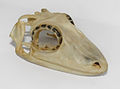 A skull of an extant satanic leaf-tailed gecko, showing large scleral rings.
 Virtually complete scleral ring of the ichthyosaur Ophthalmosaurus
 Partial scleral ring of Mosasaurus .
 Scleral ring of the hadrosaur Prosaurolophus .


