Tendinosis

Last updated
Tendinosis
Classification and external resources
Specialty rheumatology
ICD-10 M67.9
MeSH D052256

Tendinosis means and is sometimes called chronic tendinitis, chronic tendinopathy, or chronic tendon injury. It is damage to a tendon at a cellular level (the suffix "osis" implies a pathology of chronic degeneration without inflammation). It is thought to be caused by microtears in the connective tissue in and around the tendon, leading to an increase in tendon repair cells. This may lead to reduced tensile strength, thus increasing the chance of tendon rupture. Tendinosis is often misdiagnosed as tendinitis because of the limited understanding of tendinopathies by the medical community. [1] Classic characteristics of "tendinosis" include degenerative changes in the collagenous matrix, hypercellularity, hypervascularity, and a lack of inflammatory cells which has challenged the original misnomer "tendinitis". [2] [3]

Tendinopathy Bruised tendon

Tendinopathy, also known as tendinitis or tendinosis, is a type of tendon disorder that results in pain, swelling, and impaired function. The pain is typically worse with movement. It most common occurs around the shoulder, elbow, wrist, hip, knee, or ankle.

Tendon type of tissue that connects muscle to bone

A tendon or sinew is a tough band of fibrous connective tissue that usually connects muscle to bone and is capable of withstanding tension.

Contents

Causes

Tendinosis of the common extensor tendon of the elbow (“tennis elbow”), as of the Rotator Cuff, is a common cause of pain in the elbow or shoulder. [4] The general opinion is that tendinosis is due to tendon overuse, and failed healing of the tendon. [5] In addition, the extensor carpi radialis brevis muscle plays a key role. [6]

Tennis elbow condition in which the outer part of the elbow becomes sore and tender

Tennis elbow, also known as lateral epicondylitis, is a condition in which the outer part of the elbow becomes painful and tender. The pain may also extend into the back of the forearm and grip strength may be weak. Onset of symptoms is generally gradual. Golfer's elbow is a similar condition that affects the inside of the elbow.

Pathophysiology

Histological findings include granulation tissue, microrupture, degenerative changes, and there is no traditional inflammation. As a consequence, “lateral elbow tendinopathy or tendinosis” is used instead of “lateral epicondylitis”. [7]

Examination of tennis elbow tissue reveals noninflammatory tissue, so the term “angiofibroblastic tendinosis” is used. [8]

Cultures from tendinopathic tendons contain an increased production of type III collagen. [9] [10]

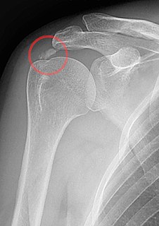
Longitudinal sonogram of the lateral elbow displays thickening and heterogeneity of the common extensor tendon that is consistent with tendinosis, as the ultrasound reveals calcifications, intrasubstance tears, and marked irregularity of the lateral epicondyle. Although the term “epicondylitis” is frequently used to describe this disorder, most histopathologic findings of studies have displayed no evidence of an acute, or a chronic inflammatory process. Histologic studies have demonstrated that this condition is the result of tendon degeneration, which causes normal tissue to be replaced by a disorganized arrangement of collagen. Therefore, the disorder is more appropriately referred to as “tendinosis” or “tendinopathy” rather than “tendinitis.” [11]

Colour Doppler ultrasound reveals structural tendon changes, with vascularity and hypo-echoic areas that correspond to the areas of pain in the extensor origin. [12]

Load-induced non-rupture tendinopathy in humans is associated with an increase in the ratio of collagen III:I proteins, a shift from large to small diameter collagen fibrils, buckling of the collagen fascicles in the tendon extracellular matrix, and buckling of the tenocyte cells and their nuclei. [13]

Diagnosis

Symptoms can vary from an ache or pain and stiffness to the local area of the tendon, or a burning that surrounds the whole joint around the affected tendon. With this condition, the pain is usually worse during and after activity, and the tendon and joint area can become stiffer the following day as swelling impinges on the movement of the tendon. Many patients report stressful situations in their life in correlation with the beginnings of pain, which may contribute to the symptoms.

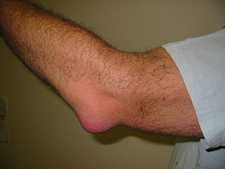
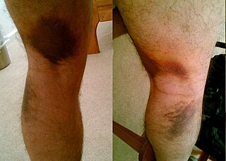
Swelling in a region of micro damage or partial tear may be detected visually or by touch.

Medical imaging

Ultrasound imaging can be used to evaluate tissue strain, as well as other mechanical properties. [14]

Ultrasound-based techniques are becoming more popular because of its affordability, safety, and speed. Ultrasound can be used for imaging tissues, and the sound waves can also provide information about the mechanical state of the tissue. [15]

Increased water content and disorganized collagen matrix in tendon lesions may be detected by ultrasonography or magnetic resonance imaging.

Treatment

An injured tendon is very slow to heal. Partial tears heal by the rapid production of disorganized type-III collagen which is weaker than normal tendon.[ citation needed ] Recurrence of injury in the damaged region of the tendon is common.

Rehabilitation, rest, and gradual return to the activity in which tendinosis was experienced is a common therapy. There is evidence to suggest that tendinosis is not an inflammatory disorder; anti-inflammatory drugs are not an effective treatment; [16] and inflammation is not the cause of this type of tendon dysfunction. [17] There is a variety of treatment options but more research is necessary to determine their effectiveness. Initial recovery is usually within 2 to 3 months, and full recovery usually within 3 to 6 months. About 80% of patients will fully recover within 12 months. [18] If the conservative therapy doesn't work, then surgery can be an option. This surgery consists of the excision of abnormal tissue. Time required to recover from surgery is about 4 to 6 months. [19] Studies with a rat model of fatigue-damaged tendons suggested that delaying exercise until after the initial inflammatory stage of repair could promote remodelling more rapidly. [20]

Research

Eccentric loading and extracorporeal shockwave therapy are currently being researched as possible treatments for tendinosis. One study found the two modalities to be equally effective in treating tendinosis of the Achilles tendon and more effective than a 'wait and see' approach. [21] Other treatments for which research is on-going include vitamin E, vitamin B6, nitric oxide, Platelet Rich Plasma (PRP), [22] and stem cell injections.

Vitamin C

One study found increased Achilles tendon healing in rats supplemented with high doses of vitamin C, which is needed for collagen synthesis. [23]

Vitamin E

Vitamin E has been found to increase the activity of fibroblasts, leading to increased collagen fibrils and synthesis, which seems to speed up the regeneration and increase the regenerative capacity of tendons. [24] [25]

Nitric oxide

Nitric oxide (NO) also appears to play a role in tendon healing [26] and inhibition of its synthesis impairs tendon healing. [27] The use of a nitric oxide delivery system (glyceryl trinitrate patches) applied over the area of maximal tenderness was tested in three clinical trials for the treatment of tendinopathies and was found to significantly reduce pain and increase range of motion and strength. [28]

One study found that arginine supplementation, which may possibly increase NO signaling, [29] was able to increase the speed of tendon healing [30]

Soft tissue mobilization

Augmented Soft Tissue Mobilization (ASTM) is a form of manual therapy that has been shown in studies on rats to speed the healing of tendons by increasing fibroblast activity. [31] [32] One case study showed ASTM resulting in full recovery in the case of an athlete suffering from chronic ankle pain and fibrosis, after an unsuccessful course of surgery and conventional physical therapy. [33]

Eccentric loading

A promising line of therapy involves eccentric loading exercises involving lengthening muscular contractions. [34]

Inflatable brace

The use of an inflatable brace (AirHeel) was shown to be as effective as eccentric loading in the treatment of chronic Achilles tendinopathy. Both modalities produced significant reduction in pain scores, but their combination was no more effective than either treatment alone. [35]

Shock-wave therapy

Shock-wave therapy (SWT) may be effective in treating calcific tendinosis in both humans [36] and rats. [37] In rat subjects, SWT increased levels of healing hormones and proteins leading to increased cell proliferation and tissue regeneration in tendons. Another study found no evidence that SWT was useful in treating chronic pain in the Achilles tendon. [38]

Tendon bioengineering

The future of non-surgical care for tendinosis is likely bioengineering. Ligament reconstruction is possible using mesenchymal stem cells and a silk scaffold. [39] These same stem cells were capable of seeding repair of damaged animal tendons. [40] Autologous tenocyte implantation is currently being tested for tendinosis. There is a large randomized, double-blind, placebo controlled trial being conducted in the Netherlands to test the safety and efficacy of tenocyte therapy. Results from the trial are expected by April 2013.

Autologous tenocyte injection

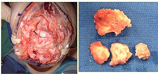
A study investigated autologous tenocyte injection for the treatment of severe, chronic resistant lateral epicondylitis. A needle biopsy was used on the patellar tendon, and the extracted tendon cells were expanded by in vitro culture. The autologous tenocytes were sorted and purified by real-time polymerase chain reaction, and amplified by flow cytometry. The tenocytes were then injected into the injured tendinopathy site, which was the origin of the extensor carpi radialis brevis tendon, under the guidance of an ultrasound. After the autologous tenocyte injection treatment, patients with chronic lateral epicondylitis showed improved clinical function and structural repair at the origin of the common extensor tendon. [41]

Nonbulbar dermal sheath cells

RepliCel has planned a Phase 1/2 Achilles tendinosis clinical trial using fibroblasts that are isolated from the nonbulbar dermal sheath cells of a hair follicle, and is recruiting in Q1 2015. The tendon treatment will be tested in approximately 28 subjects. Nonbulbar dermal sheath cells are used because they produce more type I collagen than fibroblasts that are derived from adipose tissue. Type I collagen is the primary collagen in tendons. Nonbulbar dermal sheath cells will be replicated, and then reintroduced into the wounded tendons with ultrasound. After the injections, subjects will be assessed for pain, safety, and function, as well as changes in interstitial tears, tendon thickness, echotexture, and neovascularity. [42] [43]

Injections of microRNA

Scientists found that mir-29a, a single microRNA, through its interaction with a protein, interleukin 33, plays a key role in regulating the production of collagens in tendon disease. Loss of miR29a from human tendons results in an increase in collagen type-3 production, which is a key feature of tendon disease. The replacement of miR-29a in the damaged tendon cells in the laboratory restores collagen production to pre injury levels. A trial will put injections of microRNA – small molecules that help regulate gene expression – into the tendon to decrease the production of type 3 collagen and switch to type-1. [44]

Allogenic adipose-derived mesenchymal stem cells

As of November 2013, researchers at the Seoul National University Hospital will be looking to recruit participants into a clinical trial to evaluate the efficacy of allogenic adipose-derived mesenchymal stem cells (ALLO-ASC) for the treatment of a lateral epicondylitis tendon injury where the duration of the symptoms is over six months. Adipose-derived mesenchymal stem cells will be administrated to the patients with lateral epicondylitis (tennis elbow) by an ultrasonographic-guided injection. [45]

Elastography ultrasound

Researchers have tried to analyze tissue strain and mechanical properties using elastography, which is an acoustical imaging technique that measures strain distributions in tissues that result from stress or compression of the tissue. Strain is inversely related to stiffness, so under a given amount of stress, tissue that displays less strain is assumed to be stiffer than tissue that exhibits more strain. Elastography is therefore an indirect method to estimate tissue stiffness. [14]

One limitation of elastography is that it is inherently linear when ultrasound wave velocity and the material properties do not change during the strain measurement. This is a problem in soft tissues like tendons, as they are nonlinear in stiffness, and can undergo large deformations in activity. Elastography measures strain, and to more completely described the mechanical behavior, more data, like stiffness or stress would be required. [14]

Acoustoelastography ultrasound

Acoustoelastic theory is based on the principle that the acoustic properties of a material are altered as the material is loaded and deformed. The properties can be measured as a change in amplitude and wave velocity. [15]

With the use of A-mode ultrasound, researchers have derived an acoustic relationship between reflected wave amplitude, and strain-dependent stiffness and stress in a deformed material. [15]

As tendon tension increases, the intensity of reflected ultrasonic echoes increases. The increased intensity results in a brighter B-mode ultrasound image. [14]

Acoustoelastography is an ultrasound technique that relates ultrasonic wave amplitude changes to the mechanical properties of a tendon. [46]

It is an ultrasound-based model that can be used evaluate tendon function. Gradual deformations of a tendon can produce cine loops, where changes in echo intensity can be observed. By analyzing the echo intensity changes with Acoustoelastography, one can deduce the stiffness gradient, which is the rate of change of normalised stiffness as a function of strain. [47]

EchoSoft ultrasound software from the Wisconsin Alumni Research Foundation and Echometrix applies the theory of acoustoelasticity to measure musculoskeletal (tendon and ligament) tissue. The software examines previously unused information found by ultrasound waves to quantify the extent of musculoskeletal injuries, or a patient's progress in a healing process. [48]

Ultrasonic Percutaneous Tenotomy

Percutaneous ultrasonic tenotomy provided continued pain relief and functional improvement for recalcitrant tennis elbow at a 3-year follow-up. [49]

Other animals

Bowed tendon is a horseman's term for tendinitis (inflammation) and tendinosis (degeneration), most commonly seen in the superficial digital flexor tendon in the front leg of horses.

Diagnosis

When the superficial digital flexor tendon of horses is damaged, there is a thickening of the tendon, giving it a bowed appearance when the leg is viewed from the side.

Medical imaging

A study tested the repeatability and feasibility of using acoustoelastography for in vivo measurement of stiffness gradients in the superficial digital flexor tendons (SDFTs) of clinically normal horses. The results show that acoustoelastography is a repeatable and feasible technique for measuring stiffness gradients of superficial digital flexor tendons in clinically normal horses, and acoustoelastography has the potential to be used for comparing diseased and healthy tendon states. [50]

A report describes the use of acoustoelastography to monitor the mechanical healing of an Achilles tendon laceration in a dog after suture repair. Serial acoustoelastography examinations of the tendon showed that mechanical properties improved throughout the recovery period. At 29 weeks, the mechanical properties of the repaired tendon were similar to that of the normal contralateral Achilles mechanism. [46]

A study shows that acoustoelastography is a repeatable and feasible method for measuring stiffness gradients in equine superficial digital flexor tendons. [47]

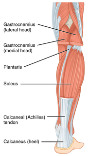
The acoustoelastic strain gauge is an ultrasound-based tissue evaluation technique that relates echo intensity changes that are observed during the stretching or relaxation tendons to the mechanical properties of the tissue. The method deduces stiffness gradient (the rate of change of normalized stiffness as a function of tissue strain) by evaluating the ultrasound dynamic images that are captured from tissue as it gradually deforms. Acoustoelastic strain gauge has been shown to accurately model stiffness and strain within tendons in vitro. To determine the repeatability and feasibility of in vivo ASG measurements of canine tendon function, stiffness gradients for the gastrocnemius tendons of dogs were recorded. Findings indicate that acoustoelastic strain gauge is a repeatable and feasible technique for measuring stiffness gradients in canine tendons. [51]

Pathophysiology

Achilles tendons in rats were studied with acoustoelastography ultrasound. After a tendon injury, and during tendon healing, vascularity changes and cellular activity are vital to the formation of granular tissue in the tendon gap, and the subsequent development of neo-tendinous tissue that replaces damaged native tissue. Normal, intact tendon is composed mainly of type I collagen, and type III collagen increases after injury. Another study has recorded an association between collagen fibers and echogenicity tendon during Achilles tendon healing. [52]

This report further indicates that a decrease in type I procollagen (and increase in type III collagen and periostin) correlates to reduced strength, echo intensity, and normalized stiffness. It shows that tissue normalized stiffness is linearly correlated to procollagen I, while echo intensity is seen to be nonlinearly correlated to type I procollagen. Furthermore, the increase in M1 macrophages, blood vessels, and proliferating cells that occur within two weeks of the injury are associated with a formation of granulation tissue. Ultimate stress, echo intensity, and normalized stiffness are all their lowest during these times. The results assert that the reduction in stress and normalized stiffness that is measured by ultrasonic and mechanical methods correlate well with the biological aspect of tendon healing. [52]

Treatment

Mesenchymal stem cells, derived from a horse's bone marrow or fat, are currently being used for tendon repair in horses. [53]

Confusion between tendonitis and tendinosis [54]

Tendonitis is a very common, but misleading term. By definition, the suffix "-itis" means "inflammation of". Inflammation [55] is the body's local response to tissue damage which involves red blood cells, white blood cells, blood proteins with dilation of blood vessels around the site of injury. Tendons are relatively avascular. [56] [54] Corticosteroids are drugs that reduce inflammation. Corticosteroids can be useful to relieve chronic tendinopathy pain, improve function, and reduce swelling in the short term. However, there is a greater risk of long-term recurrence. [57] They are typically injected along with a small amount of a numbing drug called lidocaine. Research shows that tendons are weaker following corticosteroid injections. Tendinitis is still a very common diagnosis, though research increasingly documents that what is thought to be tendinitis is usually tendinosis. [58]

See also

Related Research Articles

Rheumatology is a branch of medicine devoted to the diagnosis and therapy of rheumatic diseases. Physicians who have undergone formal training in rheumatology are called rheumatologists. Rheumatologists deal mainly with immune-mediated disorders of the musculoskeletal system, soft tissues, autoimmune diseases, vasculitides, and heritable connective tissue disorders.

De Quervain syndrome human disease

De Quervain syndrome is inflammation of two tendons that control movement of the thumb and their tendon sheath. This results in pain at the outside of the wrist. Pain is typically increased with gripping or rotating the wrist. The thumb may also be difficult to move smoothly. Onset of symptoms is gradual.

Shoulder problem

Shoulder problems including pain, are one of the more common reasons for physician visits for musculoskeletal symptoms. The shoulder is the most movable joint in the body. However, it is an unstable joint because of the range of motion allowed. This instability increases the likelihood of joint injury, often leading to a degenerative process in which tissues break down and no longer function well.

Achilles tendon type of tendon in the lower leg

The Achilles tendon or heel cord, also known as the calcaneal tendon, is a tendon of the back of the leg, and the thickest in the human body. It serves to attach the plantaris, gastrocnemius (calf) and soleus muscles to the calcaneus (heel) bone. These muscles, acting via the tendon, cause plantar flexion of the foot at the ankle, and flexion at the knee.

Bursitis human disease

Bursitis is the inflammation of one or more bursae of synovial fluid in the body. They are lined with a synovial membrane that secretes a lubricating synovial fluid. There are more than 150 bursae in the human body. The bursae rest at the points where internal functionaries, such as muscles and tendons, slide across bone. Healthy bursae create a smooth, almost frictionless functional gliding surface making normal movement painless. When bursitis occurs, however, movement relying on the inflamed bursa becomes difficult and painful. Moreover, movement of tendons and muscles over the inflamed bursa aggravates its inflammation, perpetuating the problem. Muscle can also be stiffened.

Plantar fasciitis Human disease

Plantar fasciitis is a disorder of the connective tissue which supports the arch of the foot. It results in pain in the heel and bottom of the foot that is usually most severe with the first steps of the day or following a period of rest. Pain is also frequently brought on by bending the foot and toes up towards the shin. The pain typically comes on gradually, and it affects both feet in about one third of cases.

Achilles tendinitis tendonitis of the Achilles tendon

Achilles tendinitis, also known as achilles tendinopathy, is when the Achilles tendon, found at the back of the ankle, becomes irritated. The most common symptoms are pain and swelling around the affected tendon. The pain is typically worse at the start of exercise and decreases thereafter. Stiffness of the ankle may also be present. Onset is generally gradual.

Rotator cuff tear Rotator cuff tear

A rotator cuff tear is an injury of one or more of the tendons or muscles of the rotator cuff of the shoulder. Symptoms may include shoulder pain, which is often worse with movement, or weakness. This may limit peoples’ ability to brush their hair or put on clothing. Clicking may also occur with movement of the arm.

Adhesive capsulitis of shoulder Human disease:  painful and disabling disorder of unclear cause in which the shoulder capsule

Adhesive capsulitis is a painful and disabling disorder of unclear cause in which the shoulder capsule, the connective tissue surrounding the glenohumeral joint of the shoulder, becomes inflamed and stiff, greatly restricting motion and causing chronic pain. Pain is usually constant, worse at night, and with cold weather. Certain movements or bumps can provoke episodes of tremendous pain and cramping. The condition is thought to be caused by injury or trauma to the area and may have an autoimmune component.

Prolotherapy, also called proliferation therapy is an injection-based treatment used in chronic musculoskeletal conditions. It has been characterised as an alternative medicine practice.

Achilles tendon rupture

Achilles tendon rupture is when the Achilles tendon, at the back of the ankle, breaks. Symptoms include the sudden onset of sharp pain in the heel. A snapping sound may be heard as the tendon breaks and walking becomes difficult.

Patellar tendinitis Human disease

Patellar tendinitis, also known as jumper's knee, is a common cause of knee pain among athletes. It is a repetitive strain injury to the tendon connecting the patella, or kneecap, with the shinbone. Jumper's knee commonly occurs in athletes who are involved in jumping sports such as basketball and volleyball, but can affect any athlete.

Synovitis is the medical term for inflammation of the synovial membrane. This membrane lines joints that possess cavities, known as synovial joints. The condition is usually painful, particularly when the joint is moved. The joint usually swells due to synovial fluid collection.

Strain (injury) injury to a muscle in which the muscle fibers tear as a result of overstretching

A strain is an acute or chronic soft tissue injury that occurs to a muscle, tendon, or both. The equivalent injury to a ligament is a sprain.

Subacromial bursitis

Subacromial bursitis is a condition caused by inflammation of the bursa that separates the superior surface of the supraspinatus tendon from the overlying coraco-acromial ligament, acromion, and coracoid and from the deep surface of the deltoid muscle. The subacromial bursa helps the motion of the supraspinatus tendon of the rotator cuff in activities such as overhead work.

Calcific tendinitis disorder characterized by deposits of hydroxyapatite (a crystalline calcium phosphate) in any tendon of the body

Calcific tendinitis is a form of tendinitis, a disorder characterized by deposits of hydroxyapatite in any tendon of the body, but most commonly in the tendons of the rotator cuff (shoulder), causing pain and inflammation. The condition is related to and may cause adhesive capsulitis.

Extracorporeal shockwave therapy

Extracorporeal shockwave therapy (ESWT) is a treatment mostly used to treat kidney stones and in physical therapy and orthopedics.

Post-traumatic arthritis

Post-traumatic arthritis (PTA) is a form of osteoarthritis following an injury to a joint.

References

  1. Murrell GA (December 2002). "Understanding tendinopathies". Br J Sports Med. 36 (6): 392–3. doi:10.1136/bjsm.36.6.392. PMC   1724561 Lock-green.svg. PMID   12453831.
  2. Fu SC, Rolf C, Cheuk YC, Lui PP, Chan KM (2010). "Deciphering the pathogenesis of tendinopathy: a three-stages process". Sports Med Arthrosc Rehabil Ther Technol. 2: 30. doi:10.1186/1758-2555-2-30. PMC   3006368 Lock-green.svg. PMID   21144004.
  3. Abate M, Silbernagel KG, Siljeholm C, Di Iorio A, De Amicis D, Salini V, Werner S, Paganelli R (2009). "Pathogenesis of tendinopathies: inflammation or degeneration?". Arthritis Research & Therapy . 11 (3): 235. doi:10.1186/ar2723. PMC   2714139 Lock-green.svg. PMID   19591655.
  4. Mohamadi, Amin; Chan, Jimmy J.; Claessen, Femke M. A. P.; Ring, David; Chen, Neal C. (January 2017). "Corticosteroid Injections Give Small and Transient Pain Relief in Rotator Cuff Tendinosis: A Meta-analysis". Clinical Orthopaedics and Related Research. 475 (1): 232–243. doi:10.1007/s11999-016-5002-1. ISSN   1528-1132. PMC   5174041 Lock-green.svg. PMID   27469590.
  5. Arroll B, Goodyear-Smith F (Mar 2005). "Corticosteroid injections for painful shoulder: a meta-analysis". Br J Gen Pract. 55 (512): 224–8. PMC   1463095 Lock-green.svg. PMID   15808040.
  6. Nirschl RP, Ashman ES (2004). "Tennis elbow tendinosis (epicondylitis)". Instr Course Lect. 53: 587–98. PMID   15116648.
  7. du Toit, C; Stieler, M; Saunders, R; Bisset, L; Vicenzino, B (2008). "Diagnostic accuracy of power Doppler ultrasound in patients with chronic tennis elbow". British Journal of Sports Medicine. 42 (11): 572–576. doi:10.1136/bjsm.2007.043901. ISSN   0306-3674.
  8. Nirschl RP (October 1992). "Elbow tendinosis/tennis elbow". Clin Sports Med. 11 (4): 851–70. PMID   1423702.
  9. Maffulli N, Ewen SW, Waterston SW, Reaper J, Barrass V (2000). "Tenocytes from ruptured and tendinopathic achilles tendons produce greater quantities of type III collagen than tenocytes from normal achilles tendons. An in vitro model of human tendon healing". Am J Sports Med. 28 (4): 499–505. doi:10.1177/03635465000280040901. PMID   10921640.
  10. Ho JO, Sawadkar P, Mudera V (2014). "A review on the use of cell therapy in the treatment of tendon disease and injuries". J Tissue Eng. 5: 2041731414549678. doi:10.1177/2041731414549678. PMC   4221986 Lock-green.svg. PMID   25383170.
  11. McShane JM, Nazarian LN, Harwood MI (October 2006). "Sonographically guided percutaneous needle tenotomy for treatment of common extensor tendinosis in the elbow". J Ultrasound Med. 25 (10): 1281–9. PMID   16998100.
  12. Zeisig, Eva; Öhberg, Lars; Alfredson, Håkan (2006). "Sclerosing polidocanol injections in chronic painful tennis elbow-promising results in a pilot study". Knee Surgery, Sports Traumatology, Arthroscopy. 14 (11): 1218–1224. doi:10.1007/s00167-006-0156-0. ISSN   0942-2056.
  13. Pingel J, Lu Y, Starborg T, Fredberg U, Langberg H, Nedergaard A, et al. (2014). "3-D ultrastructure and collagen composition of healthy and overloaded human tendon: evidence of tenocyte and matrix buckling". J Anat. 224 (5): 548–55. doi:10.1111/joa.12164. PMC   3981497 Lock-green.svg. PMID   24571576.
  14. 1 2 3 4 Duenwald S, Kobayashi H, Frisch K, Lakes R, Vanderby R (February 2011). "Ultrasound echo is related to stress and strain in tendon". J Biomech. 44 (3): 424–9. doi:10.1016/j.jbiomech.2010.09.033. PMC   3022962 Lock-green.svg. PMID   21030024.
  15. 1 2 3 Duenwald-Kuehl S, Lakes R, Vanderby R (June 2012). "Strain-induced damage reduces echo intensity changes in tendon during loading". J Biomech. 45 (9): 1607–11. doi:10.1016/j.jbiomech.2012.04.004. PMC   3358489 Lock-green.svg. PMID   22542220.
  16. Khan, K.M.; Cook, J.L.; Kannus, P.; Maffulli, N.; Bonar, S.F. (2002-03-16). "Time to abandon the "tendinitis" myth : Painful, overuse tendon conditions have a non-inflammatory pathology". British Medical Journal. 324 (7338): 626–7. doi:10.1136/bmj.324.7338.626. PMC   1122566 Lock-green.svg. PMID   11895810 . Retrieved 2007-04-02.
  17. Marsolais D, Duchesne E, Côté CH, Frenette J (2007). "Inflammatory cells do not decrease the ultimate tensile strength of intact tendons in vivo and in vitro: protective role of mechanical loading". J Appl Physiol. 102 (1): 3–4. doi:10.1152/japplphysiol.00162.2006. PMID   16916923.
  18. Wilson, J.J.; Best, T.M. (2005). "Common overuse tendon problems: A review and recommendations for treatment" (PDF). American Family Physician. American Academy of Family Physicians. 72 (5): 811–8. PMID   16156339. Archived from the original (PDF) on 2007-09-29. Retrieved 2007-04-02.
  19. David J. Magee, James E. Zachazewski, William S. Quillen Pathology and intervention in musculoskeletal rehabilitation
  20. Bell R, Boniello MR, Gendron NR, Flatow EL, Andarawis-Puri N (2015). "Delayed exercise promotes remodeling in sub-rupture fatigue damaged tendons". J Orthop Res. 33 (6): 919–25. doi:10.1002/jor.22856. PMC   4417076 Lock-green.svg. PMID   25732052.
  21. Rompe JD, Nafe B, Furia JP, Maffulli N (2007). "Eccentric loading, shock-wave treatment, or a wait-and-see policy for tendinopathy of the main body of tendo Achillis: a randomized controlled trial". Am J Sports Med. 35 (3): 374–83. doi:10.1177/0363546506295940. PMID   17244902.
  22. "What treatments are there for elbow pain?". Arthritis Research UK.
  23. Omeroğlu, S; Peker; Türközkan; Omeroğlu, H (Feb 2009). "High-dose vitamin C supplementation accelerates the Achilles tendon healing in healthy rats". Arch Orthop Trauma Surg. 129 (2).
  24. González Santander R, Plasencia Arriba MA, Martínez Cuadrado G, González-Santander Martinez M, Monteagudo de la Rosa M (1996). "Effects of "in situ" vitamin E on fibroblast differentiation and on collagen fibril development in the regenerating tendon". The International Journal of Developmental Biology. University Of The Basque Country Press. 1 (Supplemental): 181–2. PMID   9087752.
  25. Plasencia MA, Ortiz C, Vázquez B, San Román J, López-Bravo A, López-Alonso A (1999). "Resorbable polyacrylic hydrogels derived from vitamin E and their application in the healing of tendons". Journal of Materials Science: Materials in Medicine. Kluwer Academic Publishers. 10 (10/11): 641–8. doi:10.1023/A:1008991825657. PMID   15347979.
  26. Xia, W.; Szomor Z.; Wang Y.; Murrell G.A. (2006). "Nitric oxide enhances collagen synthesis in cultured human tendon cells". Journal of Orthopaedic Research. Wiley. 24 (2): 159–72. doi:10.1002/jor.20060. PMID   16435353.
  27. Darmani, H.; Crossan J.C.; Curtis A. (2004). "Single dose of inducible nitric oxide synthase inhibitor induces prolonged inflammatory cell accumulation and fibrosis around injured tendon and synovium". Mediators of Inflammation. Hindawi Pub. Corp. 13 (3): 157–64. doi:10.1080/09511920410001713556. PMC   1781556 Lock-green.svg. PMID   15223606.
  28. Murrell GA. (2007). "Using nitric oxide to treat tendinopathy". Br J Sports Med. 41 (4): 227–31. doi:10.1136/bjsm.2006.034447. PMC   2658939 Lock-green.svg. PMID   17289859.
  29. Bailey; Winyard; Vanhatalo; Blackwell; DiMenna; Wilkerson; Jones (Nov 2010). "Acute L-arginine supplementation reduces the O2 cost of moderate-intensity exercise and enhances high-intensity exercise tolerance". J Appl Physiol. 109 (5): 1394–403. doi:10.1152/japplphysiol.00503.2010. PMID   20724562.
  30. Moraes; Oliveira; Crespo-Lopez; Picanco-Diniz; Herculano (2013). "Local NO synthase inhibition produces histological and functional recovery in Achilles tendon of rats after tenotomy: tendon repair and local NOS inhibition". Cell and Tissue Research. 353 (3): 457–463. doi:10.1007/s00441-013-1662-2.
  31. Craig J. Davidson et. al., "Rat tendon morphologic and functional changes resulting from soft tissue mobilization", Medicine & Science in Sports & Exercise, Mar. 1997, Vol. 29, No. 3, pp. 313-319.
  32. Gale M. Gehlsen, "Fibroblast responses to variation in soft tissue mobilization pressure", Medicine & Science in Sports & Exercise, Apr. 1999, Vol. 31, No. 4, pp. 531-535.
  33. Thomas J. Melham et. al., "Chronic ankle pain and fibrosis successfully treated with a new noninvasive augmented soft tissue mobilization technique (ASTM): a case report", Medicine & Science in Sports & Exercise, Jun. 1998, Vol. 30, No. 6, pp. 801-804.
  34. Rowe V, Hemmings S, Barton C, Malliaras P, Maffulli N, Morrissey D (November 2012). "Conservative management of midportion Achilles tendinopathy: a mixed methods study, integrating systematic review and clinical reasoning". Sports Med. 42 (11): 941–67. doi:10.2165/11635410-000000000-00000. PMID   23006143.
  35. Petersen W, Welp R, Rosenbaum D (June 14, 2007). "Chronic Achilles Tendinopathy: A Prospective Randomized Study Comparing the Therapeutic Effect of Eccentric Training, the AirHeel Brace, and a Combination of Both". Am J Sports Med. 35 (10): 1659–67. doi:10.1177/0363546507303558. PMID   17569792.
  36. Cacchio A, Paoloni M, Barile A, Don R, de Paulis F, Calvisi V, Ranavolo A, Frascarelli M, Santilli V, Spacca G (2006). "Effectiveness of radial shock-wave therapy for calcific tendinosis of the shoulder: single-blind, randomized clinical study". Phys Ther. 86 (5): 672–82. PMID   16649891.
  37. Chen YJ, Wang CJ, Yang KD, Kuo YR, Huang HC, Huang YT, Sun YC, Wang FS (2004). "Extracorporeal shock waves promote healing of collagenase-induced Achilles tendinosis and increase TGF-beta1 and IGF-I expression". J Orthop Res. 22 (4): 854–61. doi:10.1016/j.orthres.2003.10.013. PMID   15183445.
  38. Costa ML, Shepstone L, Donell ST, Thomas TL (2005). "Shock wave therapy for chronic Achilles tendon pain: a randomized placebo-controlled trial". Clin Orthop Relat Res. 440: 199–204. doi:10.1097/01.blo.0000180451.03425.48. PMID   16239807.
  39. Fan H, Liu H, Wong EJ, Toh SL, Goh JC (August 2008). "In vivo study of anterior cruciate ligament regeneration using mesenchymal stem cells and silk scaffold". Biomaterials. 29 (23): 3324–37. doi:10.1016/j.biomaterials.2008.04.012. PMID   18462787.
  40. Long JH, Qi M, Huang XY, Lei SR, Ren LC (June 2005). "[Repair of rabbit tendon by autologous bone marrow mesenchymal stem cells]". Zhonghua Shao Shang Za Zhi (in Chinese). 21 (3): 210–2. PMID   15996290.
  41. Wang, A.; Breidahl, W.; Mackie, K. E.; Lin, Z.; Qin, A.; Chen, J.; Zheng, M. H. (2013). "Autologous Tenocyte Injection for the Treatment of Severe, Chronic Resistant Lateral Epicondylitis: A Pilot Study". The American Journal of Sports Medicine. 41 (12): 2925–2932. doi:10.1177/0363546513504285. ISSN   0363-5465.
  42. Safety and Efficacy of RCT-01 in Men and Women With Unilateral, Chronic Achilles Tendinosis (ReaCT) In: ClinicalTrials.gov [Internet]. Last updated: March 12, 2015. Available from: https://clinicaltrials.gov/ct2/show/NCT02330146 NLM Identifier: NCT02330146.
  43. Ilic D (2013). "Industry Update: Latest developments in stem cell research and regenerative medicine". Regenerative Medicine. 8 (5): 535–542. doi:10.2217/rme.13.56. ISSN   1746-0751.
  44. Millar NL, Gilchrist DS, Akbar M, Reilly JH, Kerr SC, Campbell AL, et al. (2015). "MicroRNA29a regulates IL-33-mediated tissue remodelling in tendon disease". Nat Commun. 6: 6774. doi:10.1038/ncomms7774. PMC   4403384 Lock-green.svg. PMID   25857925.
  45. Seoul National University Hospital. Treatment of Tendon Injury Using Mesenchymal Stem Cells (ALLO-ASC). In: ClinicalTrials.gov [Internet]. Last updated: November 22, 2013. Available from: http://clinicaltrials.gov/show/NCT01856140 NLM Identifier: NCT01856140.
  46. 1 2 Hans, E. C.; Sample, S. J.; Duenwald-Kuehl, S. E.; Vanderby, R.; Muir, P. (2014). "Use of acoustoelastography to evaluate tendon healing after surgical repair of an Achilles mechanism laceration and rehabilitation with a custom tarsal orthotic splint in a dog". Veterinary Record Case Reports. 2 (1): e000046–e000046. doi:10.1136/vetreccr-2014-000046. ISSN   2052-6121.
  47. 1 2 Brounts, S.H.; Ellison, M.E.; Duenwald-Kuehl, S.; Forrest, L.; Vanderby Jr, R. (2013). "In vivo Evaluation of Acoustoelastography in the Normal Equine Superficial Digital Flexor Tendon". Equine Veterinary Journal. 45: 7–7. doi:10.1111/evj.12145_17. ISSN   0425-1644.
  48. "September 2010 New Products". Journal of Orthopaedic & Sports Physical Therapy. 40 (9): 598–601. 2010. doi:10.2519/jospt.2010.40.9.598. ISSN   0190-6011.
  49. Seng, C.; Mohan, P. C.; Koh, S. B. J.; Howe, T. S.; Lim, Y. G.; Lee, B. P.; Morrey, B. F. (2015). "Ultrasonic Percutaneous Tenotomy for Recalcitrant Lateral Elbow Tendinopathy: Sustainability and Sonographic Progression at 3 Years". The American Journal of Sports Medicine. 44 (2): 504–510. doi:10.1177/0363546515612758. ISSN   0363-5465.
  50. Ellison ME, Duenwald-Kuehl S, Forrest LJ, Vanderby R, Brounts SH (June 2014). "Reproducibility and feasibility of acoustoelastography in the superficial digital flexor tendons of clinically normal horses". Am. J. Vet. Res. 75 (6): 581–7. doi:10.2460/ajvr.75.6.581. PMID   24866514.
  51. Ellison M, Kobayashi H, Delaney F, et al. (2013). "Feasibility and repeatability for in vivo measurements of stiffness gradients in the canine gastrocnemius tendon using an acoustoelastic strain gauge". Vet Radiol Ultrasound. 54 (5): 548–54. doi:10.1111/vru.12052. PMC   3962655 Lock-green.svg. PMID   23663072.
  52. 1 2 Chamberlain CS, Duenwald-Kuehl SE, Okotie G, Brounts SH, Baer GS, Vanderby R (March 2013). "Temporal healing in rat achilles tendon: ultrasound correlations". Ann Biomed Eng. 41 (3): 477–87. doi:10.1007/s10439-012-0689-y. PMC   3600106 Lock-green.svg. PMID   23149902.
  53. Koch TG, Berg LC, Betts DH (2009). "Current and future regenerative medicine - principles, concepts, and therapeutic use of stem cell therapy and tissue engineering in equine medicine". Can Vet J. 50 (2): 155–65. PMC   2629419 Lock-green.svg. PMID   19412395.
  54. 1 2 "tendinosis". The Free Dictionary.
  55. "Inflammation". The Free Dictionary.
  56. "avascular". The Free Dictionary.
  57. Rees, J. D.; Stride, M.; Scott, A. (2013). "Tendons - time to revisit inflammation". British Journal of Sports Medicine. 48 (21): 1553–1557. doi:10.1136/bjsports-2012-091957. ISSN   0306-3674. PMC   4215290 Lock-green.svg.
  58. Bass, Lmt (2012). "Tendinopathy: Why the Difference Between Tendinitis and Tendinosis Matters". International Journal of Therapeutic Massage & Bodywork: Research, Education, & Practice. 5. doi:10.3822/ijtmb.v5i1.153.