Adhesive capsulitis of the shoulder | |
---|---|
Other names | Frozen shoulder |
The right shoulder & glenohumeral joint. | |
Specialty | Orthopedics |
Symptoms | Pain, stiffness, restricted movement [1] |
Adhesive capsulitis, also known as frozen shoulder, is a condition associated with shoulder joint pain and stiffness. There is loss of both passive and active range of motion and shoulder movement is extremely painful. The pain and stiffness can impact daily life activities but tend to improve over time. It can be diagnosed by history and physical exam.
The cause of frozen shoulder is not completely understood. The condition can occur after injury or surgery to the shoulder (secondary) or may develop with no trigger (primary or idiopathic). Risk factors for frozen shoulder include diabetes mellitus, stroke, lung disease, connective tissue diseases, thyroid disease, and heart disease. It is more common in people 40–65 years of age and in women. [1]
The condition tends to be self-limiting and usually resolves over time without intervention but may take several years. Treatments can include physical therapy, occupational therapy, joint injections, massage therapy, hydrodilatation or surgery. Pain and inflammation can be controlled with pain medications such as NSAIDs.
Symptoms include shoulder pain and limited range of motion although these symptoms are common in many shoulder conditions. An important symptom of adhesive capsulitis is the severity of stiffness that often makes it nearly impossible to carry out simple arm movements. Pain due to frozen shoulder is usually dull or aching and may be worse at night and with any motion.
The symptoms of primary frozen shoulder has been described as having three [2] or four stages. [1] Sometimes a prodromal stage is described that can be present up to three months prior to the shoulder freezing. During this stage people describe sharp pain at end ranges of motion, achy pain at rest, and sleep disturbances.
Physical exam findings include restricted range of motion in all planes of movement in both active and passive range of motion. [5] This contrasts with conditions such as shoulder impingement syndrome or rotator cuff tendinitis in which the active range of motion is restricted but passive range of motion is normal. Some exam maneuvers of the shoulder may be impossible due to pain.
The causes of adhesive capsulitis are incompletely understood, however there are several factors associated with higher risk. Risk factors for secondary adhesive capsulitis include injury or surgery that lead to prolonged immobility. Risk factors for primary, or idiopathic adhesive capsulitis include many systemic diseases such as diabetes mellitus, stroke, lung disease, connective tissue diseases, thyroid disease, heart disease, autoimmune disease, and Dupuytren's contracture. [6] Both type 1 diabetes and type 2 diabetes are risk factors for the condition. [6]
Primary adhesive capsulitis, also known as idiopathic adhesive capsulitis occurs with no known trigger. It is more likely to develop in the non-dominant arm.
Secondary adhesive capsulitis develops after an injury or surgery to the shoulder.
The underlying pathophysiology is incompletely understood but is generally accepted to have both inflammatory and fibrotic components. The hardening of the shoulder joint capsule is central to the disease process. This is the result of scar tissue (adhesions) around the joint capsule. [6] There also may be reduction in synovial fluid, which normally helps the shoulder joint, a ball and socket joint, move by lubricating the gap between the humerus (upper arm bone) and the socket in the shoulder blade. In the painful stage (stage I), there is evidence of inflammatory cytokines in the joint fluid. Later stages are characterized by dense collagenous tissue in the joint capsule. [6]
Adhesive capsulitis can be diagnosed by history and physical exam. It is often a diagnosis of exclusion as other causes of shoulder pain and stiffness must first be ruled out. On physical exam, adhesive capsulitis can be diagnosed if limits of the active range of motion are the same or similar to the limits to the passive range of motion. The movement that is most severely inhibited is external rotation of the shoulder.
Imaging studies are not required for diagnosis but may be used to rule out other causes of pain. Radiographs are often normal but imaging features of adhesive capsulitis can be seen on ultrasound or non-contrast MRI. Ultrasound and MRI can help in diagnosis by assessing the coracohumeral ligament, with a width of greater than 3 mm being 60% sensitive and 95% specific for the diagnosis. Shoulders with adhesive capsulitis also characteristically fibrose and thicken at the axillary pouch and rotator interval, best seen as dark signal on T1 sequences with edema and inflammation on T2 sequences. [7] A finding on ultrasound associated with adhesive capsulitis is hypoechoic material surrounding the long head of the biceps tendon at the rotator interval, reflecting fibrosis. In the painful stage, such hypoechoic material may demonstrate increased vascularity with Doppler ultrasound. [8]
Management of this disorder focuses on restoring joint movement and reducing shoulder pain, involving medications, physical therapy, and/or surgical intervention. Treatment may continue for months; there is no strong evidence to favor any particular approach. [9]
Medications such as NSAIDs can be used for pain control. Corticosteroids are used in some cases either through local injection or systemically. Oral steroids may provide short-term benefits in range of movement and pain but have side effects such as hyperglycemia. [10] Steroid injections compared to physical therapy have similar effect in improving shoulder function and decreasing pain. [11] The benefits of steroid injections appear to be short-term. [12] [13] It is unclear whether ultrasound guided injections can improve pain or function over anatomy-guided injections. [14]
The role for physical therapy in adhesive capsulitis is not settled. Physical therapy is utilized as an initial treatment in adhesive capsulitis or frozen shoulder with the use of range of motion (ROM) exercises and manual therapy techniques of shoulder joint to restore range and function. A low-dose corticosteroid injection and home exercise programs in those with symptoms less than 6 months may be useful. There may be some benefit with manual therapy and stretching as part of a rehabilitation program but due to the time required such use should be carefully considered. [15] Physical therapists may utilize joint mobilizations directly at the glenohumeral joint to decrease pain, increase function, and increase range of motion as another form of treatment. [1] There are some studies that have shown that intensive passive stretching can promote healing. [16] Additional interventions include modalities such as ultrasound, shortwave diathermy, laser therapy and electrical stimulation. [1] [17] Another osteopathic technique used to treat the shoulder is called the Spencer technique. Mobilization techniques and other therapeutic modalities are most commonly used by physical therapist, however there is not strong evidence that these methods can change the course of the disease. [16]
If these measures are unsuccessful, more aggressive interventions such as surgery can be trialed. Manipulation of the shoulder under general anesthesia to break up the adhesions is sometimes used. [9] Hydrodilatation or distension arthrography is controversial. [18] However, some studies show that arthrographic distension may play a positive role in reducing pain and improve range of movement and function. [19] Surgery to cut the adhesions (capsular release) may be indicated in prolonged and severe cases; the procedure is usually performed by arthroscopy. [20] Surgical evaluation of other problems with the shoulder, e.g., subacromial bursitis or rotator cuff tear may be needed. Resistant adhesive capsulitis may respond to open release surgery. This technique allows the surgeon to find and correct the underlying cause of restricted glenohumeral movement such as contracture of coracohumeral ligament and rotator interval. [21] Physical therapy may achieve improved results after surgical procedure and postoperative rehabilitation. [22]
Most cases of adhesive capsulitis are self limiting but may take 1–3 years to fully resolve. Pain and stiffness may not completely resolve as 20-50% of patients may have persistent symptoms. [6]
Adhesive capsulitis newly affects approximately 0.75% to 5.0% percent of people a year. [23] Rates are higher in people with diabetes (10–46%). [24] Following breast surgery, some known complications include loss of shoulder range of motion (ROM) and reduced functional mobility in the involved arm. [25] Occurrence is rare in children and people under 40 with highest prevalence between 40 and 70 years of age. [9] The condition is more common in women than in men (70% of patients are women aged 40–60). People with diabetes, stroke, lung disease, rheumatoid arthritis, or heart disease are at a higher risk for frozen shoulder. Symptoms in people with diabetes may be more protracted than in the non-diabetic population. [26]
Arthritis is a term often used to mean any disorder that affects joints. Symptoms generally include joint pain and stiffness. Other symptoms may include redness, warmth, swelling, and decreased range of motion of the affected joints. In some types of arthritis, other organs are also affected. Onset can be gradual or sudden.
Rheumatoid arthritis (RA) is a long-term autoimmune disorder that primarily affects joints. It typically results in warm, swollen, and painful joints. Pain and stiffness often worsen following rest. Most commonly, the wrist and hands are involved, with the same joints typically involved on both sides of the body. The disease may also affect other parts of the body. This may result in a low red blood cell count, inflammation around the lungs, and inflammation around the heart. Fever and low energy may also be present. Often, symptoms come on gradually over weeks to months.
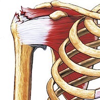
In anatomy, the rotator cuff is a group of muscles and their tendons that act to stabilize the shoulder and allow for its extensive range of motion. Of the seven scapulohumeral muscles, four make up the rotator cuff. The four muscles are the supraspinatus muscle, the infraspinatus muscle, teres minor muscle, and the subscapularis muscle.
Tendinopathy, also known as tendinitis or tendonitis, is a type of tendon disorder that results in pain, swelling, and impaired function. The pain is typically worse with movement. It most commonly occurs around the shoulder, elbow, wrist, hip, knee, or ankle.
Trigger finger, also known as stenosing tenosynovitis, is a disorder characterized by catching or locking of the involved finger. Pain may occur in the palm of the hand or knuckles. The name is due to the popping sound made by the affected finger when moved. Most commonly the ring finger or thumb is affected.
Shoulder problems including pain, are one of the more common reasons for physician visits for musculoskeletal symptoms. The shoulder is the most movable joint in the body. However, it is an unstable joint because of the range of motion allowed. This instability increases the likelihood of joint injury, often leading to a degenerative process in which tissues break down and no longer function well.
Osteoarthritis (OA) is a type of joint disease that results from breakdown of joint cartilage and underlying bone. The most common symptoms are joint pain and stiffness. Usually the symptoms progress slowly over years. Initially they may only occur after exercise, but can become constant over time. Other symptoms may include joint swelling, decreased range of motion, and, when the back is affected, weakness or numbness of the arms and legs. The most commonly involved joints are the two near the ends of the fingers and the joint at the base of the thumbs; the knee and hip joints; and the joints of the neck and lower back. Joints on one side of the body are often more affected than those on the other. The symptoms can interfere with work and normal daily activities. Unlike some other types of arthritis, only the joints, not internal organs, are affected.
Diabetic neuropathy refers to various types of nerve damage associated with diabetes mellitus. Symptoms depend on the site of nerve damage and can include motor changes such as weakness; sensory symptoms such as numbness, tingling, or pain; or autonomic changes such as urinary symptoms. These changes are thought to result from microvascular injury involving small blood vessels that supply nerves. Relatively common conditions which may be associated with diabetic neuropathy include distal symmetric polyneuropathy; third, fourth, or sixth cranial nerve palsy; mononeuropathy; mononeuropathy multiplex; diabetic amyotrophy; and autonomic neuropathy.
Tennis elbow, also known as lateral epicondylitis, is a condition in which the outer part of the elbow becomes painful and tender. The pain may also extend into the back of the forearm and grip strength may be weak. Onset of symptoms is generally gradual. Golfer's elbow is a similar condition that affects the inside of the elbow.
Spondylosis is the degeneration of the vertebral column from any cause. In the more narrow sense it refers to spinal osteoarthritis, the age-related wear and tear of the spinal column, which is the most common cause of spondylosis. The degenerative process in osteoarthritis chiefly affects the vertebral bodies, the neural foramina and the facet joints. If severe, it may cause pressure on the spinal cord or nerve roots with subsequent sensory or motor disturbances, such as pain, paresthesia, imbalance, and muscle weakness in the limbs.
A rotator cuff tear is an injury of one or more of the tendons or muscles of the rotator cuff of the shoulder. Symptoms may include shoulder pain, which is often worse with movement, or weakness. This may limit people’s ability to brush their hair or put on clothing. Clicking may also occur with movement of the arm.
Electrotherapy is the use of electrical energy as a medical treatment. In medicine, the term electrotherapy can apply to a variety of treatments, including the use of electrical devices such as deep brain stimulators for neurological disease. The term has also been applied specifically to the use of electric current to speed wound healing. Additionally, the term "electrotherapy" or "electromagnetic therapy" has also been applied to a range of alternative medical devices and treatments.
Myofascial pain syndrome (MPS), also known as chronic myofascial pain (CMP), is a syndrome characterized by chronic pain in multiple myofascial trigger points ("knots") and fascial constrictions. It can appear in any body part. Symptoms of a myofascial trigger points include: focal point tenderness, reproduction of pain upon trigger point palpation, hardening of the muscle upon trigger point palpation, pseudo-weakness of the involved muscle, referred pain, and limited range of motion following approximately 5 seconds of sustained trigger point pressure.
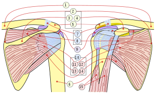
Greater trochanteric pain syndrome (GTPS), is inflammation of the trochanteric bursa, a part of the hip.
Subacromial bursitis is a condition caused by inflammation of the bursa that separates the superior surface of the supraspinatus tendon from the overlying coraco-acromial ligament, acromion, and coracoid and from the deep surface of the deltoid muscle. The subacromial bursa helps the motion of the supraspinatus tendon of the rotator cuff in activities such as overhead work.
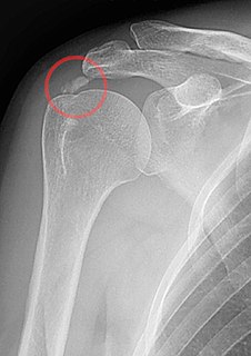
Calcific tendinitis is a form of tendinitis, a disorder characterized by deposits of hydroxyapatite in any tendon of the body, but most commonly in the tendons of the rotator cuff (shoulder), causing pain and inflammation. The condition is related to and may cause adhesive capsulitis.
Radiculopathy, also commonly referred to as pinched nerve, refers to a set of conditions in which one or more nerves are affected and do not work properly. This can result in pain, weakness, numbness, or difficulty controlling specific muscles.
Arthrofibrosis has been described in most joints like knee, hip, ankle, foot joints, shoulder, elbow, wrist, hand joints as well as spinal vertebrae. In the knee, it can happen after knee injury or surgery. There is excessive scar tissue formation within the joint and surrounding soft tissues leading to painful restriction of joint motion that persists despite physical therapy and rehabilitation. The scar tissue can involve only a part of the knee joint or the whole knee. The scar tissue may be located inside the knee joint or may involve the soft tissue structures around the knee joint. Arthrofibrosis may arise without an obvious cause or it may follow a known cause.
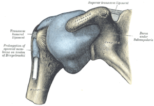
The capsule of the glenohumeral (shoulder) joint is the articular capsule of the shoulder. It completely surrounds the joint. It is attached above to the circumference of the glenoid cavity beyond the glenoidal labrum, and below to the anatomical neck of the humerus, approaching nearer to the articular cartilage above than in the rest of its extent.
Shoulder impingement syndrome is a syndrome involving tendonitis of the rotator cuff muscles as they pass through the subacromial space, the passage beneath the acromion. It is particularly associated with tendonitis of the supraspinatus muscle. This can result in pain, weakness, and loss of movement at the shoulder.
This article contains text from the public domain document "Frozen Shoulder", American Academy of Orthopaedic Surgeons.
Classification | |
---|---|
External resources |