Tumor suppressor

Last updated
The cell cycle. Many tumor suppressors work to regulate the cycle at specific checkpoints in order to prevent damaged cells from replicating. Cell Cycle 3-3.svg
The cell cycle. Many tumor suppressors work to regulate the cycle at specific checkpoints in order to prevent damaged cells from replicating.

A tumor suppressor gene, or anti-oncogene, is a gene that regulates a cell during cell division and replication. [1] If the cell grows uncontrollably, it will result in cancer. When a tumor suppressor gene is mutated, it results in a loss or reduction in its function. In combination with other genetic mutations, this could allow the cell to grow abnormally. The loss of function for these genes may be even more significant in the development of human cancers, compared to the activation of oncogenes. [2]

Contents

Tumor suppressor genes (TSGs) can be grouped into the following categories: caretaker genes, gatekeeper genes, and more recently landscaper genes. Caretaker genes ensure stability of the genome via DNA repair and subsequently when mutated allow mutations to accumulate. [3] Meanwhile gatekeeper genes directly regulate cell growth by either inhibiting cell cycle progression or inducing apoptosis. [3] Lastly landscaper genes regulate growth by contributing to the surrounding environment, when mutated can cause an environment that promotes unregulated proliferation. [4] The classification schemes are evolving as medical advances are being made from fields including molecular biology, genetics, and epigenetics.

History

The discovery of oncogenes and their ability to deregulate cellular processes related to cell proliferation and development appeared first in the literature as opposed to the idea of tumor suppressor genes. [5] However, the idea of genetic mutation leading to increased tumor growth gave way to another possible genetic idea of genes playing a role in decreasing cellular growth and development of cells. This idea was not solidified until experiments byHenry Harris were conducted with somatic cell hybridization in 1969. [6]

Within Dr. Harris’s experiments, tumor cells were fused with normal somatic cells to make hybrid cells. Each cell had chromosomes from both parents and upon growth, a majority of these hybrid cells did not have the capability of developing tumors within animals. [6] The suppression of tumorigenicity in these hybrid cells prompted researchers to hypothesize that genes within the normal somatic cell had inhibitory actions to stop tumor growth. [6] This initial hypothesis eventually lead to the discovery of the first classic tumor suppressor gene by Alfred Knudson, known as the Rb gene, which codes for the retinoblastoma tumor suppressor protein. [5]

Alfred Knudson, a pediatrician and cancer geneticist, proposed that in order to develop retinoblastoma, two allelic mutations are required to lose functional copies of both the Rb genes to lead to tumorigenicity. [6] Knudson observed that retinoblastoma often developed early in life for younger patients in both eyes, while in some rarer cases retinoblastoma would develop later in life and only be unilateral. [5] This unique development pattern allowed Knudson and several other scientific groups in 1971 to correctly hypothesize that the early development of retinoblastoma was caused by inheritance of one loss of function mutation to an RB germ-line gene followed by a later de novo mutation on its functional Rb gene allele. The more sporadic occurrence of unilateral development of retinoblastoma was hypothesized to develop much later in life due to two de novo mutations that were needed to fully lose tumor suppressor properties. [5] This finding formed the basis of the two-hit hypothesis. In order to verify that the loss of function of tumor suppressor genes causes increased tumorigenicity, interstitial deletion experiments on chromosome 13q14 were conducted to observe the effect of deleting the loci for the Rb gene. This deletion caused increased tumor growth in retinoblastoma, suggesting thatloss or inactivation of a tumor suppressor gene can increase tumorigenicity. [6]

Two-hit hypothesis

Unlike oncogenes, tumor suppressor genes generally follow the two-hit hypothesis, which states both alleles that code for a particular protein must be affected before an effect is manifested. [7] If only one allele for the gene is damaged, the other can still produce enough of the correct protein to retain the appropriate function. In other words, mutant tumor suppressor alleles are usually recessive, whereas mutant oncogene alleles are typically dominant.

Models of tumor suppression Models of tumour suppression.svg
Models of tumor suppression
Illustration of two-hit hypothesis Two-hit.jpg
Illustration of two-hit hypothesis

Proposed by A.G. Knudson for cases of retinoblastoma. [7] He observed that 40% of U.S cases were caused by a mutation in the germ-line. However, affected parents could have children without the disease, but the unaffected children became parents of children with retinoblastoma. [8] This indicates that one could inherit a mutated germ-line but not display the disease. Knudson observed that the age of onset of retinoblastoma followed 2nd order kinetics, implying that two independent genetic events were necessary. He recognized that this was consistent with a recessive mutation involving a single gene, but requiring bi-allelic mutation. Hereditary cases involve an inherited mutation and a single mutation in the normal allele. [8] Non-hereditary retinoblastoma involves two mutations, one on each allele. [8] Knudson also noted that hereditary cases often developed bilateral tumors and would develop them earlier in life, compared to non-hereditary cases where individuals were only affected by a single tumor. [8]

There are exceptions to the two-hit rule for tumor suppressors, such as certain mutations in the p53 gene product. p53 mutations can function as a dominant negative, meaning that a mutated p53 protein can prevent the function of the natural protein produced from the non-mutated allele. [9] Other tumor-suppressor genes that do not follow the two-hit rule are those that exhibit haploinsufficiency, including PTCH in medulloblastoma and NF1 in neurofibroma. Another example is p27, a cell-cycle inhibitor, that when one allele is mutated causes increased carcinogen susceptibility. [10]

Functions

The proteins encoded by most tumor suppressor genes inhibit cell proliferation or survival. Inactivation of tumor suppressor genes therefore leads to tumor development by eliminating negative regulatory proteins. In most cases, tumor suppressor proteins inhibit the same cell regulatory pathways that are stimulated by the products of oncogenes. [11] While tumor suppressor genes have the same main function, they have various mechanisms of action, that their transcribed products perform, which include the following: [12]

  1. Intracellular proteins, that control gene expression of a specific stage of the cell cycle. If these genes are not expressed, the cell cycle does not continue, effectively inhibiting cell division. (e.g., pRB and p16) [13]
  2. Receptors or signal transducers for secreted hormones or developmental signals that inhibit cell proliferation (e.g., transforming growth factor (TGF)-β and adenomatous polyposis coli (APC)). [14]
  3. Checkpoint-control proteins that trigger cell cycle arrest in response to DNA damage or chromosomal defects (e.g., breast cancer type 1 susceptibility protein (BRCA1), p16, and p14). [15]
  4. Proteins that induce apoptosis. If damage cannot be repaired, the cell initiates programmed cell death to remove the threat it poses to the organism as a whole. (e.g., p53). [16]
  5. Cell adhesion. Some proteins involved in cell adhesion prevent tumor cells from dispersing, block loss of contact inhibition, and inhibit metastasis. These proteins are known as metastasis suppressors. (eg., CADM1) [17] [18]
  6. Proteins involved in repairing mistakes in DNA. Caretaker genes encode proteins that function in repairing mutations in the genome, preventing cells from replicating with mutations. Furthermore, increased mutation rate from decreased DNA repair leads to increased inactivation of other tumor suppressors and activation of oncogenes. [19] (e.g., p53 and DNA mismatch repair protein 2 (MSH2)). [20]
  7. Certain genes can also act as tumor suppressors and oncogenes. Dubbed Proto-oncogenes with Tumor suppressor function, these genes act as “double agents” that both positively and negatively regulate transcription. (e.g., NOTCH receptors,TP53 and FAS). [21]

Epigenetic Influences on Tumor Suppressors

Scientists Shahjehan A. Wajed et al. state the expression of genes, including tumor suppressors, can be altered through biochemical alterations known as DNA methylation. [22] Methylation is an example of epigenetic modifications, which commonly regulate expression in mammalian genes. The addition of a methyl group to either histone tails or directly on DNA causes the nucleosome to pack tightly together restricting the transcription of any genes in this region. This process not only has the capabilities to inhibit gene expression, it can also increase the chance of mutations. Stephen Baylin observed that if promoter regions experience a phenomenon known as hypermethylation, it could result in later transcriptional errors, tumor suppressor gene silencing,  protein misfolding, and eventually cancer growth. Baylin et al. found methylation inhibitors known as azacitidine and decitabine. These compounds can actually help prevent cancer growth by inducing re-expression of previously silenced genes, arresting the cell cycle of the tumor cell and forcing it into apoptosis. [23]

There are further clinical trials under current investigation regarding treatments for hypermethylation as well as alternate tumor suppression therapies that include prevention of tissue hyperplasia, tumor development, or metastatic spread of tumors. [24] The team working with Wajed have investigated neoplastic tissue methylation in order to one day identify early treatment options for gene modification that can silence the tumor suppressor gene. [25] In addition to DNA methylation, other epigenetic modifications like histone deacetylation or chromatin-binding proteins can prevent DNA polymerase from effectively transcribing desired sequences, such as ones containing tumor suppressor genes.

Clinical Interests

Gene therapy is used to reinstate the function of a mutated or deleted gene type. When tumor suppressor genes are altered in a way that results in less or no expression, several severe problems can arise for the host. This is why tumor suppressor genes have commonly been studied and used for gene therapy. The two main approaches used currently to introduce genetic material into cells are viral and non-viral delivery methods. [25]

Viral Methods

The viral method of transferring genetic material harnesses the power of viruses. [25] By using viruses that are durable to genetic material alterations, viral methods of gene therapy for tumor suppressor genes have shown to be successful. [26] In this method, vectors from viruses are used. The two most commonly used vectors are adenoviral vectors and adeno-associated vectors. In vitro genetic manipulation of these types of vectors is easy and in vivo application is relatively safe compared to other vectors. [25] [27] Before the vectors are inserted into the tumors of the host, they are prepared by having the parts of their genome that control replication either mutated or deleted. This makes them safer for insertion. Then, the desired genetic material is inserted and ligated to the vector. [26] In the case with tumor suppressor genes, genetic material which encodes p53 has been used successfully, which after application, has shown reduction in tumor growth or proliferation. [27] [28]

Non-Viral Methods

The non-viral method of transferring genetic material is used less often than the viral method. [25] [27] However, the non-viral method is a more cost-effective, safer, available method of gene delivery not to mention that non-viral methods have shown to induce fewer host immune responses and possess no restrictions on size or length of the transferable genetic material. [25] Non-viral gene therapy uses either chemical or physical methods to introduce genetic material to the desired cells. [25] [27] The chemical methods are used primarily for tumor suppressor gene introduction and are divided into two categories which are naked plasmid or liposome-coated plasmids. [27] The naked plasmid strategy has garnered interest because of its easy to use methods. [25] Direct injection into the muscles allows for the plasmid to be taken up into the cell of possible tumors where the genetic material of the plasmid can be incorporated into the genetic material of the tumor cells and revert any previous damage done to tumor suppressor genes. [25] [27] The liposome-coated plasmid method has recently also been of interest since they produce relatively low host immune response and are efficient with cellular targeting. [27] The positively charged capsule in which the genetic material is packaged helps with electrostatic attraction to the negatively charged membranes of the cells as well as the negatively charged DNA of the tumor cells. [25] [27] In this way, non-viral methods of gene therapy are highly effective in restoring tumor suppressor gene function to tumor cells that have either partially or entirely lost this function.

Limitations

The viral and non-viral gene therapies mentioned above are commonly used but each has some limitations which must be considered. The most important limitation these methods have is the efficacy at which the adenoviral and adeno-associated vectors, naked plasmids, or liposome-coated plasmids are taken in by the host’s tumor cells. If proper uptake by the host’s tumor cells is not achieved, re-insertion introduces problems such as the host’s immune system recognizing these vectors or plasmids and destroying them which impairs the overall effectiveness of the gene therapy treatment further. [28]

Examples

GeneOriginal FunctionTwo-Hit?Associated Carcinomas
Rb DNA Replication, cell division and deathYesRetinoblastoma [5]
p53 ApoptosisNoHalf of all known malignancies [5]
VHL Cell division, death, and differentiationYesKidney Cancer [25]
APC DNA damage, cell division, migration, adhesion, deathYesColorectal Cancer [25]
BRAC2 Cell division and death, and repair of double-stranded DNA breaksYesBreast/Ovarian Cancer [5]
NF1 Cell differentiation, division, development, RAS signal transductionNoNerve tumors, Neuroblastoma [25]
PTCH Hedgehog signalingNoMedulloblastoma, Basal Cell Carcinoma [5]

[25]

As the cost of DNA sequencing continues to diminish, more cancers can be sequenced. This allows for the discovery of novel tumor suppressors and can give insight on how to treat and cure different cancers in the future. Other examples of tumor suppressors include pVHL, APC, CD95, ST5, YPEL3, ST7, and ST14, p16, BRCA2. [34]

See also

Related Research Articles

Cell cycle Series of events and stages that result in cell division

The cell cycle, or cell-division cycle, is the series of events that take place in a cell that cause it to divide into two daughter cells. These events include the duplication of its DNA and some of its organelles, and subsequently the partitioning of its cytoplasm and other components into two daughter cells in a process called cell division.

Oncogene

An oncogene is a gene that has the potential to cause cancer. In tumor cells, these genes are often mutated, or expressed at high levels.

p53

Tumor protein P53, also known as p53, cellular tumor antigen p53, the Guardian of the Genome, phosphoprotein p53, tumor suppressor p53, antigen NY-CO-13, or transformation-related protein 53 (TRP53), is any isoform of a protein encoded by homologous genes in various organisms, such as TP53 (humans) and Trp53 (mice). This homolog is crucial in multicellular vertebrates, where it prevents cancer formation, and thus functions as a tumor suppressor. As such, p53 has been described as "the guardian of the genome" because of its role in conserving stability by preventing genome mutation. Hence TP53 is classified as a tumor suppressor gene.

Retinoblastoma

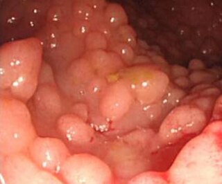
Retinoblastoma (Rb) is a rare form of cancer that rapidly develops from the immature cells of a retina, the light-detecting tissue of the eye. It is the most common primary malignant intraocular cancer in children, and it is almost exclusively found in young children.

Li–Fraumeni syndrome

Li–Fraumeni syndrome is a rare, autosomal dominant, hereditary disorder that predisposes carriers to cancer development. It was named after two American physicians, Frederick Pei Li and Joseph F. Fraumeni, Jr., who first recognized the syndrome after reviewing the medical records and death certificates of 648 childhood rhabdomyosarcoma patients. This syndrome is also known as the sarcoma, breast, leukaemia and adrenal gland (SBLA) syndrome.

An oncovirus is a virus that can cause cancer. This term originated from studies of acutely transforming retroviruses in the 1950–60s, when the term "oncornaviruses" was used to denote their RNA virus origin. With the letters "RNA" removed, it now refers to any virus with a DNA or RNA genome causing cancer and is synonymous with "tumor virus" or "cancer virus". The vast majority of human and animal viruses do not cause cancer, probably because of longstanding co-evolution between the virus and its host. Oncoviruses have been important not only in epidemiology, but also in investigations of cell cycle control mechanisms such as the retinoblastoma protein.

The Knudson hypothesis, also known as the two-hit hypothesis, is the hypothesis that most tumor suppressor genes require both alleles to be inactivated, either through mutations or through epigenetic silencing, to cause a phenotypic change. It was first formulated by Alfred G. Knudson in 1971 and led indirectly to the identification of cancer-related genes. Knudson won the 1998 Albert Lasker Clinical Medical Research Award for this work.

Alfred G. Knudson

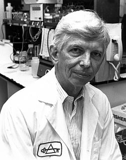
Alfred George Knudson, Jr. was an American physician and geneticist specializing in cancer genetics. Among his many contributions to the field was the formulation of the Knudson hypothesis in 1971, which explains the effects of mutation on carcinogenesis.

p73

p73 is a protein related to the p53 tumor protein. Because of its structural resemblance to p53, it has also been considered a tumor suppressor. It is involved in cell cycle regulation, and induction of apoptosis. Like p53, p73 is characterized by the presence of different isoforms of the protein. This is explained by splice variants, and an alternative promoter in the DNA sequence.

Carcinogenesis, also called oncogenesis or tumorigenesis, is the formation of a cancer, whereby normal cells are transformed into cancer cells. The process is characterized by changes at the cellular, genetic, and epigenetic levels and abnormal cell division. Cell division is a physiological process that occurs in almost all tissues and under a variety of circumstances. Normally the balance between proliferation and programmed cell death, in the form of apoptosis, is maintained to ensure the integrity of tissues and organs. According to the prevailing accepted theory of carcinogenesis, the somatic mutation theory, mutations in DNA and epimutations that lead to cancer disrupt these orderly processes by disrupting the programming regulating the processes, upsetting the normal balance between proliferation and cell death. This results in uncontrolled cell division and the evolution of those cells by natural selection in the body. Only certain mutations lead to cancer whereas the majority of mutations do not.

SV40 large T antigen

SV40 large T antigen is a hexamer protein that is a dominant-acting oncoprotein derived from the polyomavirus SV40. TAg is capable of inducing malignant transformation of a variety of cell types. The transforming activity of TAg is due in large part to its perturbation of the retinoblastoma (pRb) and p53 tumor suppressor proteins. In addition, TAg binds to several other cellular factors, including the transcriptional co-activators p300 and CBP, which may contribute to its transformation function.

p14ARF is an alternate reading frame protein product of the CDKN2A locus. p14ARF is induced in response to elevated mitogenic stimulation, such as aberrant growth signaling from MYC and Ras (protein). It accumulates mainly in the nucleolus where it forms stable complexes with NPM or Mdm2. These interactions allow p14ARF to act as a tumor suppressor by inhibiting ribosome biogenesis or initiating p53-dependent cell cycle arrest and apoptosis, respectively. p14ARF is an atypical protein, in terms of its transcription, its amino acid composition, and its degradation: it is transcribed in an alternate reading frame of a different protein, it is highly basic, and it is polyubiquinated at the N-terminus.

Cyclin D

Cyclin D is a member of the cyclin protein family that is involved in regulating cell cycle progression. The synthesis of cyclin D is initiated during G1 and drives the G1/S phase transition. Cyclin D protein is anywhere from 155 to 477 amino acids in length.

Changes in the genome that allow uncontrolled cell proliferation or cell immortality are responsible for cancer. It is believed that the major changes in the genome that lead to cancer arise from mutations in tumor suppressor genes. In 1997, Kinzler and Bert Vogelstein grouped these cancer susceptibility genes into two classes: "caretakers" and "gatekeepers". In 2004, a third classification of tumor suppressor genes was proposed by Franziska Michor, Yoh Iwasa, and Martin Nowak; "landscaper" genes.

CDKN2A

CDKN2A, also known as cyclin-dependent kinase inhibitor 2A, is a gene which in humans is located at chromosome 9, band p21.3. It is ubiquitously expressed in many tissues and cell types. The gene codes for two proteins, including the INK4 family member p16 and p14arf. Both act as tumor suppressors by regulating the cell cycle. p16 inhibits cyclin dependent kinases 4 and 6 and thereby activates the retinoblastoma (Rb) family of proteins, which block traversal from G1 to S-phase. p14ARF activates the p53 tumor suppressor. Somatic mutations of CDKN2A are common in the majority of human cancers, with estimates that CDKN2A is the second most commonly inactivated gene in cancer after p53. Germline mutations of CDKN2A are associated with familial melanoma, glioblastoma and pancreatic cancer. The CDKN2A gene also contains one of 27 SNPs associated with increased risk of coronary artery disease.

LATS2

Large tumor suppressor kinase 2 (LATS2) is an enzyme that in humans is encoded by the LATS2 gene.

Retinoblastoma protein

The retinoblastoma protein is a tumor suppressor protein that is dysfunctional in several major cancers. One function of Rb is to prevent excessive cell growth by inhibiting cell cycle progression until a cell is ready to divide. When the cell is ready to divide, Rb is phosphorylated to pRb, leading to the inactivation of Rb. This process allows cells to enter into the cell cycle state. It is also a recruiter of several chromatin remodeling enzymes such as methylases and acetylases.

Antineoplastic resistance, often used interchangeably with chemotherapy resistance, is the resistance of neoplastic (cancerous) cells, or the ability of cancer cells to survive and grow despite anti-cancer therapies. In some cases, cancers can evolve resistance to multiple drugs, called multiple drug resistance.

Cancer syndrome Genetic condition that predisposes a person to cancer

A cancer syndrome, or family cancer syndrome, is a genetic disorder in which inherited genetic mutations in one or more genes predispose the affected individuals to the development of cancers and may also cause the early onset of these cancers. Cancer syndromes often show not only a high lifetime risk of developing cancer, but also the development of multiple independent primary tumors.

CpG island hypermethylation is an epigenetic control aberration that is important for gene inactivation in cancer cells. Hypermethylation of CpG islands has been described in almost every type of tumor. Many important cellular pathways, such as DNA repair, cell cycle (p14ARF), apoptosis (DAPK), cell adherence, are inactivated by this epigenetic lesion. Hypermethylation is linked to methyl-binding proteins, DNA methyltransferases and histone deacetylase, but the degree to which this process selectively silences tumor suppressor genes remains a vibrant field of study. The list for hypermethylated genes is growing and functional and genetic studies are being performed to determine which hypermethylation events are relevant for tumorigenesis. Basic as well as translational research will be needed to understand the mechanisms and roles of CpG island hypermethylation in cancer.

References

  1. "Oncogenes and tumor suppressor genes | American Cancer Society". www.cancer.org. Retrieved 2019-09-26.
  2. Weinberg, Robert A (2014). "The Biology of Cancer." Garland Science, page 231.
  3. 1 2 "Glossary of Cancer Genetics (side-frame)". www.cancerindex.org. Retrieved 2019-11-19.
  4. "Cancer Genetics - CuboCube". www.cubocube.com. Retrieved 2019-11-19.
  5. 1 2 3 4 5 6 7 8 Sherr, C. J. (2004). Principles of Tumor Suppression. Cell, 116(2), 235–246. https://doi.org/10.1016/S0092-8674(03)01075-4
  6. 1 2 3 4 5 Cooper, G. M. (2000). Tumor Suppressor Genes. The Cell: A Molecular Approach. 2nd Edition. https://www.ncbi.nlm.nih.gov/books/NBK9894/
  7. 1 2 Knudson AG (1971). "Mutation and Cancer: Statistical Study of Retinoblastoma". Proc Natl Acad Sci USA. 68 (4): 820–3. Bibcode:1971PNAS...68..820K. doi:10.1073/pnas.68.4.820. PMC   389051 . PMID   5279523.
  8. 1 2 3 4 5 "Tumor Suppressor (TS) Genes and the Two-Hit Hypothesis | Learn Science at Scitable". www.nature.com. Retrieved 2019-10-06.
  9. Baker SJ, Markowitz S, Fearon ER, Willson JK, Vogelstein B (1990). "Suppression of human colorectal carcinoma cell growth by wild-type p53". Science. 249 (4971): 912–5. Bibcode:1990Sci...249..912B. doi:10.1126/science.2144057. PMID   2144057.
  10. Fero ML, Randel E, Gurley KE, Roberts JM, Kemp CJ (1998). "The murine gene p27Kip1 is haplo-insufficient for tumour suppression". Nature. 396 (6707): 177–80. Bibcode:1998Natur.396..177F. doi:10.1038/24179. PMC   5395202 . PMID   9823898.
  11. Cooper, GM (2000). "Tumor Suppressor Genes". The Cell: A Molecular Approach. 2nd edition.
  12. Shin, Young Kee; Rajasekaran, Nirmal (2018). "Loss of Tumor Suppressor Gene Function in Human Cancer: An Overview". Cell Physiol Biochem. 51 (6). doi:10.1159/000495956.
  13. Leiderman, YI; Kiss, Slizard; Mukai, Shizuo (2007). "Molecular genetics of RB1–the retinoblastoma gene". Seminars in ophthalmology.
  14. Smith, Anna; Robin, Tyler; Ford, Heide (2012). "Molecular Pathways: Targeting the TGF-β Pathway for Cancer Therapy". Clinical Cancer Research. 18 (17).
  15. Savage, Kienan; Harkin, Paul (2015). "BRCA1, a 'complex' protein involved in the maintenance of genomic stability". The FEBS Journal. 282 (4): 630–646. doi:10.1111/febs.13150.
  16. Nayak, Surendra; Panesar, Paramjit; Kumar, Harish (2009). "p53-Induced apoptosis and inhibitors of p53". Current Medical Chemistry. 16 (21): 2627–2640. doi:10.2174/092986709788681976.
  17. Yoshida BA, Sokoloff MM, Welch DR, Rinker-Schaeffer CW (November 2000). "Metastasis-suppressor genes: a review and perspective on an emerging field". J. Natl. Cancer Inst. 92 (21): 1717–30. doi: 10.1093/jnci/92.21.1717 . PMID   11058615.
  18. Hirohashi S, Kanai Y (2003). "Cell adhesion system and human cancer morphogenesis". Cancer Sci . 94 (7): 575–81. doi:10.1111/j.1349-7006.2003.tb01485.x. PMID   12841864. S2CID   22154824.
  19. Markowitz S (November 2000). "DNA repair defects inactivate tumor suppressor genes and induce hereditary and sporadic colon cancers". J. Clin. Oncol. 18 (21 Suppl): 75S–80S. PMID   11060332.
  20. Scott, Richard; Rahman, Nazneen (15 April 2007). "Cancer genes associated with phenotypes in monoallelic and biallelic mutation carriers: new lessons from old players". Human Molecular Genetics. 16 (R1): R60 - R66. doi:10.1093/hmg/ddm026.
  21. Shen, Libing; Shi, Qili; Wang, Wenyuan (13 March 2018). "Double agents: genes with both oncogenic and tumor-suppressor functions". Oncogenesis. 7 (25). doi:10.1038/s41389-018-0034-x.
  22. Wajed, Shahjehan A.; Laird, Peter W.; DeMeester, Tom R. (2001–2007). "DNA Methylation: An Alternative Pathway to Cancer". Annals of Surgery. 234 (1): 10–20. ISSN   0003-4932. PMC   1421942 . PMID   11420478.
  23. Baylin, Stephen B. (2005). "DNA methylation and gene silencing in cancer". Nature Clinical Practice. Oncology. 2 Suppl 1: S4–11. doi:10.1038/ncponc0354. ISSN   1743-4254. PMID   16341240.
  24. Delbridge, Alex R.D.; Valente, Liz J.; Strasser, Andreas (November 2012). "The Role of the Apoptotic Machinery in Tumor Suppression". Cold Spring Harbor Perspectives in Biology. 4 (11). doi:10.1101/cshperspect.a008789. ISSN   1943-0264. PMC   3536334 . PMID   23125015.
  25. 1 2 3 4 5 6 7 8 9 10 11 12 13 14 "Tumor Suppressor (TS) Genes and the Two-Hit Hypothesis | Learn Science at Scitable". www.nature.com. Retrieved 2020-10-27.
  26. 1 2 Nayerossadat, Nouri; Maedeh, Talebi; Ali, Palizban Abas (2012-07-06). "Viral and nonviral delivery systems for gene delivery". Advanced Biomedical Research. 1. doi:10.4103/2277-9175.98152. ISSN   2277-9175. PMC   3507026 . PMID   23210086.
  27. 1 2 3 4 5 6 7 8 Guo, Xuning Emily; Ngo, Bryan; Modrek, Aram Sandaldjian; Lee, Wen-Hwa (2014). "Targeting Tumor Suppressor Networks for Cancer Therapeutics". Current drug targets. 15 (1): 2–16. ISSN   1389-4501. PMC   4032821 . PMID   24387338.
  28. 1 2 Morris, Luc G. T.; Chan, Timothy A. (2015-05-01). "Therapeutic Targeting of Tumor Suppressor Genes". Cancer. 121 (9): 1357–1368. doi:10.1002/cncr.29140. ISSN   0008-543X. PMC   4526158 . PMID   25557041.
  29. "RETINOBLASTOMA: Protein". dpuadweb.depauw.edu. Retrieved 2019-11-21.
  30. 1 2 Harris, Curtis C. (October 16, 1996). "Structure and Function of p53 Tumor Suppressor Gene: Clues for Rational Cancer Therapeutic Strategies". Journal of the National Cancer Institute. 88 (20): 1442–1455. doi: 10.1093/jnci/88.20.1442 . PMID   8841019.
  31. 1 2 "BCL2 (B-Cell Leukemia/Lymphoma 2)". atlasgeneticsoncology.org. Retrieved 2019-11-21.
  32. Goodsell, David S. (2004-04-01). "The Molecular Perspective: Cytochrome c and Apoptosis". The Oncologist. 9 (2): 226–227. doi:10.1634/theoncologist.9-2-226. ISSN   1083-7159. PMID   15047927.
  33. 1 2 3 Shain, AH; Pollack, JR (2013). "The spectrum of SWI/SNF mutations, ubiquitous in human cancers". PLOS ONE. 8 (1): e55119. Bibcode:2013PLoSO...855119S. doi:10.1371/journal.pone.0055119. PMC   3552954 . PMID   23355908.
  34. "TUMOUR SUPPRESSOR GENES IN CANCER". www.letstalkacademy.com. Retrieved 2019-11-21.