P16

Last updated
CDKN2A
Protein CDKN2A PDB 1a5e.png
Available structures
PDB Ortholog search: PDBe RCSB
Identifiers
Aliases CDKN2A , CDK4I, CDKN2, CMM2, INK4, INK4A, MLM, MTS-1, MTS1, P14, P14ARF, P16, P16-INK4A, P16INK4, P16INK4A, P19, P19ARF, TP16, cyclin-dependent kinase inhibitor 2A, cyclin dependent kinase inhibitor 2A, Genes, p16, ARF.
External IDs OMIM: 600160 MGI: 104738 HomoloGene: 55430 GeneCards: CDKN2A
Orthologs
SpeciesHumanMouse
Entrez
Ensembl
UniProt
RefSeq (mRNA)

NM_001040654
NM_009877

RefSeq (protein)

NP_001035744
NP_034007
NP_034007.1

Location (UCSC) Chr 9: 21.97 – 22 Mb Chr 4: 89.19 – 89.21 Mb
PubMed search [3] [4]
Wikidata
View/Edit Human View/Edit Mouse
Cyclin-dependent kinase inhibitor 2a p19Arf N-terminus
PDB 1hn3 EBI.jpg
solution structure of the n-terminal 37 amino acids of the mouse arf tumor suppressor protein
Identifiers
SymbolP19Arf_N
Pfam PF07392
InterPro IPR010868
SCOP2 1hn3 / SCOPe / SUPFAM
Available protein structures:
Pfam   structures / ECOD  
PDB RCSB PDB; PDBe; PDBj
PDBsum structure summary

p16 (also known as p16INK4a, cyclin-dependent kinase inhibitor 2A, CDKN2A, multiple tumor suppressor 1 and numerous other synonyms), is a protein that slows cell division by slowing the progression of the cell cycle from the G1 phase to the S phase, thereby acting as a tumor suppressor. It is encoded by the CDKN2A gene. A deletion (the omission of a part of the DNA sequence during replication) in this gene can result in insufficient or non-functional p16, accelerating the cell cycle and resulting in many types of cancer. [5] [6] [7]

Contents

p16 can be used as a biomarker to improve the histological diagnostic accuracy of grade 3 cervical intraepithelial neoplasia (CIN). p16 is also implicated in the prevention of melanoma, oropharyngeal squamous cell carcinoma, cervical cancer, vulvar cancer and esophageal cancer.

p16 was discovered in 1993. It is a protein with 148 amino acids and a molecular weight of 16 kDa that comprises four ankyrin repeats. [8] The name of p16 is derived from its molecular weight, and the alternative name p16INK4a refers to its role in inhibiting cyclin-dependent kinase CDK4. [8]

Nomenclature

p16 is also known as:

Gene

In humans, p16 is encoded by the CDKN2A gene, located on chromosome 9 (9p21.3). This gene generates several transcript variants that differ in their first exons. At least three alternatively spliced variants encoding distinct proteins have been reported, two of which encode structurally related isoforms known to function as inhibitors of CDK4. The remaining transcript includes an alternate exon 1 located 20 kb upstream of the remainder of the gene; this transcript contains an alternate open reading frame (ARF) that specifies a protein that is structurally unrelated to the products of the other variants. [9] The ARF product functions as a stabilizer of the tumor suppressor protein p53, as it can interact with and sequester MDM2, a protein responsible for the degradation of p53. [10] [11] In spite of their structural and functional differences, the CDK inhibitor isoforms and the ARF product encoded by this gene, through the regulatory roles of CDK4 and p53 in cell cycle G1 progression, share a common functionality in controlling the G1 phase of the cell cycle. This gene is frequently mutated or deleted in a wide variety of tumors and is known to be an important tumor suppressor gene. [5]

When organisms age, the expression of p16 increases to reduce the proliferation of stem cells. [12] This reduction in the division and production of stem cells protects against cancer while increasing the risks associated with cellular senescence.

Function

p16 is an inhibitor of cyclin-dependent kinases (CDK). It slows down the cell cycle by prohibiting progression from G1 phase to S phase. Otherwise, CDK4/6 binds cyclin D and forms an active protein complex that phosphorylates retinoblastoma protein (pRB). Once phosphorylated, pRB dissociates from the transcription factor E2F1. This liberates E2F1 from its bound state in the cytoplasm and allows it to enter the nucleus. Once in the nucleus, E2F1 promotes the transcription of target genes that are essential for transition from G1 to S phase. [13] [14]

This pathway connects the processes of tumor oncogenesis and senescence, fixing them on opposite ends of a spectrum. On one end, p16 hypermethylation, mutation, or deletion leads to downregulation of the gene and can lead to cancer through the dysregulation of cell cycle progression. Conversely, activation of p16 through reactive oxygen species, DNA damage, or senescence leads to the buildup of p16 in tissues and is implicated in the aging of cells. [13]

Regulation

Regulation of p16 is complex and involves the interaction of several transcription factors, as well as several proteins involved in epigenetic modification through methylation and repression of the promoter region. [13]

PRC1 and PRC2 are two protein complexes that modify the expression of p16 through the interaction of various transcription factors that execute methylation patterns that can repress transcription of p16. These pathways are activated in the cellular response to reduce senescence. [15] [16]

Clinical significance

Role in carcinogenesis

Mutations resulting in deletion or reduction of function of the CDKN2A gene are associated with increased risk of a wide range of cancers, and alterations of the gene are frequently seen in cancer cell lines. [17] [18] Examples include:

Pancreatic adenocarcinoma is often associated with mutations in the CDKN2A gene. [19] [20] [21]

Carriers of germline mutations in CDKN2A have, besides their high risks of melanoma, also increased risks of pancreatic, lung, laryngeal and oropharyngeal cancers. Tobacco smoking increases the carriers’ susceptibility for such non-melanoma cancers. [22]

Homozygous deletions of p16 are frequently found in esophageal cancer and gastric cancer cell lines. [23]

Germline mutations in CDKN2A are associated with an increased susceptibility to develop skin cancer. [24]

Hypermethylation of tumor suppressor genes has been implicated in various cancers. In 2013, a meta-analysis revealed an increased frequency of DNA methylation of the p16 gene in esophageal cancer. As the degree of tumor differentiation increased, so did the frequency of p16 DNA methylation.

Tissue samples of primary oral squamous cell carcinoma (OSCC) often display hypermethylation in the promoter regions of p16. Cancer cells show a significant increase in the accumulation of methylation in CpG islands in the promoter region of p16. This epigenetic change leads to loss of the tumor suppressor gene function through two possible mechanisms: first, methylation can physically inhibit the transcription of the gene, and second, methylation can lead to the recruitment of transcription factors that repress transcription. Both mechanisms cause the same end result: downregulation of gene expression that leads to decreased levels of the p16 protein. It has been suggested that this process is responsible for the development of various forms of cancer serving as an alternative process to gene deletion or mutation. [25] [26] [27] [28] [29] [30]

p16 positivity has been shown to be favorably prognostic in oropharyngeal squamous cell carcinoma. [31] In a retrospective trial analysis of patients with Stage III and IV oropharyngeal cancer, HPV status was assessed and it was found that the 3-year rates of overall survival were 82.4% (95% CI, 77.2 to 87.6) in the HPV-positive subgroup and 57.1% (95% CI, 48.1 to 66.1) in the HPV-negative subgroup, and the 3-year rates of progression-free survival were 73.7% (95% CI, 67.7 to 79.8) and 43.4% (95% CI, 34.4 to 52.4), respectively. p16 status is so prognostic that the AJCC staging system has been revised to include p16 status in oropharyngeal squamous cell cancer group staging. [32] However, some people can have elevated levels of p16 but test negative for HPV and vice versa. This is known as discordant cancer. The 5-year survival for people who test positive for HPV and p16 is 81%, for discordant cancer it is 53 – 55%, and 40% for those who test negative for p16 and HPV. [33] [34]

Clinical use

Biomarker for cancer types

Expression of p16 is used as a prognostic biomarker for certain types of cancer. The reason for this is different types of cancer can have different effects on p16 expression: cancers that overexpress p16 are usually caused by the human papillomavirus (HPV), whereas cancers in which p16 is downregulated will usually have other causes. For patients with oropharyngeal squamous cell carcinoma, using immunohistochemistry to detect the presence of the p16 biomarker has been shown to be the strongest indicator of disease course. Presence of the biomarker is associated with a more favorable prognosis as measured by cancer-specific survival (CSS), recurrence-free survival (RFS), locoregional control (LRC), as well as other measurements. The appearance of hypermethylation of p16 is also being evaluated as a potential prognostic biomarker for prostate cancer. [35] [36] [37]

p16 FISH

p16 deletion detected by FISH in surface epithelial mesothelial proliferations is predictive of underlying invasive mesothelioma. [38]

p16 immunochemistry

High grade squamous intraepithelial lesion showing strong p16 staining. High grade squamous intraepithelial lesion - 2 - p16 -- high mag.jpg
High grade squamous intraepithelial lesion showing strong p16 staining.

As consensus grows regarding the strength of p16 as a biomarker for detecting and determining prognoses of cancer, p16 immunohistochemistry is growing in importance. [13] [35] [39]

Gynecologic cancers

p16 is a widely used immunohistochemical marker in gynecologic pathology. Strong and diffuse cytoplasmic and nuclear expression of p16 in squamous cell carcinomas (SCC) of the female genital tract is strongly associated with high-risk human papilloma virus (HPV) infection and neoplasms of cervical origin. The majority of SCCs of uterine cervix express p16. However, p16 can be expressed in other neoplasms and in several normal human tissues. [40]

Urinary bladder SCCs

More than a third of urinary bladder SCCs express p16. SCCs of urinary bladder express p16 independent of gender. p16 immunohistochemical expression alone cannot be used to discriminate between SCCs arising from uterine cervix versus urinary bladder. [40]

Role in cellular senescence

Concentrations of p16INK4a increase dramatically as tissue ages. p16INK4a, along with senescence-associated beta-galactosidase, is regarded to be a biomarker of cellular senescence. [41] Therefore, p16INK4a could potentially be used as a blood test that measures how fast the body's tissues are aging at a molecular level. [42] Notably, a recent survey of cellular senescence induced by multiple treatments to several cell lines does not identify p16 as belonging to a "core signature" of senescence markers. [43]

It has been used as a target to delay some aging changes in mice. [44]

Role in neurogenesis

Increasing p16INK4a expression during aging is associated with reduced progenitor functions from the subventricular zone, which generates throughout life new neurons migrating to the olfactory bulb, thereby reducing olfactory neurogenesis. [45] Deletion of p16INK4a does not affect neurogenesis in the other adult neurogenic niche, the dentate gyrus of the hippocampus. [45] However, recently, it has been demonstrated that p16INK4a protects from depletion after a powerful proneurogenic stimulus - i.e., running - also stem and progenitor cells of the aged dentate gyrus. [46] In fact, after deletion of p16INK4a, stem cells of the dentate gyrus are greatly activated by running, while, in wild-type p16INK4a dentate gyrus stem cells are not affected by running. [46] Therefore, p16Ink4a plays a role in the maintenance of dentate gyrus stem cells after stimulus, by keeping a reserve of their self-renewal capacity during aging. Since the dentate gyrus plays a key role in spatial and contextual memory formation, p16INK4a is implicated in the maintenance of cognitive functions during aging.

Discovery

Researchers Manuel Serrano, Gregory J. Hannon and David Beach discovered p16 in 1993 and correctly characterized the protein as a cyclin-dependent kinase inhibitor.

Role in carcinogenesis

Since its discovery, p16 has become significant in the field of cancer research. The protein was suspected to be involved in carcinogenesis due to the observation that mutation or deletion in the gene was implicated in human cancer cell lines. The detection of p16 inactivation in familial melanoma supplied further evidence. p16 deletion, mutation, hypermethylation, or overexpression is now associated with various cancers. Whether mutations in p16 can be considered to be driver mutations requires further investigation. [17]

Interactions

p16 has been shown to interact with:

See also

Related Research Articles

<span class="mw-page-title-main">Cell cycle</span> Series of events and stages that result in cell division

The cell cycle, or cell-division cycle, is the series of events that take place in a cell that causes it to divide into two daughter cells. These events include the duplication of its DNA and some of its organelles, and subsequently the partitioning of its cytoplasm, chromosomes and other components into two daughter cells in a process called cell division.
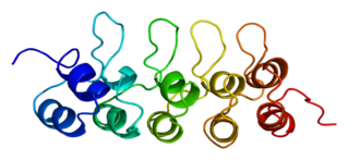
<span class="mw-page-title-main">Tumor suppressor gene</span> Gene that inhibits expression of the tumorigenic phenotype

A tumor suppressor gene (TSG), or anti-oncogene, is a gene that regulates a cell during cell division and replication. If the cell grows uncontrollably, it will result in cancer. When a tumor suppressor gene is mutated, it results in a loss or reduction in its function. In combination with other genetic mutations, this could allow the cell to grow abnormally. The loss of function for these genes may be even more significant in the development of human cancers, compared to the activation of oncogenes.

<span class="mw-page-title-main">Head and neck cancer</span> Cancer arises in the head or neck region

Head and neck cancer develops from tissues in the lip and oral cavity (mouth), larynx (throat), salivary glands, nose, sinuses, or skin of the face. The most common types of head and neck cancer occur in the lips, mouth, and larynx. Symptoms predominantly include a sore that does not heal or a change in the voice. In those with advanced disease, there may be unusual bleeding, facial pain, numbness or swelling, and visible lumps on the outside of the neck or oral cavity. Given the location of these cancers, it is possible for an afflicted individual to experience difficulty in breathing.

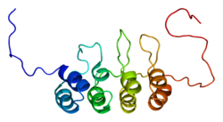
p14ARF is an alternate reading frame protein product of the CDKN2A locus. p14ARF is induced in response to elevated mitogenic stimulation, such as aberrant growth signaling from MYC and Ras (protein). It accumulates mainly in the nucleolus where it forms stable complexes with NPM or Mdm2. These interactions allow p14ARF to act as a tumor suppressor by inhibiting ribosome biogenesis or initiating p53-dependent cell cycle arrest and apoptosis, respectively. p14ARF is an atypical protein, in terms of its transcription, its amino acid composition, and its degradation: it is transcribed in an alternate reading frame of a different protein, it is highly basic, and it is polyubiquinated at the N-terminus.

INK4 is a family of cyclin-dependent kinase inhibitors (CKIs). The members of this family (p16INK4a, p15INK4b, p18INK4c, p19INK4d) are inhibitors of CDK4 (hence their name INhibitors of CDK4), and of CDK6. The other family of CKIs, CIP/KIP proteins are capable of inhibiting all CDKs. Enforced expression of INK4 proteins can lead to G1 arrest by promoting redistribution of Cip/Kip proteins and blocking cyclin E-CDK2 activity. In cycling cells, there is a resassortment of Cip/Kip proteins between CDK4/5 and CDK2 as cells progress through G1. Their function, inhibiting CDK4/6, is to block progression of the cell cycle beyond the G1 restriction point. In addition, INK4 proteins play roles in cellular senescence, apoptosis and DNA repair.

<span class="mw-page-title-main">Cyclin D</span> Member of the cyclin protein family

Cyclin D is a member of the cyclin protein family that is involved in regulating cell cycle progression. The synthesis of cyclin D is initiated during G1 and drives the G1/S phase transition. Cyclin D protein is anywhere from 155 to 477 amino acids in length.

<span class="mw-page-title-main">Cyclin-dependent kinase 4</span> Human protein

Cyclin-dependent kinase 4 also known as cell division protein kinase 4 is an enzyme that in humans is encoded by the CDK4 gene. CDK4 is a member of the cyclin-dependent kinase family.

<span class="mw-page-title-main">Cyclin-dependent kinase 6</span> Protein-coding gene in the species Homo sapiens

Cell division protein kinase 6 (CDK6) is an enzyme encoded by the CDK6 gene. It is regulated by cyclins, more specifically by Cyclin D proteins and Cyclin-dependent kinase inhibitor proteins. The protein encoded by this gene is a member of the cyclin-dependent kinase, (CDK) family, which includes CDK4. CDK family members are highly similar to the gene products of Saccharomyces cerevisiae cdc28, and Schizosaccharomyces pombe cdc2, and are known to be important regulators of cell cycle progression in the point of regulation named R or restriction point.
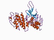
<span class="mw-page-title-main">RASSF1</span> Protein-coding gene in the species Homo sapiens

Ras association domain-containing protein 1 is a protein that in humans is encoded by the RASSF1 gene.

<span class="mw-page-title-main">CEBPA</span> Protein-coding gene in the species Homo sapiens

CCAAT/enhancer-binding protein alpha is a protein encoded by the CEBPA gene in humans. CCAAT/enhancer-binding protein alpha is a transcription factor involved in the differentiation of certain blood cells. For details on the CCAAT structural motif in gene enhancers and on CCAAT/Enhancer Binding Proteins see the specific page.

<span class="mw-page-title-main">CDKN2B</span> Protein-coding gene in the species Homo sapiens

Cyclin-dependent kinase 4 inhibitor B also known as multiple tumor suppressor 2 (MTS-2) or p15INK4b is a protein that is encoded by the CDKN2B gene in humans.

<span class="mw-page-title-main">CDKN2C</span> Protein-coding gene in the species Homo sapiens

Cyclin-dependent kinase 4 inhibitor C is an enzyme that in humans is encoded by the CDKN2C gene.

<span class="mw-page-title-main">CDKN2D</span> Protein-coding gene in the species Homo sapiens

Cyclin-dependent kinase 4 inhibitor D is an enzyme that in humans is encoded by the CDKN2D gene.

<span class="mw-page-title-main">CDKN2A</span> Protein-coding gene in the species Homo sapiens

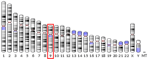
CDKN2A, also known as cyclin-dependent kinase inhibitor 2A, is a gene which in humans is located at chromosome 9, band p21.3. It is ubiquitously expressed in many tissues and cell types. The gene codes for two proteins, including the INK4 family member p16 and p14arf. Both act as tumor suppressors by regulating the cell cycle. p16 inhibits cyclin dependent kinases 4 and 6 and thereby activates the retinoblastoma (Rb) family of proteins, which block traversal from G1 to S-phase. p14ARF activates the p53 tumor suppressor. Somatic mutations of CDKN2A are common in the majority of human cancers, with estimates that CDKN2A is the second most commonly inactivated gene in cancer after p53. Germline mutations of CDKN2A are associated with familial melanoma, glioblastoma and pancreatic cancer. The CDKN2A gene also contains one of 27 SNPs associated with increased risk of coronary artery disease.

<span class="mw-page-title-main">Cellular senescence</span> Phenomenon characterized by the cessation of cell division

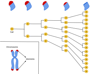
Cellular senescence is a phenomenon characterized by the cessation of cell division. In their experiments during the early 1960s, Leonard Hayflick and Paul Moorhead found that normal human fetal fibroblasts in culture reach a maximum of approximately 50 cell population doublings before becoming senescent. This process is known as "replicative senescence", or the Hayflick limit. Hayflick's discovery of mortal cells paved the path for the discovery and understanding of cellular aging molecular pathways. Cellular senescence can be initiated by a wide variety of stress inducing factors. These stress factors include both environmental and internal damaging events, abnormal cellular growth, oxidative stress, autophagy factors, among many other things.

<span class="mw-page-title-main">HPV-positive oropharyngeal cancer</span> Cancer of the throat

Human papillomavirus-positive oropharyngeal cancer, is a cancer of the throat caused by the human papillomavirus type 16 virus (HPV16). In the past, cancer of the oropharynx (throat) was associated with the use of alcohol or tobacco or both, but the majority of cases are now associated with the HPV virus, acquired by having oral contact with the genitals of a person who has a genital HPV infection. Risk factors include having a large number of sexual partners, a history of oral-genital sex or anal–oral sex, having a female partner with a history of either an abnormal Pap smear or cervical dysplasia, having chronic periodontitis, and, among men, younger age at first intercourse and a history of genital warts. HPV-positive OPC is considered a separate disease from HPV-negative oropharyngeal cancer.

<span class="mw-page-title-main">YPEL3</span> Protein-coding gene in humans

Yippee-like 3 (Drosophila) is a protein that in humans is encoded by the YPEL3 gene. YPEL3 has growth inhibitory effects in normal and tumor cell lines. One of five family members (YPEL1-5), YPEL3 was named in reference to its Drosophila melanogaster orthologue. Initially discovered in a gene expression profiling assay of p53 activated MCF7 cells, induction of YPEL3 has been shown to trigger permanent growth arrest or cellular senescence in certain human normal and tumor cell types. DNA methylation of a CpG island near the YPEL3 promoter as well as histone acetylation may represent possible epigenetic mechanisms leading to decreased gene expression in human tumors.

miR-137

In molecular biology, miR-137 is a short non-coding RNA molecule that functions to regulate the expression levels of other genes by various mechanisms. miR-137 is located on human chromosome 1p22 and has been implicated to act as a tumor suppressor in several cancer types including colorectal cancer, squamous cell carcinoma and melanoma via cell cycle control.

<span class="mw-page-title-main">Cancer epigenetics</span> Field of study in cancer research

Cancer epigenetics is the study of epigenetic modifications to the DNA of cancer cells that do not involve a change in the nucleotide sequence, but instead involve a change in the way the genetic code is expressed. Epigenetic mechanisms are necessary to maintain normal sequences of tissue specific gene expression and are crucial for normal development. They may be just as important, if not even more important, than genetic mutations in a cell's transformation to cancer. The disturbance of epigenetic processes in cancers, can lead to a loss of expression of genes that occurs about 10 times more frequently by transcription silencing than by mutations. As Vogelstein et al. points out, in a colorectal cancer there are usually about 3 to 6 driver mutations and 33 to 66 hitchhiker or passenger mutations. However, in colon tumors compared to adjacent normal-appearing colonic mucosa, there are about 600 to 800 heavily methylated CpG islands in the promoters of genes in the tumors while these CpG islands are not methylated in the adjacent mucosa. Manipulation of epigenetic alterations holds great promise for cancer prevention, detection, and therapy. In different types of cancer, a variety of epigenetic mechanisms can be perturbed, such as the silencing of tumor suppressor genes and activation of oncogenes by altered CpG island methylation patterns, histone modifications, and dysregulation of DNA binding proteins. There are several medications which have epigenetic impact, that are now used in a number of these diseases.

<span class="mw-page-title-main">DIRAS3 (gene)</span> Mammalian protein found in Homo sapiens

GTP-binding protein Di-Ras3 (DIRAS3) also known as aplysia ras homology member I (ARHI) is a protein that in humans is encoded by the DIRAS3 gene.

References

  1. 1 2 3 GRCh38: Ensembl release 89: ENSG00000147889 - Ensembl, May 2017
  2. 1 2 3 GRCm38: Ensembl release 89: ENSMUSG00000044303 - Ensembl, May 2017
  3. "Human PubMed Reference:". National Center for Biotechnology Information, U.S. National Library of Medicine.
  4. "Mouse PubMed Reference:". National Center for Biotechnology Information, U.S. National Library of Medicine.
  5. 1 2 "Entrez Gene: CDKN2A cyclin-dependent kinase inhibitor 2A (melanoma, p16, inhibits CDK4)".
  6. Nobori T, Miura K, Wu DJ, Lois A, Takabayashi K, Carson DA (April 1994). "Deletions of the cyclin-dependent kinase-4 inhibitor gene in multiple human cancers". Nature. 368 (6473): 753–756. Bibcode:1994Natur.368..753N. doi:10.1038/368753a0. PMID   8152487. S2CID   13199401.
  7. Stone S, Jiang P, Dayananth P, Tavtigian SV, Katcher H, Parry D, et al. (July 1995). "Complex structure and regulation of the P16 (MTS1) locus". Cancer Research. 55 (14): 2988–2994. PMID   7606716.
  8. 1 2 3 Serrano M, Hannon GJ, Beach D (December 1993). "A new regulatory motif in cell-cycle control causing specific inhibition of cyclin D/CDK4". Nature. 366 (6456): 704–707. Bibcode:1993Natur.366..704S. doi:10.1038/366704a0. PMID   8259215. S2CID   4368128.
  9. Hamosh, Ada. "Cyclin-dependent kinase inhibitor 2A; CDKN2A". OMIM. Retrieved 10 December 2013.
  10. "Molecular biology of cancer", Oxford University Press, 2005, ISBN   978-0-19-926472-8, Section 5.3
  11. Roussel MF (September 1999). "The INK4 family of cell cycle inhibitors in cancer". Oncogene. 18 (38): 5311–5317. doi: 10.1038/sj.onc.1202998 . PMID   10498883.
  12. Krishnamurthy J, Ramsey MR, Ligon KL, Torrice C, Koh A, Bonner-Weir S, Sharpless NE (September 2006). "p16INK4a induces an age-dependent decline in islet regenerative potential". Nature. 443 (7110): 453–457. Bibcode:2006Natur.443..453K. doi:10.1038/nature05092. PMID   16957737. S2CID   4402013.
  13. 1 2 3 4 Rayess H, Wang MB, Srivatsan ES (April 2012). "Cellular senescence and tumor suppressor gene p16". International Journal of Cancer. 130 (8): 1715–1725. doi:10.1002/ijc.27316. PMC   3288293 . PMID   22025288.
  14. Hara E, Smith R, Parry D, Tahara H, Stone S, Peters G (March 1996). "Regulation of p16CDKN2 expression and its implications for cell immortalization and senescence". Molecular and Cellular Biology. 16 (3): 859–867. doi:10.1128/mcb.16.3.859. PMC   231066 . PMID   8622687.
  15. Cao R, Wang L, Wang H, Xia L, Erdjument-Bromage H, Tempst P, et al. (November 2002). "Role of histone H3 lysine 27 methylation in Polycomb-group silencing". Science. 298 (5595): 1039–1043. Bibcode:2002Sci...298.1039C. doi:10.1126/science.1076997. PMID   12351676. S2CID   6265267.
  16. Bracken AP, Kleine-Kohlbrecher D, Dietrich N, Pasini D, Gargiulo G, Beekman C, et al. (March 2007). "The Polycomb group proteins bind throughout the INK4A-ARF locus and are disassociated in senescent cells". Genes & Development. 21 (5): 525–530. doi:10.1101/gad.415507. PMC   1820894 . PMID   17344414.
  17. 1 2 Liggett WH, Sidransky D (March 1998). "Role of the p16 tumor suppressor gene in cancer". Journal of Clinical Oncology. 16 (3): 1197–1206. doi:10.1200/JCO.1998.16.3.1197. PMID   9508208.
  18. Rocco JW, Sidransky D (March 2001). "p16(MTS-1/CDKN2/INK4a) in cancer progression". Experimental Cell Research. 264 (1): 42–55. doi:10.1006/excr.2000.5149. PMID   11237522.
  19. Caldas C, Hahn SA, da Costa LT, Redston MS, Schutte M, Seymour AB, et al. (September 1994). "Frequent somatic mutations and homozygous deletions of the p16 (MTS1) gene in pancreatic adenocarcinoma". Nature Genetics. 8 (1): 27–32. doi:10.1038/ng0994-27. PMID   7726912. S2CID   23195660.
  20. Bartsch D, Shevlin DW, Tung WS, Kisker O, Wells SA, Goodfellow PJ (November 1995). "Frequent mutations of CDKN2 in primary pancreatic adenocarcinomas". Genes, Chromosomes & Cancer. 14 (3): 189–195. doi:10.1002/gcc.2870140306. PMID   8589035. S2CID   22823227.
  21. Liu L, Lassam NJ, Slingerland JM, Bailey D, Cole D, Jenkins R, Hogg D (July 1995). "Germline p16INK4A mutation and protein dysfunction in a family with inherited melanoma". Oncogene. 11 (2): 405–412. PMID   7624155.
  22. Helgadottir H, Höiom V, Jönsson G, Tuominen R, Ingvar C, Borg A, et al. (August 2014). "High risk of tobacco-related cancers in CDKN2A mutation-positive melanoma families". Journal of Medical Genetics. 51 (8): 545–552. doi:10.1136/jmedgenet-2014-102320. PMC   4112445 . PMID   24935963.
  23. Igaki H, Sasaki H, Kishi T, Sakamoto H, Tachimori Y, Kato H, et al. (September 1994). "Highly frequent homozygous deletion of the p16 gene in esophageal cancer cell lines". Biochemical and Biophysical Research Communications. 203 (2): 1090–1095. doi:10.1006/bbrc.1994.2294. PMID   8093026.
  24. Puig-Butille JA, Escámez MJ, Garcia-Garcia F, Tell-Marti G, Fabra À, Martínez-Santamaría L, et al. (March 2014). "Capturing the biological impact of CDKN2A and MC1R genes as an early predisposing event in melanoma and non melanoma skin cancer". Oncotarget. 5 (6): 1439–1451. doi:10.18632/oncotarget.1444. PMC   4039222 . PMID   24742402.
  25. Khor GH, Froemming GR, Zain RB, Abraham MT, Omar E, Tan SK, et al. (2013). "DNA methylation profiling revealed promoter hypermethylation-induced silencing of p16, DDAH2 and DUSP1 in primary oral squamous cell carcinoma". International Journal of Medical Sciences. 10 (12): 1727–1739. doi:10.7150/ijms.6884. PMC   3805925 . PMID   24155659.
  26. Demokan S, Chuang A, Suoğlu Y, Ulusan M, Yalnız Z, Califano JA, Dalay N (October 2012). "Promoter methylation and loss of p16(INK4a) gene expression in head and neck cancer". Head & Neck. 34 (10): 1470–1475. doi:10.1002/hed.21949. PMID   22106032. S2CID   11512476.
  27. Shaw RJ, Liloglou T, Rogers SN, Brown JS, Vaughan ED, Lowe D, et al. (February 2006). "Promoter methylation of P16, RARbeta, E-cadherin, cyclin A1 and cytoglobin in oral cancer: quantitative evaluation using pyrosequencing". British Journal of Cancer. 94 (4): 561–568. doi:10.1038/sj.bjc.6602972. PMC   2361183 . PMID   16449996.
  28. Sharma G, Mirza S, Prasad CP, Srivastava A, Gupta SD, Ralhan R (April 2007). "Promoter hypermethylation of p16INK4A, p14ARF, CyclinD2 and Slit2 in serum and tumor DNA from breast cancer patients". Life Sciences. 80 (20): 1873–1881. doi:10.1016/j.lfs.2007.02.026. PMID   17383681.
  29. Jabłonowski Z, Reszka E, Gromadzińska J, Wąsowicz W, Sosnowski M (June 2011). "Hypermethylation of p16 and DAPK promoter gene regions in patients with non-invasive urinary bladder cancer". Archives of Medical Science. 7 (3): 512–516. doi:10.5114/aoms.2011.23421. PMC   3258754 . PMID   22295037.
  30. Xu R, Wang F, Wu L, Wang J, Lu C (January 2013). "A systematic review of hypermethylation of p16 gene in esophageal cancer". Cancer Biomarkers. 13 (4): 215–226. doi:10.3233/CBM-130355. PMID   24240582.
  31. Ang KK, Harris J, Wheeler R, Weber R, Rosenthal DI, Nguyen-Tân PF, et al. (July 2010). "Human papillomavirus and survival of patients with oropharyngeal cancer". The New England Journal of Medicine. 363 (1): 24–35. doi:10.1056/NEJMoa0912217. PMC   2943767 . PMID   20530316.
  32. Lydiatt WM, Patel SG, O'Sullivan B, Brandwein MS, Ridge JA, Migliacci JC, et al. (March 2017). "Head and Neck cancers-major changes in the American Joint Committee on cancer eighth edition cancer staging manual". CA. 67 (2): 122–137. doi: 10.3322/caac.21389 . PMID   28128848.
  33. Mehanna H, Taberna M, von Buchwald C, Tous S, Brooks J, Mena M, et al. (March 2023). "Prognostic implications of p16 and HPV discordance in oropharyngeal cancer (HNCIG-EPIC-OPC): a multicentre, multinational, individual patient data analysis". The Lancet. Oncology. 24 (3): 239–251. doi:10.1016/s1470-2045(23)00013-x. hdl:2445/198366. PMID   36796393. S2CID   256860640.
  34. "Testing for both HPV and p16 can give more precise prognoses in oropharyngeal (throat) cancer". NIHR Evidence. 2023. doi:10.3310/nihrevidence_59749. S2CID   261791439.
  35. 1 2 Oguejiofor KK, Hall JS, Mani N, Douglas C, Slevin NJ, Homer J, et al. (November 2013). "The prognostic significance of the biomarker p16 in oropharyngeal squamous cell carcinoma". Clinical Oncology. 25 (11): 630–638. doi:10.1016/j.clon.2013.07.003. PMID   23916365.
  36. Balgkouranidou I, Liloglou T, Lianidou ES (February 2013). "Lung cancer epigenetics: emerging biomarkers". Biomarkers in Medicine. 7 (1): 49–58. doi: 10.2217/bmm.12.111 . PMID   23387484.
  37. Sinha P, Thorstad WT, Nussenbaum B, Haughey BH, Adkins DR, Kallogjeri D, Lewis JS (January 2014). "Distant metastasis in p16-positive oropharyngeal squamous cell carcinoma: a critical analysis of patterns and outcomes". Oral Oncology. 50 (1): 45–51. doi:10.1016/j.oraloncology.2013.10.007. PMC   3942323 . PMID   24211084.
  38. Hwang H, Tse C, Rodriguez S, Gown A, Churg A (May 2014). "p16 FISH deletion in surface epithelial mesothelial proliferations is predictive of underlying invasive mesothelioma". The American Journal of Surgical Pathology. 38 (5): 681–688. doi:10.1097/PAS.0000000000000176. PMID   24503757. S2CID   28068784.
  39. Dreyer JH, Hauck F, Oliveira-Silva M, Barros MH, Niedobitek G (April 2013). "Detection of HPV infection in head and neck squamous cell carcinoma: a practical proposal". Virchows Archiv. 462 (4): 381–389. doi:10.1007/s00428-013-1393-5. PMID   23503925. S2CID   7469046.
  40. 1 2 Cioffi-Lavina M, Chapman-Fredricks J, Gomez-Fernandez C, Ganjei-Azar P, Manoharan M, Jorda M (July 2010). "P16 expression in squamous cell carcinomas of cervix and bladder". Applied Immunohistochemistry & Molecular Morphology. 18 (4): 344–347. doi:10.1097/PAI.0b013e3181d2bbd7. PMID   20571342. S2CID   5065484.
  41. Hall BM, Balan V, Gleiberman AS, Strom E, Krasnov P, Virtuoso LP, et al. (July 2016). "Aging of mice is associated with p16(Ink4a)- and β-galactosidase-positive macrophage accumulation that can be induced in young mice by senescent cells". Aging. 8 (7): 1294–1315. doi:10.18632/aging.100991. PMC   4993332 . PMID   27391570.
  42. Liu Y, Sanoff HK, Cho H, Burd CE, Torrice C, Ibrahim JG, et al. (August 2009). "Expression of p16(INK4a) in peripheral blood T-cells is a biomarker of human aging". Aging Cell. 8 (4): 439–448. doi:10.1111/j.1474-9726.2009.00489.x. PMC   2752333 . PMID   19485966.
  43. Hernandez-Segura A, de Jong TV, Melov S, Guryev V, Campisi J, Demaria M (September 2017). "Unmasking Transcriptional Heterogeneity in Senescent Cells". Current Biology. 27 (17): 2652–2660.e4. doi:10.1016/j.cub.2017.07.033. PMC   5788810 . PMID   28844647.
  44. Baker DJ, Wijshake T, Tchkonia T, LeBrasseur NK, Childs BG, van de Sluis B, et al. (November 2011). "Clearance of p16Ink4a-positive senescent cells delays ageing-associated disorders". Nature. 479 (7372): 232–236. Bibcode:2011Natur.479..232B. doi:10.1038/nature10600. PMC   3468323 . PMID   22048312.
  45. 1 2 Molofsky AV, Slutsky SG, Joseph NM, He S, Pardal R, Krishnamurthy J, et al. (September 2006). "Increasing p16INK4a expression decreases forebrain progenitors and neurogenesis during ageing". Nature. 443 (7110): 448–452. Bibcode:2006Natur.443..448M. doi:10.1038/nature05091. PMC   2586960 . PMID   16957738.
  46. 1 2 Micheli L, D'Andrea G, Ceccarelli M, Ferri A, Scardigli R, Tirone F (2019). "p16Ink4a Prevents the Activation of Aged Quiescent Dentate Gyrus Stem Cells by Physical Exercise". Frontiers in Cellular Neuroscience. 13: 10. doi: 10.3389/fncel.2019.00010 . PMC   6374340 . PMID   30792628.
  47. Zhao L, Samuels T, Winckler S, Korgaonkar C, Tompkins V, Horne MC, Quelle DE (January 2003). "Cyclin G1 has growth inhibitory activity linked to the ARF-Mdm2-p53 and pRb tumor suppressor pathways". Molecular Cancer Research. 1 (3): 195–206. PMID   12556559.
  48. 1 2 Li J, Melvin WS, Tsai MD, Muscarella P (April 2004). "The nuclear protein p34SEI-1 regulates the kinase activity of cyclin-dependent kinase 4 in a concentration-dependent manner". Biochemistry. 43 (14): 4394–4399. CiteSeerX   10.1.1.386.140 . doi:10.1021/bi035601s. PMID   15065884.
  49. 1 2 Sugimoto M, Nakamura T, Ohtani N, Hampson L, Hampson IN, Shimamoto A, et al. (November 1999). "Regulation of CDK4 activity by a novel CDK4-binding protein, p34(SEI-1)". Genes & Development. 13 (22): 3027–3033. doi:10.1101/gad.13.22.3027. PMC   317153 . PMID   10580009.
  50. Ewing RM, Chu P, Elisma F, Li H, Taylor P, Climie S, et al. (2007). "Large-scale mapping of human protein-protein interactions by mass spectrometry". Molecular Systems Biology. 3: 89. doi:10.1038/msb4100134. PMC   1847948 . PMID   17353931.
  51. 1 2 Fåhraeus R, Paramio JM, Ball KL, Laín S, Lane DP (January 1996). "Inhibition of pRb phosphorylation and cell-cycle progression by a 20-residue peptide derived from p16CDKN2/INK4A". Current Biology. 6 (1): 84–91. doi:10.1016/S0960-9822(02)00425-6. PMID   8805225. S2CID   23024663.
  52. Coleman KG, Wautlet BS, Morrissey D, Mulheron J, Sedman SA, Brinkley P, et al. (July 1997). "Identification of CDK4 sequences involved in cyclin D1 and p16 binding". The Journal of Biological Chemistry. 272 (30): 18869–18874. doi: 10.1074/jbc.272.30.18869 . PMID   9228064.
  53. Russo AA, Tong L, Lee JO, Jeffrey PD, Pavletich NP (September 1998). "Structural basis for inhibition of the cyclin-dependent kinase Cdk6 by the tumour suppressor p16INK4a". Nature. 395 (6699): 237–243. Bibcode:1998Natur.395..237R. doi:10.1038/26155. PMID   9751050. S2CID   204997058.
  54. Kaldis P, Ojala PM, Tong L, Mäkelä TP, Solomon MJ (December 2001). "CAK-independent activation of CDK6 by a viral cyclin". Molecular Biology of the Cell. 12 (12): 3987–3999. doi:10.1091/mbc.12.12.3987. PMC   60770 . PMID   11739795.
  55. 1 2 Ivanchuk SM, Mondal S, Rutka JT (June 2008). "p14ARF interacts with DAXX: effects on HDM2 and p53". Cell Cycle. 7 (12): 1836–1850. doi: 10.4161/cc.7.12.6025 . PMID   18583933.
  56. 1 2 Rizos H, Diefenbach E, Badhwar P, Woodruff S, Becker TM, Rooney RJ, Kefford RF (February 2003). "Association of p14ARF with the p120E4F transcriptional repressor enhances cell cycle inhibition". The Journal of Biological Chemistry. 278 (7): 4981–4989. doi: 10.1074/jbc.M210978200 . PMID   12446718.
  57. 1 2 3 Zhang Y, Wolf GW, Bhat K, Jin A, Allio T, Burkhart WA, Xiong Y (December 2003). "Ribosomal protein L11 negatively regulates oncoprotein MDM2 and mediates a p53-dependent ribosomal-stress checkpoint pathway". Molecular and Cellular Biology. 23 (23): 8902–8912. doi:10.1128/MCB.23.23.8902-8912.2003. PMC   262682 . PMID   14612427.
  58. 1 2 Zhang Y, Xiong Y, Yarbrough WG (March 1998). "ARF promotes MDM2 degradation and stabilizes p53: ARF-INK4a locus deletion impairs both the Rb and p53 tumor suppression pathways". Cell. 92 (6): 725–734. doi: 10.1016/S0092-8674(00)81401-4 . PMID   9529249. S2CID   334187.
  59. Clark PA, Llanos S, Peters G (July 2002). "Multiple interacting domains contribute to p14ARF mediated inhibition of MDM2". Oncogene. 21 (29): 4498–4507. doi: 10.1038/sj.onc.1205558 . PMID   12085228.
  60. Pomerantz J, Schreiber-Agus N, Liégeois NJ, Silverman A, Alland L, Chin L, et al. (March 1998). "The Ink4a tumor suppressor gene product, p19Arf, interacts with MDM2 and neutralizes MDM2's inhibition of p53". Cell. 92 (6): 713–723. doi: 10.1016/S0092-8674(00)81400-2 . PMID   9529248. S2CID   17190271.
  61. Vivo M, Calogero RA, Sansone F, Calabrò V, Parisi T, Borrelli L, et al. (April 2001). "The human tumor suppressor arf interacts with spinophilin/neurabin II, a type 1 protein-phosphatase-binding protein". The Journal of Biological Chemistry. 276 (17): 14161–14169. doi: 10.1074/jbc.M006845200 . PMID   11278317.