Chondroblastoma

Last updated
Chondroblastoma
Chondroblastoma - very high mag.jpg
Micrograph of a chondroblastoma. H&E stain.
Specialty Oncology   OOjs UI icon edit-ltr-progressive.svg

Chondroblastoma is a rare, benign, locally aggressive bone tumor that typically affects the epiphyses or apophyses of long bones. [1] [2] It is thought to arise from an outgrowth of immature cartilage cells (chondroblasts) from secondary ossification centers, originating from the epiphyseal plate or some remnant of it. [2] [3]

Contents

Chondroblastoma is very uncommon, accounting less than 1% of all bone tumors. (The chances of having this condition are roughly one in a million.) [1] [3] It affects mostly children and young adults with most patients being less than 20 years of age. [1] [4] Chondroblastoma shows a predilection towards the male sex, with a ratio of male to female patients of 2:1. [1] [4] [5] The most commonly affected site is the femur, followed by the humerus and tibia. [1] [3] [4] [6] Less commonly affected sites include the talus and calcaneus of the foot and flat bones. [1] [6]

Signs and symptoms

The most common symptom is mild to severe pain that is gradually progressive in the affected region and may be initially attributed to a minor injury or sports-related injury. [1] [3] [5] [6] Pain may be present for several weeks, months, or years. [1] [5] Other symptoms in order of most common to least commonly observed include swelling, a limp (when affected bone is in the lower extremity), joint stiffness, and a soft tissue mass. [1] [6]

Physical findings include localized tenderness and a decreased range of motion in the involved bone and nearby joint, muscle atrophy, a palpable mass, soft tissue swelling, and joint effusion in the affected area. [1] [5] [6] Less commonly, pathological fractures can be found, especially in cases involving the foot. [1] In cases involving the temporal bone, tinnitus, dizziness, and hearing loss have been reported. [6]

In a publication by Turcotte et al. it was found that the average duration of symptoms for patients with chondroblastoma was about 20 months, ranging from 5 weeks to 16 years. [5] [6]

Risk factors

Currently, the genetic or environmental factors that predispose an individual for chondroblastoma are not well known or understood. [1] Chondroblastoma affects males more often than females at a ratio of 2:1 in most clinical reports. [1] [3] [4] [5] [6] Furthermore, it is most often observed in young patients that are skeletally immature, with most cases diagnosed in the second decade of life. [2] [5] Approximately 92% of patients presenting with chondroblastoma are younger than 30 years. There is no indication of a racial predilection for chondroblastoma. [5]

Pathogenesis

The etiology of chondroblastoma is uncertain, as there is no specific characteristic abnormality or chromosomal breaking point observed, despite cytogenetic abnormalities being highly specific for some tumors. [1] [5]

Romeo et al has noted that chondroblastoma arising in long bones mainly affects the epiphyses, while in other locations it is close to ossification centers. Additionally, rare prevalence of chondroblastoma in intra-membranous ossification suggests a close relationship with growth plate cartilage. In chondroblastoma, growth signaling molecules may be present due to the pre-pubertal signaling network as well as cartilage growth. Sex hormones are thought to be linked to this process because of the spatial relationship of chondroblastoma with the growth plate and its typical occurrence before growth plate fusion. [2] Both Indian Hedgehog/parathyroid hormone-related protein (IHh/PtHrP) and fibroblast growth factor (FGF) signaling pathways, important for development of the epiphyseal growth plate, are active in chondroblastoma leading to greater proliferation among the cells in the proliferating/pre-hypertrophic zone (cellular-rich area) versus the hypertrophic/calcifying zone (matrix-rich area). These findings suggest that chondroblastoma is derived from a mesenchymal cell undergoing chondrogenesis via active growth-plate signaling pathways (see Endochondral ossification). [1] [2]

The highly heterogeneous nature of the tumor makes classification particularly difficult especially considering the origins of chondroblastoma. [2] There are two opposing views on the nature of chondroblastoma, one favoring an osseous origin and the other favoring a cartilaginous origin. The work of Aigner et al suggests that chondroblastoma should be reclassified as a bone-forming neoplasm versus a cartilaginous neoplasm due to the presence of osteoid matrix, type I collagen, and absence of true cartilage matrix (collagen II). [2] [5] However, Edel et al found that collagen II, a marker for mature chondrocytes, was expressed in chondroblastoma, supporting the chondroid nature of the neoplasm. The results of Romeo and colleagues favor the view of Edel et al of chondroblastoma being cartilaginous in nature but recognize that any definitive determinations regarding the origin of this neoplasm are not possible because of the plasticity of mesenchymal cells when set into different microenvironments and static approaches used in literature. Romeo et al have observed chondroblastoma neoplasms to be composed of mesenchymal cells that have completed normal chondrogenesis along with the production of osteoid and collagen I that could be the result of transdifferentiation of chondrocytes towards osteoblasts. [2]

Diagnosis

Imaging studies

A variety of imaging studies can be used to diagnose chondroblastoma, with radiographs being the most common. [1] [5] Laboratory studies are not considered useful. [5] Classical chondroblastoma (appearing on long bones) appears as a well-defined eccentric oval or round lytic lesion that usually involves the adjacent bone cortex without periosteal reaction. A sclerotic margin can be seen in some cases. For long bone chondroblastomas the tumor is typically contained to the epiphysis or apophysis but may extend through the epiphyseal plate. [1] [6] Chondroblastomas are usually located in the medullary portion of bones and can, in some cases, include the metaphysis. [1] [6] However, true metaphyseal chondroblastomas are rare and are typically the result of an extension from a neighboring epiphyseal legion. [1] [6] Most lesions are less than 4 cm. A mottled appearance on the radiograph is not atypical and indicates areas of calcification which is commonly associated with skeletally immature patients. Additionally, one-third of all cases involve aneurysmal bone cysts which are thought to be the result of stress, trauma or hemorrhage. [1] In cases involving older patients or flat bones, typical radiographic presentation is not as common and may mimic aggressive processes. [1] [6]

Other imaging techniques involve computed tomography (CT), magnetic resonance imaging (MRI), and bone scans, which may be helpful in determining the anatomical boundaries, associated edema, or biological activity of the chondroblastoma, respectively. [1] [5] MRI studies may show extensive oedema around the lesion and show variable T2 signal intensity. [7]

Histological findings

Chondroid differentiation is a common feature of chondroblastoma. [1] [4] [6] A typical histological appearance consists of a combination of oval mononuclear and multi-nucleated osteoclast-type giant cells. [1] [3] [4] However this is not a prerequisite for diagnosis, as cells with epithelioid characteristics have been observed in lesions of the skull and facial bones. [4] A "chicken-wire" appearance is characteristic of chondroblastoma cells and is the result of dystrophic calcification that may surround individual cells. [1] [5] Although, calcification may not be present and is not a prerequisite for diagnosis. [1] [3] [4] Mitotic figures can be observed in chondroblastoma tissue but are not considered atypical in nature, and therefore, should not be viewed as a sign of a more serious pathology. [1] [4] There is no correlation between mitotic activity and location of the lesion. [4] Furthermore, the presence of atypical cells is rare and is not associated with malignant chondroblastoma. [1] [6]

Differential diagnosis

Chondromyxoid fibromas can share characteristics with chondroblastomas with regards to histologic and radiographic findings. However they more commonly originate from the metaphysis, lack calcification and have a different histologic organization pattern. [5] Other differential diagnoses for chondroblastoma consist of giant cell tumors, bone cysts, eosinophilic granulomas, clear cell chondrosarcomas, and enchondromas (this list is not exhaustive). [1] [5]

Treatment

Chondroblastoma has not been known to spontaneously heal and the standard treatment is surgical curettage of the lesion with bone grafting. [1] To prevent recurrence or complications it is important to excise the entire tumor following strict oncologic criteria. [1] [5] However, in skeletally immature patients intraoperative fluoroscopy may be helpful to avoid destruction of the epiphyseal plate. In patients who are near the end of skeletal growth, complete curettage of the growth plate is an option. [1] In addition to curettage, electric or chemical cauterization (via phenol) can be used as well as cryotherapy and wide or marginal resection. Depending on the size of the subsequent defect, autograft or allograft bone grafts are the preferred filling materials. [1] [5] Other options include substituting polymethylmethacrylate (PMMA) or fat implantation in place of the bone graft. [1] [3] [5] The work of Ramappa et al suggests that packing with PMMA may be a more optimal choice because the heat of polymerization of the cement is thought to kill any remaining lesion. [1] [3]

Both radiotherapy and chemotherapy are not commonly used. Radiotherapy has been implemented in chondroblastoma cases that are at increased risk of being more aggressive and are suspected of malignant transformation. [1] [5] Furthermore, radiofrequency ablation has been used, but is typically most successful for small chondroblastoma lesions (approximately 1.5 cm). [1] Treatment with radiofrequency ablation is highly dependent on size and location due to the increased risk of larger, weight-bearing lesions being at an increased risk for articular collapse and recurrence. [1] [5] Overall, the success and method of treatment is highly dependent upon the location and size of the chondroblastoma. [1] [4] [5]

Prognosis

Although not specific to one mode of management, lesion size, patient sex, or follow-up, the recurrence rate for chondroblastoma is relatively high, and has been shown in select studies to be dependent upon the anatomical location, method of treatment, and biological aggressiveness of the initial lesion. [1] [3] [5] The rate of recurrence is highly variable, ranging between 5% and 40%, as study results are generally inconclusive. [1] However, local recurrence for long bone lesions is around 10%, with chondroblastoma in flat bones having higher recurrence and more complications. Recurrences are more common in cases involving an open epiphyseal plate where they can be attributed to inadequate curettage to avoid damage. [1] [5] Lesions of the proximal femur are particularly problematic because of difficulties accessing the femoral head for complete excision. [1] Chondroblastoma may recur in the soft tissue surrounding the initial lesion, especially in the case of incomplete curettage. [1] Recurrences have been shown to occur between 5 months and 7 years after initial treatment and are generally treated with repeat curettage and excision of affected soft-tissue. [1] [5] No histological differences have been seen between recurrent and non-recurrent chondroblastomas. [1] [4] [6]

Rarely, more aggressive chondroblastomas can metastasize. [1] The most common location for metastases is the lung, with some cases also involving secondary bone sites, soft tissue, skin, or the liver. [1] [5] The prevalence of metastatic chondroblastoma, however, is quite low and is believed to be less than 0.5%. There is no relationship established between metastasis and previous surgery, non-surgical treatment, anatomical location, or patient age. Survival of patients with metastatic lesions is better when the metastases are surgically resectable, as chemotherapy has been shown to have little to no benefit. [1] Prognosis is bleak for patients with malignant chondroblastomas that are resistant to surgery, radiation, and chemotherapy. [5] However, patients with resectable metastases have survived for several years following diagnosis. [1]

While recurrence is the most common complication of chondroblastoma other issues include post-surgery infection, degenerative joint disease, pathological fractures, failure of bone grafts, pre-mature epiphyseal closure, functional impairment, and malignant transformation. [1] [5] Complications are less common in patients presenting with chondroblastoma in accessible areas. [1] Overall, patients with more classical chondroblastoma (appearing in long bones, typical presentation) have better prognoses than patients with atypical chondroblastoma (flat bones, skull, etc.). [1] [3] [4] [5] [6]

History

Chondroblastoma was first described in 1927 as a cartilage-containing giant cell tumor by Kolodny but later characterized by Codman in 1931. [1] [4] Codman believed chondroblastoma to be an "epiphyseal chondromatous giant cell tumor" in the proximal humerus. [1] [3] This view was changed later by a comprehensive review completed by Jaffe and Lichtenstein in 1942 of similar tumors in other locations than the proximal humerus. [1] [4] They re-defined the tumor as a benign chondroblastoma of the bone that is separate from giant cell tumors. [1] [5] However, chondroblastoma of the proximal humerus is still sometimes referred to as Codman's Tumor. [1] [3] [4]

Related Research Articles

<span class="mw-page-title-main">Epiphysis</span> End of a long bone that ossifies from a secondary center

An epiphysis is one of the rounded ends or tips of a long bone that ossify from a secondary center of ossification. Between the epiphysis and diaphysis lies the metaphysis, including the epiphyseal plate. At the joint, the epiphysis is covered with articular cartilage; below that covering is a zone similar to the epiphyseal plate, known as subchondral bone. In evolution, reptiles do not have epiphyses and diaphyses, being restricted to mammals.

<span class="mw-page-title-main">Endochondral ossification</span> Cartilaginous bone development that forms the long bones

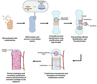
Endochondral ossification is one of the two essential pathways by which bone tissue is produced during fetal development of the mammalian skeletal system, the other pathway being intramembranous ossification. Both endochondral and intramembranous processes initiate from a precursor mesenchymal tissue, but their transformations into bone are different. In intramembranous ossification, mesenchymal tissue is directly converted into bone. On the other hand, endochondral ossification starts with mesenchymal tissue turning into an intermediate cartilage stage, which is eventually substituted by bone.

<span class="mw-page-title-main">Benign tumor</span> Mass of cells which cannot spread throughout the body

A benign tumor is a mass of cells (tumor) that does not invade neighboring tissue or metastasize. Compared to malignant (cancerous) tumors, benign tumors generally have a slower growth rate. Benign tumors have relatively well differentiated cells. They are often surrounded by an outer surface or stay contained within the epithelium. Common examples of benign tumors include moles and uterine fibroids.

<span class="mw-page-title-main">Ossification</span> Development process in bones

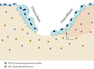
Ossification in bone remodeling is the process of laying down new bone material by cells named osteoblasts. It is synonymous with bone tissue formation. There are two processes resulting in the formation of normal, healthy bone tissue: Intramembranous ossification is the direct laying down of bone into the primitive connective tissue (mesenchyme), while endochondral ossification involves cartilage as a precursor.

<span class="mw-page-title-main">Ameloblastoma</span> Medical condition

Ameloblastoma is a rare, benign or cancerous tumor of odontogenic epithelium much more commonly appearing in the lower jaw than the upper jaw. It was recognized in 1827 by Cusack. This type of odontogenic neoplasm was designated as an adamantinoma in 1885 by the French physician Louis-Charles Malassez. It was finally renamed to the modern name ameloblastoma in 1930 by Ivey and Churchill.

<span class="mw-page-title-main">Enchondroma</span> Medical condition

Enchondroma is a type of benign bone tumor belonging to the group of cartilage tumors. There may be no symptoms, or it may present typically in the short tubular bones of the hands with a swelling, pain or pathological fracture.

<span class="mw-page-title-main">Osteochondroma</span> Medical condition

Osteochondromas are the most common benign tumors of the bones. The tumors take the form of cartilage-capped bony projections or outgrowth on the surface of bones exostoses. It is characterized as a type of overgrowth that can occur in any bone where cartilage forms bone. Tumors most commonly affect long bones about the knee and in the forearm. Additionally, flat bones such as the pelvis and scapula may be affected. Hereditary multiple exostoses usually present during childhood. Yet, the vast majority of affected individuals become clinically manifest by the time they reach adolescence. Osteochondromas occur in 3% of the general population and represent 35% of all benign tumors and 8% of all bone tumors. The majority of these tumors are solitary non-hereditary lesions and approximately 15% of osteochondromas occur as hereditary multiple exostoses preferably known as hereditary multiple osteochondromas (HMOs). Osteochondromas do not result from injury and the exact cause remains unknown. Recent research has indicated that multiple osteochondromas is an autosomal dominant inherited disease. Germ line mutations in EXT1 and EXT2 genes located on chromosomes 8 and 11 have been associated with the cause of the disease. The treatment choice for osteochondroma is surgical removal of solitary lesion or partial excision of the outgrowth, when symptoms cause motion limitations or nerve and blood vessel impingements. In hereditary multiple exostoses the indications of surgery are based upon multiple factors that are taken collectively, namely: patient's age, tumor location and number, accompanying symptomatology, esthetic concerns, family history and underlying gene mutation. A variety of surgical procedures have been employed to remedy hereditary multiple exostoses such as osteochondroma excision, bone lengthening, corrective osteotomy and hemiepiphysiodesis. Sometimes a combination of the previous procedures is used. The indicators of surgical success in regard to disease and patient characteristics are greatly disputable. Because most studies of hereditary multiple exostoses are retrospective and of limited sample size with missing data, the best evidence for each of the currently practiced surgical procedures is lacking.

<span class="mw-page-title-main">Myositis ossificans</span> Medical condition

Myositis ossificans comprises two syndromes characterized by heterotopic ossification (calcification) of muscle. The World Health Organization, 2020, has grouped myositis ossificans together with fibro-osseous pseudotumor of digits as a single specific entity in the category of fibroblastic and myofibroblastic tumors.

<span class="mw-page-title-main">Giant-cell tumor of bone</span> Medical condition

Giant-cell tumor of the bone (GCTOB), is a relatively uncommon tumor of the bone. It is characterized by the presence of multinucleated giant cells. Malignancy in giant-cell tumor is uncommon and occurs in about 2% of all cases. However, if malignant degeneration does occur, it is likely to metastasize to the lungs. Giant-cell tumors are normally benign, with unpredictable behavior. It is a heterogeneous tumor composed of three different cell populations. The giant-cell tumour stromal cells (GCTSC) constitute the neoplastic cells, which are from an osteoblastic origin and are classified based on expression of osteoblast cell markers such as alkaline phosphatase and osteocalcin. In contrast, the mononuclear histiocytic cells (MNHC) and multinucleated giant cell (MNGC) fractions are secondarily recruited and comprise the non-neoplastic cell population. They are derived from an osteoclast-monocyte lineage determined primarily by expression of CD68, a marker for monocytic precursor cells. In most patients, the tumors are slow to develop, but may recur locally in as many as 50% of cases.

<span class="mw-page-title-main">Aneurysmal bone cyst</span> Medical condition

Aneurysmal bone cyst (ABC) is a non-cancerous bone tumor composed of multiple varying sizes of spaces in a bone which are filled with blood. The term is a misnomer, as the lesion is neither an aneurysm nor a cyst. It generally presents with pain and swelling in the affected bone. Pressure on neighbouring tissues may cause compression effects such as neurological symptoms.

<span class="mw-page-title-main">Epiphyseal plate</span> Cartilage plate in the neck of a long bone

The epiphyseal plate, epiphysial plate, physis, or growth plate is a hyaline cartilage plate in the metaphysis at each end of a long bone. It is the part of a long bone where new bone growth takes place; that is, the whole bone is alive, with maintenance remodeling throughout its existing bone tissue, but the growth plate is the place where the long bone grows longer.

<span class="mw-page-title-main">Ollier disease</span> Medical condition

Ollier disease is a rare sporadic nonhereditary skeletal disorder in which typically benign cartilaginous tumors (enchondromas) develop near the growth plate cartilage. This is caused by cartilage rests that grow and reside within the metaphysis or diaphysis and eventually mineralize over time to form multiple enchondromas. Key signs of the disorder include asymmetry and shortening of the limb as well as an increased thickness of the bone margin. These symptoms are typically first visible during early childhood with the mean age of diagnosis being 13 years of age. Many patients with Ollier disease are prone to develop other malignancies including bone sarcomas that necessitate treatment and the removal of malignant bone neoplasm. Cases in patients with Ollier disease has shown a link to IDH1, IDH2, and PTH1R gene mutations. Currently, there are no forms of treatment for the underlying condition of Ollier disease but complications such as fractures, deformities, malignancies that arise from it can be treated through surgical procedures. The prevalence of this condition is estimated at around 1 in 100,000. It is unclear whether the men or women are more affected by this disorder due to conflicting case studies.

<span class="mw-page-title-main">Calcifying odontogenic cyst</span> Medical condition

Calcifying odontogenic cyst (COC) is a rare developmental lesion that comes from odontogenic epithelium. It is also known as a calcifying cystic odontogenic tumor, which is a proliferation of odontogenic epithelium and scattered nest of ghost cells and calcifications that may form the lining of a cyst, or present as a solid mass.

<span class="mw-page-title-main">Ameloblastic fibroma</span> Medical condition

An ameloblastic fibroma is a fibroma of the ameloblastic tissue, that is, an odontogenic tumor arising from the enamel organ or dental lamina. It may be either truly neoplastic or merely hamartomatous. In neoplastic cases, it may be labeled an ameloblastic fibrosarcoma in accord with the terminological distinction that reserves the word fibroma for benign tumors and assigns the word fibrosarcoma to malignant ones. It is more common in the first and second decades of life, when odontogenesis is ongoing, than in later decades. In 50% of cases an unerupted tooth is involved.

The calcifying epithelial odontogenic tumor (CEOT), also known as a Pindborg tumor, is an odontogenic tumor first recognized by the Danish pathologist Jens Jørgen Pindborg in 1955. It was previously described as an adenoid adamantoblastoma, unusual ameloblastoma and a cystic odontoma. Like other odontogenic neoplasms, it is thought to arise from the epithelial element of the enamel origin. It is a typically benign and slow growing, but invasive neoplasm.

<span class="mw-page-title-main">Pseudoachondroplasia</span> Inherited disorder of bone growth

Pseudoachondroplasia is an inherited disorder of bone growth. It is a genetic autosomal dominant disorder. It is generally not discovered until 2–3 years of age, since growth is normal at first. Pseudoachondroplasia is usually first detected by a drop of linear growth in contrast to peers, a waddling gait or arising lower limb deformities.

<span class="mw-page-title-main">Aponeurotic fibroma</span> Medical condition

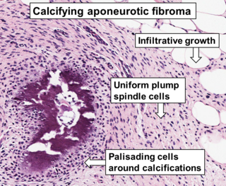
Aponeurotic fibroma, also known as calcifying aponeurotic fibroma, and juvenile aponeurotic fibroma is characterized by a lesion that usually presents as a painless, solitary, deep fibrous nodule, often adherent to tendon, fascia, or periosteum, on the hands and feet. The World Health Organization in 2020 reclassified aponeurotic fibroma nodules as a specific benign type of the fibroblastic and myofibroblastic tumors. Aponeurotic fibromas are diagnosed based on histopathology and treated by surgical excision. They are more common in males than females.

Extraskeletal myxoid chondrosarcoma (EMC) is a rare low-grade malignant mesenchymal neoplasm of the soft tissues, that differs from other sarcomas by unique histology and characteristic chromosomal translocations. There is an uncertain differentiation and neuroendocrine differentiation is even possible.

Fibrocartilaginous mesenchymoma of bone (FCMB) is an extremely rare tumor first described in 1984. About 26 cases have been reported in literature, with patient ages spanning from 9 to 25 years, though a case in a male infant aged 1 year and 7 months has been reported. Quick growth and bulky size are remarkable features of this tumor.

A juvenile active ossifying fibroma is a benign fibro-osseous neoplasm composed of mixture of stroma and bone characterized by rapid and destructive growth.

References

  1. 1 2 3 4 5 6 7 8 9 10 11 12 13 14 15 16 17 18 19 20 21 22 23 24 25 26 27 28 29 30 31 32 33 34 35 36 37 38 39 40 41 42 43 44 45 46 47 48 49 50 51 52 53 54 55 56 57 58 De Mattos, Camila B. R.; Angsanuntsukh, Chanika; Arkader, Alexandre; Dormans, John P. (2013). "Chondroblastoma and Chondromyxoid Fibroma". Journal of the American Academy of Orthopaedic Surgeons. 21 (4). Ovid Technologies (Wolters Kluwer Health): 225–233. doi:10.5435/jaaos-21-04-225. ISSN   1067-151X.
  2. 1 2 3 4 5 6 7 8 Romeo, S; Bovée, JVMG; Jadnanansing, NAA; Taminiau, AHM; Hogendoorn, PCW (2003-11-27). "Expression of cartilage growth plate signalling molecules in chondroblastoma". The Journal of Pathology. 202 (1). Wiley: 113–120. doi:10.1002/path.1501. ISSN   0022-3417.
  3. 1 2 3 4 5 6 7 8 9 10 11 12 13 Ramappa, A. J.; Lee, F. Y.; Tang, P.; Carlson, J. R.; Gebhardt, M. C.; Mankin, H. J. (2000). "Chondroblastoma of bone". The Journal of Bone and Joint Surgery. American Volume. 82 (8): 1140–1145. ISSN   0021-9355. PMID   10954104.
  4. 1 2 3 4 5 6 7 8 9 10 11 12 13 14 15 16 Kurt, Anne-Marie; Krishnan Unni, K.; Sim, Franklin H.; McLeod, Richard A. (1989). "Chondroblastoma of bone". Human Pathology. 20 (10). Elsevier BV: 965–976. doi:10.1016/0046-8177(89)90268-2. ISSN   0046-8177.
  5. 1 2 3 4 5 6 7 8 9 10 11 12 13 14 15 16 17 18 19 20 21 22 23 24 25 26 27 28 29 30 Damron, Timothy A (2022-10-14). "Chondroblastoma: Practice Essentials, Pathophysiology, Etiology". Medscape Reference. Retrieved 2024-06-22.
  6. 1 2 3 4 5 6 7 8 9 10 11 12 13 14 15 16 Turcotte, Robert E.; Kurt, Anne-Marie; Sim, Franklin H.; Krishnan Unni, K.; McLeod, Richard A. (1993). "Chondroblastoma". Human Pathology. 24 (9). Elsevier BV: 944–949. doi:10.1016/0046-8177(93)90107-r. ISSN   0046-8177.
  7. Chen, Wenqian; DiFrancesco, Lisa M. (June 2017). "Chondroblastoma: An Update". Archives of Pathology & Laboratory Medicine. 141 (6): 867–871. doi: 10.5858/arpa.2016-0281-RS . ISSN   0003-9985.