Chondrosarcoma

Last updated
Chondrosarcoma
Chondrosarcoma (1).jpg
Histopathologic image of chondrosarcoma of the chest wall. Surgical resection of recurrent mass. H & E stain.
Specialty Oncology   OOjs UI icon edit-ltr-progressive.svg

Chondrosarcoma is a bone sarcoma, a primary cancer composed of cells derived from transformed cells that produce cartilage. [1] A chondrosarcoma is a member of a category of tumors of bone and soft tissue known as sarcomas. About 30% of bone sarcomas are chondrosarcomas. [2] It is resistant to chemotherapy and radiotherapy. Unlike other primary bone sarcomas that mainly affect children and adolescents, a chondrosarcoma can present at any age. It more often affects the axial skeleton than the appendicular skeleton. [3]

Contents

Types

Table 1: Types of Chondrosarcoma
SubtypePercentage of cases %
ConventionalPrimary Central~75% [4]
Secondary Peripheral~10% [4]
Periosteal (aka juxtacortical) [5] [6] <1% [4]
RareDedifferentiated~10% [4]
Mesenchymal~2% [4]
Clear-cell~2% [6]

Symptoms and signs

Causes

The cause is unknown. There may be a history of enchondroma or osteochondroma. A small minority of secondary chondrosarcomas occur in people with Maffucci syndrome and Ollier disease. [7]

It has been associated with faulty isocitrate dehydrogenase 1 and 2 enzymes, which are also associated with gliomas and leukemias. [8]

Diagnosis

MRI of a left-pelvis chondrosarcoma in a 26-year-old male Mrichondrosarcoma.jpg
MRI of a left-pelvis chondrosarcoma in a 26-year-old male
Metastatic chondrosarcoma at the lower lip Metastatic chondrosarcoma at the lower lip.jpg
Metastatic chondrosarcoma at the lower lip

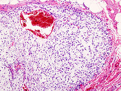
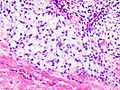
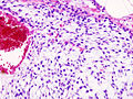
Imaging studies – including radiographs ("x-rays"), computerized tomography (CT), and magnetic resonance imaging (MRI) – are often used to make a presumptive diagnosis of chondrosarcoma. [9] However, a definitive diagnosis depends on the identification of malignant cancer cells producing cartilage in a biopsy specimen that has been examined by a pathologist. In a few cases, usually of highly anaplastic tumors, immunohistochemistry (IHC) is required.[ citation needed ]

There are no blood tests currently available to enable an oncologist to render a diagnosis of chondrosarcoma. The most characteristic imaging findings are usually obtained with CT. [10]

Nearly all chondrosarcoma patients appear to be in good health. Often, patients are not aware of the growing tumor until there is a noticeable lump or pain. Earlier diagnosis is generally accidental when a patient undergoes testing for another problem and physicians discover cancer. Occasionally the first symptom will be a broken bone at the cancerous site. Any broken bone that occurs from mild trauma warrants further investigation, although there are many conditions that can lead to weak bones, and this form of cancer is not a common cause of such breaks.[ citation needed ]

Treatment

Treatment depends on the location of the disease and the aggressiveness of the tumors. [11] Because chondrosarcomas are rare, they are treated at specialist hospitals with Sarcoma Centers.[ citation needed ]

Surgery is the main form of treatment for chondrosarcoma. Musculoskeletal tumor specialists or orthopedic oncologists are usually chosen to treat chondrosarcoma, unless it is located in the skull, spine, or chest cavity, in which case, a neurosurgeon or thoracic surgeon experienced with sarcomas is chosen. Often, a limb-sparing operation can be performed, but in some cases amputation is unavoidable. Amputation of the arm, leg, jaw, or half of the pelvis (called a hemipelvectomy) may be necessary in some cases.[ citation needed ]

There are two kinds of hemipelvectomy – internal and external.

Amputation at the hip is called hip disarticulation and amputees who have had this amputation are also called hip disartics.

Chemotherapy or traditional radiotherapy are not very effective for most chondrosarcomas, although proton therapy is showing promise with local tumor control at over 80%. [12]

Complete surgical ablation is the most effective treatment, but sometimes this is difficult. Proton therapy radiation can be useful in awkward locations to make surgery more effective.

Recent studies have shown that induction of apoptosis in high-grade chondrosarcoma, both directly and by enhancement of response to chemotherapy and radiation, is a valid therapeutic strategy. [13]

Prognosis

Prognosis depends on how early the cancer is discovered and treated. For the least aggressive grade, about 90% of patients survive more than five years after diagnosis. People usually have a good survival rate at the low-grade volume of cancer. [3] For the most aggressive grade, only 10% of patients will survive one year. Tumors may recur in the future. Follow up scans are extremely important for chondrosarcoma to make sure there has been no recurrence or metastasis, which usually occurs in the lungs. [14]

Related Research Articles

<span class="mw-page-title-main">Brain tumor</span> Neoplasm in the brain

A brain tumor occurs when abnormal cells form within the brain. There are two main types of tumors: malignant tumors and benign (non-cancerous) tumors. These can be further classified as primary tumors, which start within the brain, and secondary tumors, which most commonly have spread from tumors located outside the brain, known as brain metastasis tumors. All types of brain tumors may produce symptoms that vary depending on the size of the tumor and the part of the brain that is involved. Where symptoms exist, they may include headaches, seizures, problems with vision, vomiting and mental changes. Other symptoms may include difficulty walking, speaking, with sensations, or unconsciousness.

<span class="mw-page-title-main">Sarcoma</span> Medical condition

A sarcoma is a malignant tumor, a type of cancer that arises from transformed cells of mesenchymal origin. Connective tissue is a broad term that includes bone, cartilage, fat, vascular, or hematopoietic tissues, and sarcomas can arise in any of these types of tissues. As a result, there are many subtypes of sarcoma, which are classified based on the specific tissue and type of cell from which the tumor originates. Sarcomas are primary connective tissue tumors, meaning that they arise in connective tissues. This is in contrast to secondary connective tissue tumors, which occur when a cancer from elsewhere in the body spreads to the connective tissue. The word sarcoma is derived from the Greek σάρκωμα sarkōma "fleshy excrescence or substance", itself from σάρξsarx meaning "flesh".

<span class="mw-page-title-main">Bone tumor</span> Medical condition

A bone tumor is an abnormal growth of tissue in bone, traditionally classified as noncancerous (benign) or cancerous (malignant). Cancerous bone tumors usually originate from a cancer in another part of the body such as from lung, breast, thyroid, kidney and prostate. There may be a lump, pain, or neurological signs from pressure. A bone tumor might present with a pathologic fracture. Other symptoms may include fatigue, fever, weight loss, anemia and nausea. Sometimes there are no symptoms and the tumour is found when investigating another problem.

<span class="mw-page-title-main">Osteosarcoma</span> Cancerous tumour in a bone

An osteosarcoma (OS) or osteogenic sarcoma (OGS) is a cancerous tumor in a bone. Specifically, it is an aggressive malignant neoplasm that arises from primitive transformed cells of mesenchymal origin and that exhibits osteoblastic differentiation and produces malignant osteoid.

<span class="mw-page-title-main">Rhabdomyosarcoma</span> Medical condition

Rhabdomyosarcoma (RMS) is a highly aggressive form of cancer that develops from mesenchymal cells that have failed to fully differentiate into myocytes of skeletal muscle. Cells of the tumor are identified as rhabdomyoblasts.

<span class="mw-page-title-main">Proton therapy</span> Medical Procedure

In medicine, proton therapy, or proton radiotherapy, is a type of particle therapy that uses a beam of protons to irradiate diseased tissue, most often to treat cancer. The chief advantage of proton therapy over other types of external beam radiotherapy is that the dose of protons is deposited over a narrow range of depth; hence in minimal entry, exit, or scattered radiation dose to healthy nearby tissues.

<span class="mw-page-title-main">Synovial sarcoma</span> Medical condition

A synovial sarcoma is a rare form of cancer which occurs primarily in the extremities of the arms or legs, often in proximity to joint capsules and tendon sheaths. It is a type of soft-tissue sarcoma.

<span class="mw-page-title-main">Malignant peripheral nerve sheath tumor</span> Medical condition

A malignant peripheral nerve sheath tumor (MPNST) is a form of cancer of the connective tissue surrounding nerves. Given its origin and behavior it is classified as a sarcoma. About half the cases are diagnosed in people with neurofibromatosis; the lifetime risk for an MPNST in patients with neurofibromatosis type 1 is 8–13%. MPNST with rhabdomyoblastomatous component are called malignant triton tumors.

<span class="mw-page-title-main">Desmoplastic small-round-cell tumor</span> Aggressive and rare cancer

Desmoplastic small-round-cell tumor (DSRCT) is an aggressive and rare cancer that primarily occurs as masses in the abdomen. Other areas affected may include the lymph nodes, the lining of the abdomen, diaphragm, spleen, liver, chest wall, skull, spinal cord, large intestine, small intestine, bladder, brain, lungs, testicles, ovaries, and the pelvis. Reported sites of metastatic spread include the liver, lungs, lymph nodes, brain, skull, and bones. It is characterized by the EWS-WT1 fusion protein.

<span class="mw-page-title-main">Hemangiopericytoma</span> Medical condition

A hemangiopericytoma is a type of soft-tissue sarcoma that originates in the pericytes in the walls of capillaries. When inside the nervous system, although not strictly a meningioma tumor, it is a meningeal tumor with a special aggressive behavior. It was first characterized in 1942.

<span class="mw-page-title-main">Undifferentiated pleomorphic sarcoma</span> Medical condition

Undifferentiated pleomorphic sarcoma (UPS), also termed pleomorphic myofibrosarcoma, high-grade myofibroblastic sarcoma, and high-grade myofibrosarcoma, is characterized by the World Health Organization (WHO), 2020, as a rare, poorly differentiated neoplasm, i.e. an abnormal growth of cells that have an unclear identity and/or cell of origin. WHO classified it as one of the undifferentiated/unclassified sarcomas in the category of tumors of uncertain differentiation. Sarcomas are cancers known or thought to derive from mesenchymal stem cells that typically develop in bone, muscle, fat, blood vessels, lymphatic vessels, tendons, and ligaments. More than 70 sarcoma subtypes have been described. The UPS subtype of these sarcomas consists of tumor cells that are poorly differentiated and may appear as spindle-shaped cells, histiocytes, and giant cells. UPS is considered a diagnosis that defies formal sub-classification after thorough histologic, immunohistochemical, and ultrastructural examinations fail to identify the type of cells involved.

<span class="mw-page-title-main">Ollier disease</span> Medical condition

Ollier disease is a rare sporadic nonhereditary skeletal disorder in which typically benign cartilaginous tumors (enchondromas) develop near the growth plate cartilage. This is caused by cartilage rests that grow and reside within the metaphysis or diaphysis and eventually mineralize over time to form multiple enchondromas. Key signs of the disorder include asymmetry and shortening of the limb as well as an increased thickness of the bone margin. These symptoms are typically first visible during early childhood with the mean age of diagnosis being 13 years of age. Many patients with Ollier disease are prone to develop other malignancies including bone sarcomas that necessitate treatment and the removal of malignant bone neoplasm. Cases in patients with Ollier disease has shown a link to IDH1, IDH2, and PTH1R gene mutations. Currently, there are no forms of treatment for the underlying condition of Ollier disease but complications such as fractures, deformities, malignancies that arise from it can be treated through surgical procedures. The prevalence of this condition is estimated at around 1 in 100,000. It is unclear whether the men or women are more affected by this disorder due to conflicting case studies.

<span class="mw-page-title-main">Mixed Müllerian tumor</span> Medical condition

A malignant mixed Müllerian tumor, also known as malignant mixed mesodermal tumor (MMMT) is a cancer found in the uterus, the ovaries, the fallopian tubes and other parts of the body that contains both carcinomatous and sarcomatous components. It is divided into two types, homologous and a heterologous type. MMMT account for between two and five percent of all tumors derived from the body of the uterus, and are found predominantly in postmenopausal women with an average age of 66 years. Risk factors are similar to those of adenocarcinomas and include obesity, exogenous estrogen therapies, and nulliparity. Less well-understood but potential risk factors include tamoxifen therapy and pelvic irradiation.

<span class="mw-page-title-main">Ewing sarcoma</span> Type of cancer

Ewing sarcoma is a type of cancer that forms in bone or soft tissue. Symptoms may include swelling and pain at the site of the tumor, fever, and a bone fracture. The most common areas where it begins are the legs, pelvis, and chest wall. In about 25% of cases, the cancer has already spread to other parts of the body at the time of diagnosis. Complications may include a pleural effusion or paraplegia.

<span class="mw-page-title-main">Atypical teratoid rhabdoid tumor</span> Medical condition

An atypical teratoid rhabdoid tumor (AT/RT) is a rare tumor usually diagnosed in childhood. Although usually a brain tumor, AT/RT can occur anywhere in the central nervous system (CNS), including the spinal cord. About 60% will be in the posterior cranial fossa. One review estimated 52% in the posterior fossa, 39% are supratentorial primitive neuroectodermal tumors (sPNET), 5% are in the pineal, 2% are spinal, and 2% are multifocal.

The Ewing family of tumors (EFTs) is a group of small cell sarcomas including Ewing sarcoma of the bone, extra osseous Ewing tumors, and primitive neuroectodermal tumors. They are rare cancers, usually diagnosed in peoples' twenties. The sarcoma of bone is the most common of the variants. All forms are predisposed to metastasis and have had historically high rates of mortality. The family of tumors shares a common translocation mutation of the EWS gene on chromosome 22 to an ETS-type gene, most commonly the FLI1 gene. EFTs are highly malignant, with 5-year survival for patients with metastatic disease at 20%. The current standard of care includes resection, radiation, and chemotherapy.

<span class="mw-page-title-main">Bone metastasis</span> Medical condition

Bone metastasis, or osseous metastatic disease, is a category of cancer metastases that results from primary tumor invasion to bone. Bone-originating primary tumors such as osteosarcoma, chondrosarcoma, and Ewing's sarcoma are rare; the most common bone tumor is a metastasis Bone metastases can be classified as osteolytic, osteoblastic, or both. Unlike hematologic malignancies which originate in the blood and form non-solid tumors, bone metastases generally arise from epithelial tumors and form a solid mass inside the bone. Bone metastases, especially in a state of advanced disease, can cause severe pain, characterized by a dull, constant ache with periodic spikes of incident pain.

A bone sarcoma is a primary malignant bone tumour, a type of sarcoma that starts in the bones. This is in contrast to most bone cancers that are secondary having developed as a metastasis from another cancer. Bone sarcomas are rare, and mostly affect the legs. The other type of sarcoma is a soft-tissue sarcoma.

Myxofibrosarcoma (MFS), although a rare type of tumor, is one of the most common soft tissue sarcomas, i.e. cancerous tumors, that develop in the soft tissues of elderly individuals. Initially considered to be a type of histiocytoma termed fibrous histiocytoma or myxoid variant of malignant fibrous histiocytoma, Angervall et al. termed this tumor myxofibrosarcoma in 1977. In 2020, the World Health Organization reclassified MFS as a separate and distinct tumor in the category of malignant fibroblastic and myofibroblastic tumors.

Sclerosing epithelioid fibrosarcoma (SEF) is a very rare malignant tumor of soft tissues that on microscopic examination consists of small round or ovoid neoplastic epithelioid fibroblast-like cells, i.e. cells that have features resembling both epithelioid cells and fibroblasts. In 2020, the World Health Organization classified SEF as a distinct tumor type in the category of malignant fibroblastic and myofibroblastic tumors. However, current studies have reported that low-grade fibromyxoid sarcoma (LGFMS) has many clinically and pathologically important features characteristic of SEF; these studies suggest that LGSFMS may be an early form of, and over time progress to become, a SEF. Since the World Health Organization has classified LGFMS as one of the malignant fibroblastic and myofibroblastic tumors that is distinctly different than SEF, SEF and LGFMS are here regarded as different tumor forms.

References

  1. Gelderblom H, Hogendoorn PC, Dijkstra SD, et al. (March 2008). "The clinical approach towards chondrosarcoma". Oncologist. 13 (3): 320–9. doi:10.1634/theoncologist.2007-0237. PMID   18378543. S2CID   20484590.
  2. "AANOS". Archived from the original on 2012-04-30. Retrieved 2011-10-30.
  3. 1 2 "Chondrosarcoma: Overview – eMedicine Radiology" . Retrieved 2009-02-14.
  4. 1 2 3 4 5 Gelderblom, Hans; Hogendoorn, Pancras C. W.; Dijkstra, Sander D.; van Rijswijk, Carla S.; Krol, Augustinus D.; Taminiau, Antonie H. M.; Bovée, Judith V. M. G. (March 2008). "The clinical approach towards chondrosarcoma". The Oncologist. 13 (3): 320–329. doi:10.1634/theoncologist.2007-0237. ISSN   1083-7159. PMID   18378543. S2CID   20484590.
  5. Schajowicz, F. (November 1977). "Juxtacortical chondrosarcoma". The Journal of Bone and Joint Surgery. British Volume. 59-B (4): 473–480. doi: 10.1302/0301-620X.59B4.270475 . ISSN   0301-620X. PMID   270475.
  6. 1 2 Pathology and genetics of tumours of soft tissue and bone. Fletcher, Christopher D. M., Unni, K. Krishnan, 1941–, Mertens, Fredrik., World Health Organization., International Agency for Research on Cancer. Lyon: IARC Press. 2002. ISBN   978-92-832-2413-6. OCLC   51001831.{{cite book}}: CS1 maint: others (link)
  7. Lin, Patrick P.; Moussallem, Charbel D.; Deavers, Michael T. (Oct 2010). "Secondary chondrosarcoma". The Journal of the American Academy of Orthopaedic Surgeons. 18 (10): 608–615. doi:10.5435/00124635-201010000-00004. ISSN   1067-151X. PMID   20889950. S2CID   25873835.
  8. Amary, M. Fernanda; Bacsi, Krisztian; Maggiani, Francesca; Damato, Stephen; Halai, Dina; Berisha, Fitim; Pollock, Robin; O'Donnell, Paul; Grigoriadis, Anita (Jul 2011). "IDH1 and IDH2 mutations are frequent events in central chondrosarcoma and central and periosteal chondromas but not in other mesenchymal tumours". The Journal of Pathology. 224 (3): 334–343. doi:10.1002/path.2913. ISSN   1096-9896. PMID   21598255. S2CID   24201208.
  9. CT scan and gross pathology of a chondrosarcoma 2012-12-18
  10. "Chondrosarcoma". The Lecturio Medical Concept Library. Retrieved 8 July 2021.
  11. "Chondrosarcoma : Cancerbackup". Archived from the original on 2008-09-05. Retrieved 2009-02-14.
  12. "Proton Therapy at PSI (Protonentherapie)" . Retrieved 2009-02-14.
  13. Jamil N, Howie S, Salter DM. Therapeutic molecular targets in human chondrosarcoma .Int J Exp Pathol 2010; 91:387–93
  14. Limaiem F, Davis D, Sticco K (2021). "Chondrosarcoma". National Center for Biotechnology Information, U.S. National Library of Medicine. PMID   30844159 . Retrieved 8 July 2021.