Foreign body granuloma | |
---|---|
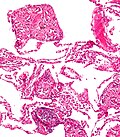
Transvaginal ultrasonography showing a foreign body granuloma at right as a hypoechoic (dark) area around a perforated intrauterine device. The uterus is at left. | |
Specialty | Dermatology |
A foreign body reaction (FBR) is a typical tissue response to a foreign body within biological tissue. [1] It usually includes the formation of a foreign body granuloma . [2] Tissue encapsulation of an implant is an example, as is inflammation around a splinter. [3] Foreign body granuloma formation consists of protein adsorption, macrophages, multinucleated foreign body giant cells (macrophage fusion), fibroblasts, and angiogenesis. It has also been proposed that the mechanical property of the interface between an implant and its surrounding tissues is critical for the host response. [4]
In the long term, the foreign body reaction results in encapsulation of the foreign body within a calcified shell. For example, a lithopedion is a rare phenomenon which occurs most commonly when a fetus dies during an abdominal pregnancy, [5] is too large to be reabsorbed by the body, and calcifies.
Following biomaterial implantation, blood and body fluids contact the implant surface. Host blood proteins adsorb onto the implant surface and a fibrin matrix forms. [6] Acute and chronic inflammation follow the initial blood protein deposition and matrix formation. [6] Macrophages at the implant site fuse to form foreign body giant cells. [6] Following the inflammatory response, granulation tissue form. The end stage of the foreign body reaction is the fibrous capsule formation around the implanted biomaterial. [6] The biocompatibility of the device affects the severity of the foreign body reaction. [7] The foreign body reaction can lead to device failure. [8]
During blood-biomaterial interaction, blood proteins spontaneously adsorb to the biomaterial surface. [9] The biomaterial surface properties affect the types, concentrations, and conformation of proteins that adsorb to the surface. [9] The Vroman effect can describe the time-dependent behavior of this protein adsorption. [9] Surface-adsorbed proteins regulate inflammatory cell interaction and adhesion. [9] The deposited proteins allow inflammatory cells to attach via integrins. [9] The biomaterial surface can also recruit and activate complement proteins. [9]
The composition and conformation of adsorbed proteins on the implant surface is critical to the foreign body reaction. For the first two days, neutrophils are the primary cell type that deposit on the implant surface. Neutrophils release degradative enzymes and reactive oxygen intermediates that damages the implant. Platelets from the blood-biomaterial interaction release inflammatory cytokines that cause monocytes and macrophages to extravasate and migrate to the implant site. [10] The degranulation and release of histamine from mast cells further recruits macrophages to the biomaterial. [10] Macrophages adhere to the biomaterial surface based on the surface protein deposits and produce cytokines that further recruit macrophages. [10] Foreign body granuloma forms as immune cells accumulate on the biomaterial surface in an attempt to eliminate the biomaterial. [8]
Adherent macrophages at the implant site can fuse into a multinucleated cell called foreign body giant cell. [11] Foreign body giant cell formation depends on the biomaterial surface properties and on the presence of interleukin-4 and interleukin-13. [11] Foreign body giant cells release reactive oxygen intermediates, degradative enzymes, and acid onto the biomaterial surface. [11] Foreign body giant cells also attempt to engulf the biomaterial for degradation. [11] Adherent macrophages and foreign body giant cells degrade biomaterials and can lead to device failure. [11] Foreign body giant cells remain on the surface of the implanted device throughout the device's lifetime. [11]
The end-stage healing response is the fibrous encapsulation of the biomaterial. [12] Macrophages and foreign body giant cells release cytokines that attract fibroblasts. Fibroblasts create a collagenous fibrous capsule to separate the biomaterial from the surrounding tissue. [12] The fibrous capsule may hinder the device's function, such as drug diffusion for drug delivery systems or normal tissue regeneration for tissue engineering implants. [12]
The foreign body giant cell formation and the fibrous encapsulation of the implanted device can affect the function of the implanted device and lead to its failure. [13] Reducing the foreign body reaction can promote better device performance and durability. [13] Even though many types of implants cannot completely escape the foreign body reaction, there are many ways to engineer biomaterials to reduce the foreign body reaction. [13] Reducing nonspecific protein adsorption is one important method to prevent fibrous encapsulation of the implant. [13]
The physical properties of the implant's surface, such as size, shape, topology, and strength, influence protein adsorption and immune cell interactions with the implant. [13] Generally, surface roughness induces greater protein adsorption and triggers greater immune response. [13] Thicker cylindrical implants on the millimeter scale were shown to produce greater fibrous encapsulation. [13] However, there may not be an absolute relationship between the modification of the biomaterial and the associated foreign body response, because some biomaterials intrinsically induce a stronger foreign body response than other biomaterials would. [13]
Implanted biomaterial can be loaded with different drugs to perform anti-inflammatory functions or promote angiogenesis to further integrate the implant with the host. [14] The sustained release of certain anti-inflammatory drugs from the biomaterial, such as dexamethasone, is shown to increase implant life by preventing inflammation and fibrosis. [14]
Coating the implant surface with biomimetic biomaterial may reduce the foreign body reaction and reduce the device failure rate. [15] Biomaterials that mimic the extracellular matrix can significantly reduce the inflammatory response as well as reduce foreign body giant cell formation. [15]
Developing materials that resist protein adsorption, fibrous encapsulation, or foreign body giant cell formation is important to resist the foreign body reaction. [13] Commonly used biomaterials like polyethylene glycol and polyhydroxyethylmethacrylate resist nonspecific protein adsorption but are easily degraded in the in vivo oxidative environment. Therefore, scientists are looking for new materials that intrinsically resist the foreign body reaction. [13]
Zwitterionic polymers are widely used in surface modification because they can inhibit non-specific protein adhesion. [16] Zwitterionic hydrogels also promote angiogenesis in surrounding tissues. [17] Commonly used zwitterionic materials include sulfobetaine, carboxybetaine, and phosphorylcholine. [13]
Zwitterionic polymers have the ability to reduce nonspecific adsorption of proteins and cells at biological interfaces. Zwitterionic coatings are often used to minimize the negative effects of foreign body response for implanted devices and improve the device's biocompatibility. The mechanism is related to surface charge balancing and creation of a strong hydration sphere. [18]
Zwitterionic coatings can be covalently attached by “grafting to” and “grafting from” methods. With “grafting to” methods, the surface is modified with the polymer after synthesis, whereas with “grafting from” methods, polymer is directly synthesized on a modified surface. [18]
Several “grafting to” methods have been used to covalently attach zwitterionic polymers to surfaces. Anderson group developed attachment to the surface via polydopamine conjugation. [19] In this case, copolymer should contain a thiol group. Nazarova and coworkers synthesized MPC copolymers with 2-methacrylamido-D-glucose, N-vinylpyrrolidone, and N-vinyl-N-methyl-acetamide and grafted them onto the surface of carbon fibre biosorbent using γ-radiation. [20] MPC copolymers with trimethoxysilylpropyl methacrylate can be thermally cured and self-crosslinked. [21]
Alginates are widely used for their low cost, low toxicity, and tunability. [13] However, implantation of only alginate will trigger serious foreign body reaction. [13] There are a few triazole-containing alginate derivative microspheres that remain clear of fibrotic deposition in mice, but more studies are required to explore the relationship between triazole groups and the inflammatory response. [22]
Polyethylene glycol peptides and zwitterionic peptides have immunomodulatory functions that help to resist the foreign body reaction. [13]
A granuloma is an aggregation of macrophages that forms in response to chronic inflammation. This occurs when the immune system attempts to isolate foreign substances that it is otherwise unable to eliminate. Such substances include infectious organisms including bacteria and fungi, as well as other materials such as foreign objects, keratin, and suture fragments.
An implant is a medical device manufactured to replace a missing biological structure, support a damaged biological structure, or enhance an existing biological structure. For example, an implant may be a rod, used to strengthen weak bones. Medical implants are human-made devices, in contrast to a transplant, which is a transplanted biomedical tissue. The surface of implants that contact the body might be made of a biomedical material such as titanium, silicone, or apatite depending on what is the most functional. In 2018, for example, American Elements developed a nickel alloy powder for 3D printing robust, long-lasting, and biocompatible medical implants. In some cases implants contain electronics, e.g. artificial pacemaker and cochlear implants. Some implants are bioactive, such as subcutaneous drug delivery devices in the form of implantable pills or drug-eluting stents.
Bioglass 45S5 or calcium sodium phosphosilicate, is a bioactive glass specifically composed of 45 wt% SiO2, 24.5 wt% CaO, 24.5 wt% Na2O, and 6.0 wt% P2O5. Typical applications of Bioglass 45S5 include: bone grafting biomaterials, repair of periodontal defects, cranial and maxillofacial repair, wound care, blood loss control, stimulation of vascular regeneration, and nerve repair.
A biomaterial is a substance that has been engineered to interact with biological systems for a medical purpose – either a therapeutic or a diagnostic one. The corresponding field of study, called biomaterials science or biomaterials engineering, is about fifty years old. It has experienced steady growth over its history, with many companies investing large amounts of money into the development of new products. Biomaterials science encompasses elements of medicine, biology, chemistry, tissue engineering and materials science.
Osteostimulation is a technique attempted for improving healing of bone injuries or defects. It has not however been found to be significantly effective in increasing bone healing.
A foreign-body giant cell is a collection of fused macrophages which are generated in response to the presence of a large foreign body. This is particularly evident with catheters, parasites, or biomaterials that are inserted into the body for replacement or regeneration of diseased or damaged tissues. Foreign body giant cells are also produced to digest foreign material that is too large for phagocytosis. The inflammatory process that creates these cells often leads to a foreign body granuloma.
Nano-scaffolding or nanoscaffolding is a medical process used to regrow tissue and bone, including limbs and organs. The nano-scaffold is a three-dimensional structure composed of polymer fibers very small that are scaled from a Nanometer scale. Developed by the American military, the medical technology uses a microscopic apparatus made of fine polymer fibers called a scaffold. Damaged cells grip to the scaffold and begin to rebuild missing bone and tissue through tiny holes in the scaffold. As tissue grows, the scaffold is absorbed into the body and disappears completely.
Acellular dermis is a type of biomaterial derived from processing human or animal tissues to remove cells and retain portions of the extracellular matrix (ECM). These materials are typically cell-free, distinguishing them from classical allografts and xenografts, can be integrated or incorporated into the body, and have been FDA approved for human use for more than 10 years in a wide range of clinical indications.
Cell encapsulation is a possible solution to graft rejection in tissue engineering applications. Cell microencapsulation technology involves immobilization of cells within a polymeric semi-permeable membrane. It permits the bidirectional diffusion of molecules such as the influx of oxygen, nutrients, growth factors etc. essential for cell metabolism and the outward diffusion of waste products and therapeutic proteins. At the same time, the semi-permeable nature of the membrane prevents immune cells and antibodies from destroying the encapsulated cells, regarding them as foreign invaders.
Adsorption is the accumulation and adhesion of molecules, atoms, ions, or larger particles to a surface, but without surface penetration occurring. The adsorption of larger biomolecules such as proteins is of high physiological relevance, and as such they adsorb with different mechanisms than their molecular or atomic analogs. Some of the major driving forces behind protein adsorption include: surface energy, intermolecular forces, hydrophobicity, and ionic or electrostatic interaction. By knowing how these factors affect protein adsorption, they can then be manipulated by machining, alloying, and other engineering techniques to select for the most optimal performance in biomedical or physiological applications.
Biomaterials are materials that are used in contact with biological systems. Biocompatibility and applicability of surface modification with current uses of metallic, polymeric and ceramic biomaterials allow alteration of properties to enhance performance in a biological environment while retaining bulk properties of the desired device.
Titanium was first introduced into surgeries in the 1950s after having been used in dentistry for a decade prior. It is now the metal of choice for prosthetics, internal fixation, inner body devices, and instrumentation. Titanium is used from head to toe in biomedical implants. One can find titanium in neurosurgery, bone conduction hearing aids, false eye implants, spinal fusion cages, pacemakers, toe implants, and shoulder/elbow/hip/knee replacements along with many more. The main reason why titanium is often used in the body is due to titanium's biocompatibility and, with surface modifications, bioactive surface. The surface characteristics that affect biocompatibility are surface texture, steric hindrance, binding sites, and hydrophobicity (wetting). These characteristics are optimized to create an ideal cellular response. Some medical implants, as well as parts of surgical instruments are coated with titanium nitride (TiN).
Surgical mesh is a medical implant made of loosely woven mesh, which is used in surgery as either a permanent or temporary structural support for organs and other tissues. Surgical mesh can be made from both inorganic and biological materials and is used in a variety of surgeries, although hernia repair is the most common application. It can also be used for reconstructive work, such as in pelvic organ prolapse or to repair physical defects created by extensive resections or traumatic tissue loss.
Biomaterials exhibit various degrees of compatibility with the harsh environment within a living organism. They need to be nonreactive chemically and physically with the body, as well as integrate when deposited into tissue. The extent of compatibility varies based on the application and material required. Often modifications to the surface of a biomaterial system are required to maximize performance. The surface can be modified in many ways, including plasma modification and applying coatings to the substrate. Surface modifications can be used to affect surface energy, adhesion, biocompatibility, chemical inertness, lubricity, sterility, asepsis, thrombogenicity, susceptibility to corrosion, degradation, and hydrophilicity.
Bovine submaxillary mucin (BSM) coatings are a surface treatment provided to biomaterials intended to reduce the growth of disadvantageous bacteria and fungi such as S. epidermidis, E. coli, and Candida albicans. BSM is a substance extracted from the fresh salivary glands of cows. It exhibits unique physical properties, such as high molecular weight and amphiphilicity, that allow it to be used for many biomedical applications.
Ultra-low fouling is a rating of a surface's ability to shed potential contamination. Surfaces are prone to contamination, which is a phenomenon known as fouling. Unwanted adsorbates caused by fouling change the properties of a surface, which is often counter-productive to the function of that surface. Consequently, a necessity for anti-fouling surfaces has arisen in many fields: blocked pipes inhibit factory productivity, biofouling increases fuel consumption on ships, medical devices must be kept sanitary, etc. Although chemical fouling inhibitors, metallic coatings, and cleaning processes can be used to reduce fouling, non-toxic surfaces with anti-fouling properties are ideal for fouling prevention. To be considered effective, an ultra-low fouling surface must be able to repel and withstand the accumulation of detrimental aggregates down to less than 5 ng/cm2. A recent surge of research has been conducted to create these surfaces in order to benefit the biological, nautical, mechanical, and medical fields.
As with any material implanted in the body, it is important to minimize or eliminate foreign body response and maximize effectual integration. Neural implants have the potential to increase the quality of life for patients with such disabilities as Alzheimer's, Parkinson's, epilepsy, depression, and migraines. With the complexity of interfaces between a neural implant and brain tissue, adverse reactions such as fibrous tissue encapsulation that hinder the functionality, occur. Surface modifications to these implants can help improve the tissue-implant interface, increasing the lifetime and effectiveness of the implant.
A chronic electrode implant is an electronic device implanted chronically into the brain or other electrically excitable tissue. It may record electrical impulses in the brain or may stimulate neurons with electrical impulses from an external source.
Tissue engineered heart valves (TEHV) offer a new and advancing proposed treatment of creating a living heart valve for people who are in need of either a full or partial heart valve replacement. Currently, there are over a quarter of a million prosthetic heart valves implanted annually, and the number of patients requiring replacement surgeries is only suspected to rise and even triple over the next fifty years. While current treatments offered such as mechanical valves or biological valves are not deleterious to one's health, they both have their own limitations in that mechanical valves necessitate the lifelong use of anticoagulants while biological valves are susceptible to structural degradation and reoperation. Thus, in situ (in its original position or place) tissue engineering of heart valves serves as a novel approach that explores the use creating a living heart valve composed of the host's own cells that is capable of growing, adapting, and interacting within the human body's biological system.
Bioinstructive materials provide instruction to biological cells or tissue, for example immune instruction when monocytes are cultured on certain polymers they polarise to pro- or anti-inflammatory macrophages with potential applications in implanted devices, or materials for the repair of musculoskeletal tissues. Due to the paucity of information on the mechanism of materials control of cells, beyond the general recognition of the important role of adsorbed biomolecules, high throughput screening of large libraries of materials, topographies, and shapes are often used to identify cell instructive material systems. Applications of bioinstructive materials as substrates for stem cell production, cell delivery and reduction of foreign body reaction and coatings to reduce infections on medical devices. This non-leaching approach is distinct from strategies of infection control relying on antibiotic release, cytokine delivery or guidance of cells by surface located epitopes inspired by nature.