Mitochondrial trifunctional protein deficiency

Last updated
Mitochondrial trifunctional protein deficiency
Other namesTFP deficiency [1]
Autorecessive.svg
Mitochondrial trifunctional protein deficiency has an autosomal recessive pattern of inheritance
Symptoms Cardiomyopathy, skeletal myopathy [2]
TypesMutations in the HADHA and HADHB gene [2]
Diagnostic method CBC, Urine test [3]
TreatmentLow fat diet, Limited exercise [3]

Mitochondrial trifunctional protein deficiency (MTP deficiency or MTPD) is an autosomal recessive fatty acid oxidation disorder [4] that prevents the body from converting certain fats to energy, particularly during periods without food. [5] [6] People with this disorder have inadequate levels of an enzyme that breaks down a certain group of fats called long-chain fatty acids. [6]

Contents

Signs and symptoms

The presentation of mitochondrial trifunctional protein deficiency may begin during infancy, features that occur are: low blood sugar, weak muscle tone, and liver problems. Infants with this disorder are at risk for heart problems, breathing difficulties, and pigmentary retinopathy. Signs and symptoms of mitochondrial trifunctional protein deficiency that may begin after infancy include hypotonia, muscle pain, a breakdown of muscle tissue, and a loss of sensation in the extremities called peripheral neuropathy. Some who have MTP deficiency show a progressive course associated with myopathy, and recurrent rhabdomyolysis. [2] [6] [7]

Genetics

HADHB function in beta-oxidation Beta-ketothiolase.png
HADHB function in beta-oxidation

The genetics of mitochondrial trifunctional protein deficiency is based on mutations in the HADHA [8] and HADHB [9] genes which cause this disorder. These genes each provide instructions for making part of an enzyme complex called mitochondrial trifunctional protein. This enzyme complex functions in mitochondria, the energy-producing centers within cells: mitochondrial trifunctional protein contains three enzymes that each perform a different function. This enzyme complex is required to metabolize a group of fats called long-chain fatty acids. These fatty acids are stored in the body's fat tissues and are a major source of energy for the heart and muscles. During periods of fasting, fatty acids are also an important energy source for the liver and other tissues. [10] [11] [12]

Mutations in the HADHA or HADHB genes that cause mitochondrial trifunctional protein deficiency disrupt all functions of this enzyme complex. [13] Without enough of this enzyme complex, long-chain fatty acids cannot be metabolized. As a result, these fatty acids are not converted to energy, which can lead to some features of this disorder. Long-chain fatty acids may also build up and damage the liver, heart, and muscles. This abnormal buildup causes other symptoms of mitochondrial trifunctional protein deficiency.[ medical citation needed ]

The mechanism of this condition indicates that the mitochondrial trifunction protein catalyzes 3 steps in mitochondrial beta-oxidation of fatty acids: long-chain 3-hydroxyacyl-CoA dehydrogenase (LCHAD), long-chain enoyl-CoA hydratase, and long-chain thiolase activities. Trifunctional protein deficiency is characterized by decreased activity of all 3 enzymes. Clinically, trifunctional protein deficiency usually results in sudden unexplained infant death, cardiomyopathy, or skeletal myopathy. [14] [11] [12]

Diagnosis

Diagnosis of mitochondrial trifunctional protein deficiency is often confirmed using tandem mass spectrometry. [4] Genetic counseling is available for this condition. Additionally the following exams are available:

Treatment

Glucose. Glucose Fisher to Haworth.gif
Glucose.

Management for mitochondrial trifunctional protein deficiency entails the following: [7]

See also

Related Research Articles

<span class="mw-page-title-main">Enoyl CoA isomerase</span> Type of enzyme

Enoyl-CoA-(∆) isomerase (EC 5.3.3.8, also known as dodecenoyl-CoA- isomerase, 3,2-trans-enoyl-CoA isomerase, ∆3 ,∆2 -enoyl-CoA isomerase, or acetylene-allene isomerase, is an enzyme that catalyzes the conversion of cis- or trans-double bonds of coenzyme A bound fatty acids at gamma-carbon to trans double bonds at beta-carbon as below:

<span class="mw-page-title-main">Long-chain 3-hydroxyacyl-coenzyme A dehydrogenase deficiency</span> Medical condition

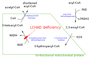
Long-chain 3-hydroxyacyl-coenzyme A dehydrogenase deficiency is a rare autosomal recessive fatty acid oxidation disorder that prevents the body from converting certain fats into energy. This can become life-threatening, particularly during periods of fasting.

In biochemistry and metabolism, beta oxidation (also β-oxidation) is the catabolic process by which fatty acid molecules are broken down in the cytosol in prokaryotes and in the mitochondria in eukaryotes to generate acetyl-CoA. Acetyl-CoA enters the citric acid cycle, generating NADH and FADH2, which are electron carriers used in the electron transport chain. It is named as such because the beta carbon of the fatty acid chain undergoes oxidation and is converted to a carbonyl group to start the cycle all over again. Beta-oxidation is primarily facilitated by the mitochondrial trifunctional protein, an enzyme complex associated with the inner mitochondrial membrane, although very long chain fatty acids are oxidized in peroxisomes.

<span class="mw-page-title-main">Inborn error of lipid metabolism</span> Medical condition

Numerous genetic disorders are caused by errors in fatty acid metabolism. These disorders may be described as fatty oxidation disorders or as a lipid storage disorders, and are any one of several inborn errors of metabolism that result from enzyme defects affecting the ability of the body to oxidize fatty acids in order to produce energy within muscles, liver, and other cell types.

<span class="mw-page-title-main">ACADVL</span> Protein-coding gene in the species Homo sapiens

Very long-chain specific acyl-CoA dehydrogenase, mitochondrial (VLCAD) is an enzyme that in humans is encoded by the ACADVL gene.

<span class="mw-page-title-main">Mitochondrial myopathy</span> Medical condition

Mitochondrial myopathies are types of myopathies associated with mitochondrial disease. Adenosine triphosphate (ATP), the chemical used to provide energy for the cell, cannot be produced sufficiently by oxidative phosphorylation when the mitochondrion is either damaged or missing necessary enzymes or transport proteins. With ATP production deficient in mitochondria, there is an over-reliance on anaerobic glycolysis which leads to lactic acidosis either at rest or exercise-induced.

<span class="mw-page-title-main">Chromosome 2</span> Human chromosome

Chromosome 2 is one of the twenty-three pairs of chromosomes in humans. People normally have two copies of this chromosome. Chromosome 2 is the second-largest human chromosome, spanning more than 242 million base pairs and representing almost eight percent of the total DNA in human cells.

<span class="mw-page-title-main">ACADM</span> Mammalian protein found in Homo sapiens

ACADM is a gene that provides instructions for making an enzyme called acyl-coenzyme A dehydrogenase that is important for breaking down (degrading) a certain group of fats called medium-chain fatty acids.

<span class="mw-page-title-main">Short-chain acyl-coenzyme A dehydrogenase deficiency</span> Medical condition

Short-chain acyl-coenzyme A dehydrogenase deficiency (SCADD) is an autosomal recessive fatty acid oxidation disorder which affects enzymes required to break down a certain group of fats called short chain fatty acids.

<span class="mw-page-title-main">ACADS</span> Protein-coding gene in humans

Acyl-CoA dehydrogenase, C-2 to C-3 short chain is an enzyme that in humans is encoded by the ACADS gene. This gene encodes a tetrameric mitochondrial flavoprotein, which is a member of the acyl-CoA dehydrogenase family. This enzyme catalyzes the initial step of the mitochondrial fatty acid beta-oxidation pathway. The ACADS gene is associated with short-chain acyl-coenzyme A dehydrogenase deficiency.

<span class="mw-page-title-main">Mitochondrial trifunctional protein</span> Inner mitochondrial membrane protein

Mitochondrial trifunctional protein (MTP) is a protein attached to the inner mitochondrial membrane which catalyzes three out of the four steps in beta oxidation. MTP is a hetero-octamer composed of four alpha and four beta subunits:
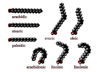
<span class="mw-page-title-main">Acyl-CoA</span> Group of coenzymes that metabolize fatty acids

Acyl-CoA is a group of coenzymes that metabolize carboxylic acids. Fatty acyl-CoA's are susceptible to beta oxidation, forming, ultimately, acetyl-CoA. The acetyl-CoA enters the citric acid cycle, eventually forming several equivalents of ATP. In this way, fats are converted to ATP, the common biochemical energy carrier.

<span class="mw-page-title-main">HADHA</span> Protein-coding gene in the species Homo sapiens

Trifunctional enzyme subunit alpha, mitochondrial also known as hydroxyacyl-CoA dehydrogenase/3-ketoacyl-CoA thiolase/enoyl-CoA hydratase, alpha subunit is a protein that in humans is encoded by the HADHA gene. Mutations in HADHA have been associated with trifunctional protein deficiency or long-chain 3-hydroxyacyl-coenzyme A dehydrogenase deficiency.

3-hydroxyacyl-coenzyme A dehydrogenase deficiency is a rare condition that prevents the body from converting certain fats to energy, particularly during fasting. Normally, through a process called fatty acid oxidation, several enzymes work in a step-wise fashion to metabolize fats and convert them to energy. People with 3-hydroxyacyl-coenzyme A dehydrogenase deficiency have inadequate levels of an enzyme required for a step that metabolizes groups of fats called medium chain fatty acids and short chain fatty acids; for this reason this disorder is sometimes called medium- and short-chain 3-hydroxyacyl-coenzyme A dehydrogenase (M/SCHAD) deficiency.

<span class="mw-page-title-main">HADHB</span> Protein-coding gene in the species Homo sapiens

Trifunctional enzyme subunit beta, mitochondrial (TP-beta) also known as 3-ketoacyl-CoA thiolase, acetyl-CoA acyltransferase, or beta-ketothiolase is an enzyme that in humans is encoded by the HADHB gene.

D-Bifunctional protein deficiency is an autosomal recessive peroxisomal fatty acid oxidation disorder. Peroxisomal disorders are usually caused by a combination of peroxisomal assembly defects or by deficiencies of specific peroxisomal enzymes. The peroxisome is an organelle in the cell similar to the lysosome that functions to detoxify the cell. Peroxisomes contain many different enzymes, such as catalase, and their main function is to neutralize free radicals and detoxify drugs. For this reason peroxisomes are ubiquitous in the liver and kidney. D-BP deficiency is the most severe peroxisomal disorder, often resembling Zellweger syndrome.

<span class="mw-page-title-main">Methylglutaconyl-CoA hydratase</span> Protein-coding gene in the species Homo sapiens

3-Methylglutaconyl-CoA hydratase, also known as MG-CoA hydratase and AUH, is an enzyme encoded by the AUH gene on chromosome 19. It is a member of the enoyl-CoA hydratase/isomerase superfamily, but it is the only member of that family that is able to bind to RNA. Not only does it bind to RNA, AUH has also been observed to be involved in the metabolic enzymatic activity, making it a dual-role protein. Mutations of this gene have been found to cause a disease called 3-Methylglutaconic Acuduria Type 1.

<span class="mw-page-title-main">HSD17B4</span> Protein-coding gene in the species Homo sapiens

D-bifunctional protein (DBP), also known as peroxisomal multifunctional enzyme type 2 (MFP-2), as well as 17β-hydroxysteroid dehydrogenase type IV is a protein that in humans is encoded by the HSD17B4 gene. It's an alcohol oxidoreductase, specifically 17β-Hydroxysteroid dehydrogenase. It is involved in fatty acid β-oxidation and steroid metabolism.

<span class="mw-page-title-main">Fatty-acid metabolism disorder</span> Medical condition

A broad classification for genetic disorders that result from an inability of the body to produce or utilize an enzyme or transport protein that is required to oxidize fatty acids. They are an inborn error of lipid metabolism, and when it affects the muscles also a metabolic myopathy.

<span class="mw-page-title-main">Hydroxyacyl-Coenzyme A dehydrogenase</span> Protein-coding gene in the species Homo sapiens

Hydroxyacyl-Coenzyme A dehydrogenase (HADH) is an enzyme which in humans is encoded by the HADH gene.

References

  1. "Mitochondrial trifunctional protein deficiency | Genetic and Rare Diseases Information Center (GARD) – an NCATS Program". rarediseases.info.nih.gov. Archived from the original on 31 July 2019. Retrieved 31 July 2019.
  2. 1 2 3 "Mitochondrial trifunctional protein deficiency | Genetic and Rare Diseases Information Center(GARD) – an NCATS Program". rarediseases.info.nih.gov. Archived from the original on 9 January 2019. Retrieved 12 November 2016.
  3. 1 2 3 4 RESERVED, INSERM US14 -- ALL RIGHTS. "Orphanet: Mitochondrial trifunctional protein deficiency". www.orpha.net. Archived from the original on 2016-11-13. Retrieved 2016-11-12.{{cite web}}: CS1 maint: numeric names: authors list (link)
  4. 1 2 Solish JO, Singh RH (2002). "Management of fatty acid oxidation disorders: a survey of current treatment strategies". J Am Diet Assoc. 102 (12): 1800–1803. doi:10.1016/S0002-8223(02)90386-X. PMID   12487544.subscription needed
  5. "OMIM Entry - # 609015 - MITOCHONDRIAL TRIFUNCTIONAL PROTEIN DEFICIENCY; MTPD". omim.org. Archived from the original on 2017-05-10. Retrieved 2016-11-05.
  6. 1 2 3 Reference, Genetics Home. "mitochondrial trifunctional protein deficiency". Genetics Home Reference. Archived from the original on 2020-04-04. Retrieved 2016-10-28.
  7. 1 2 Swaiman, Kenneth F.; Ashwal, Stephen; Ferriero, Donna M.; Schor, Nina F. (2014). Swaiman's Pediatric Neurology: Principles and Practice. Elsevier Health Sciences. pp. 461, 1638. ISBN   978-0323089111 . Retrieved 12 November 2016.
  8. Reference, Genetics Home. "HADHA gene". Genetics Home Reference. Archived from the original on 2019-10-17. Retrieved 2016-11-05.
  9. Reference, Genetics Home. "HADHB gene". Genetics Home Reference. Archived from the original on 2016-11-05. Retrieved 2016-11-05.
  10. "Long-Chain Acyl CoA Dehydrogenase Deficiency: Background, Pathophysiology, Epidemiology". eMedicine. 24 March 2016. Archived from the original on 13 March 2020. Retrieved 12 November 2016.
  11. 1 2 "HADHA hydroxyacyl-CoA dehydrogenase/3-ketoacyl-CoA thiolase/enoyl-CoA hydratase (trifunctional protein), alpha subunit [Homo sapiens (human)] - Gene - NCBI". www.ncbi.nlm.nih.gov. Archived from the original on 25 August 2018. Retrieved 12 November 2016.
  12. 1 2 "Home - Gene - NCBI". www.ncbi.nlm.nih.gov. Archived from the original on 13 November 2016. Retrieved 12 November 2016.
  13. "OMIM Entry - * 600890 - HYDROXYACYL-CoA DEHYDROGENASE/3-KETOACYL-CoA THIOLASE/ENOYL-CoA HYDRATASE, ALPHA SUBUNIT; HADHA". omim.org. Archived from the original on 31 July 2015. Retrieved 5 November 2016.
  14. Rector, R. Scott; Payne, R. Mark; Ibdah, Jamal A. (1 January 2008). "Mitochondrial Trifunctional Protein Defects: Clinical Implications and Therapeutic Approaches". Adv Drug Deliv Rev. 60 (13–14): 1488–1496. doi:10.1016/j.addr.2008.04.014. ISSN   0169-409X. PMC   2848452 . PMID   18652860.

Further reading