Sick sinus syndrome

Last updated
Sick Sinus Syndrome
Other namesSinus node dysfunction or Sinoatrial node disease
ECG Sinus Pause.jpg
Telemetry strip of a 44-year-old female with asymptomatic sinus pause found while admitted for mildly symptomatic COVID-19 pneumonia.
Specialty Cardiology   OOjs UI icon edit-ltr-progressive.svg
Complications Tachycardia-bradycardia syndrome

Sick sinus syndrome (SSS), also known as sinus node dysfunction (SND), is a group of abnormal heart rhythms (arrhythmias) presumably caused by a malfunction of the sinus node, the heart's primary pacemaker. [1] [2] Tachycardia-bradycardia syndrome is a variant of sick sinus syndrome in which the arrhythmia alternates between slow and fast heart rates. [3]

Contents

Signs and symptoms

Often sick sinus syndrome produces no symptoms, especially early in the disease course. Signs and symptoms usually appear in more advanced disease and more than 50% of patients will present with syncope or transient near-fainting spells. [4] Other presenting signs or symptoms can include confusion, fatigue, palpitations, chest pain, shortness of breath, headache, and nausea. Patients can also present with symptoms of congestive heart failure, stroke or transient ischemic attacks due to the abnormal rhythm. [4]

Complications

Electrocardiogram from a man with tachycardia-bradycardia syndrome following mitral valvuloplasty, resection of the left atrial appendage, and maze procedure. The ECG shows AV-junctional rhythm resulting in bradycardia at around 46 beats per minute. Brady-tachy syndrome AV-junctional rhythm.png
Electrocardiogram from a man with tachycardia-bradycardia syndrome following mitral valvuloplasty, resection of the left atrial appendage, and maze procedure. The ECG shows AV-junctional rhythm resulting in bradycardia at around 46 beats per minute.
This ECG from the same patient shows atrial fibrillation at around 126 beats per minute. Brady-tachy syndrome atrial fibrillation.png
This ECG from the same patient shows atrial fibrillation at around 126 beats per minute.

The most common complication of sick sinus syndrome is developing tachycardia-bradycardia syndrome with abnormal atrial rhythms such as atrial tachycardia, atrial fibrillation, and flutter. [4] [3] These rhythms increases the risk of clot formation in the atrium, embolization, and stroke. [4]

Developing sinus arrest, sinus node exit block, sinus bradycardia, atrioventricular block, and other types of abnormal rhythms are also common complications. [4] [5] Sick sinus syndrome has a close association with the presence of atrial fibrillation due to their shared etiology of remodeling. [5]

Causes

Sick sinus syndrome can be caused by intrinsic and extrinsic factors that affect the normal functioning of the sinus node. Intrinsic causes can include degeneration, dysfunction, or remodeling of the sinus node while extrinsic causes can create or worsen underly atrial arrhythmias. [4] Intrinsic causes tend to be responsible for permanent sick sinus syndrome while extrinsic causes are more commonly temporary. [4]

Intrinsic Causes

Age-related degenerative fibrosis of the sinus node is often identified as the most common intrinsic cause. [4] [5] Other intrinsic causes include inherited ion channel dysfunctions, remodeling diseases such as heart failure and atrial fibrillation, infiltrative diseases such as sarcoidosis, amyloidosis, hemochromatosis, and connective tissue diseases, inflammatory etiology such as rheumatic fever, Chagas disease, and Lyme disease, as well as atherosclerotic and ischemic changes to the sinus node artery. [4] [5]

Inherited sick sinus syndrome has been associated with mutations of the gene responsible for the formation of the Alpha subunit of the sodium channel (SCN5A). [4]

Extrinsic Causes

Common cardiac pharmacology such as beta-blockers, calcium channel blockers, digoxin, sympatholytic medication, and other antiarrhythmics can alter sinus node function to create an arrhythmia such as sick sinus syndrome. Electrolyte abnormalities such as hyperkalemia, hypokalemia, and hypocalcemia can also alter normal sinus node functioning. Hypothyroidism, hypoxia, hypothermia, and various toxins have also been associated with sinus node dysfunctions. [4] [5]

Diagnosis

Electrocardiogram (ECG) of a 64-year-old female with sinus bradycardia. Heart rate 49 bpm. ECG Sinus Bradycardia 49 bpm.jpg
Electrocardiogram (ECG) of a 64-year-old female with sinus bradycardia. Heart rate 49 bpm.

Electrocardiogram

The primary 12-lead electrocardiogram (ECG) finding in sick sinus syndrome is inappropriate sinus bradycardia. [6] Sick sinus syndrome can also present with sudden sinus arrest with or without junctional escape, sinoatrial block, prolonged asystolic period followed by tachycardias, or tachycardia-bradycardia syndrome presenting as various atrial arrhythmias such as atrial fibrillation, flutter, tachycardia, or paroxysmal supraventricular tachycardia. [6] [4]

Clinical Diagnosis

Diagnosing sick sinus syndrome requires clinical symptoms as well as ECG abnormalities. If ECG findings cannot be identified, prolonged cardiac monitoring should be pursued either with a Holter monitor in an outpatient setting or telemetry while inpatient, due to the transient nature of abnormal ECG findings. [4] If Holter or telemetry monitoring fails to identify ECG changes and suspicion of sick sinus syndrome remains high due to severe symptoms or episodes of syncope, implantable loop recorders should be considered for extended monitoring up to 24 months. [6]

Exercise stress test can be utilized to identify intrinsic causes of sick sinus syndrome. Head-up tilt test can be used to discriminate bradycardia caused by dysfunction of the autonomic nervous system. [4] [6]

Treatment

The primary reason for considering treatment is the presence of symptoms. [1] Pacemaker implantation is the primary treatment modality of symptomatic sick sinus syndrome. [4] [6] [7] The goal of this treatment modality is to relieve symptoms associated with sick sinus syndrome and improve quality of life. [4] Dual chamber pacemakers are preferred due to the possibility of developing atrioventricular block [4] as well as long term cost-effectiveness relative to single-chamber atrial pacemakers. [8]

In tachycardia-bradycardia syndrome, medication based management can treat atrial tachyarrhythmias. However, these medication may exacerbate underlying bradyarrhythmia. Therefore, specialized dual chamber pacemaker capable of managing atrial tachyarrhythmias as well as bradyarrhythmias is implanted before drug therapy is begun. [6]

Epidemiology

Overall incidence of sick sinus syndrome increases with age [9] with 1 in 1000 in adults over 45 years old [5] and 1 in 600 cardiac patients over 65 years old. [4] Sick sinus syndrome is the primary indication for approximately 30%-50% of all pacemaker implantation in the United States. [9] Sick sinus syndrome is a relatively uncommon syndrome in the young and middle-aged population.

Related Research Articles

Bradycardia Heart rate that is below the normal range

Bradycardia is a condition typically defined wherein an individual has a resting heart rate of under 60 beats per minute (BPM) in adults, although some studies use a heart rate of less than 50 BPM. Bradycardia typically does not cause symptoms until the rate drops below 50 BPM. When symptomatic, it may cause fatigue, weakness, dizziness, sweating, and at very low rates, fainting.

Electrocardiography

Electrocardiography is the process of producing an electrocardiogram. It is a graph of voltage versus time of the electrical activity of the heart using electrodes placed on the skin. These electrodes detect the small electrical changes that are a consequence of cardiac muscle depolarization followed by repolarization during each cardiac cycle (heartbeat). Changes in the normal ECG pattern occur in numerous cardiac abnormalities, including cardiac rhythm disturbances, inadequate coronary artery blood flow, and electrolyte disturbances.

Wolff–Parkinson–White syndrome

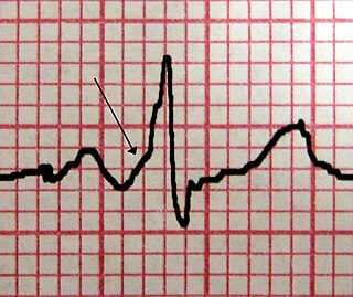
Wolff–Parkinson–White syndrome (WPWS) is a disorder due to a specific type of problem with the electrical system of the heart which has resulted in symptoms. About 40% of people with the electrical problem never develop symptoms. Symptoms can include an abnormally fast heartbeat, palpitations, shortness of breath, lightheadedness, or syncope. Rarely, cardiac arrest may occur. The most common type of irregular heartbeat that occurs is known as paroxysmal supraventricular tachycardia.

Palpitations are the perceived abnormality of the heartbeat characterized by awareness of cardiac muscle contractions in the chest, which is further characterized by the hard, fast and/or irregular beatings of the heart.

Third-degree atrioventricular block

Third-degree atrioventricular block is a medical condition in which the nerve impulse generated in the sinoatrial node in the atrium of the heart can not propagate to the ventricles.

Short QT syndrome

Short QT syndrome (SQT) is a very rare genetic disease of the electrical system of the heart, and is associated with an increased risk of abnormal heart rhythms and sudden cardiac death. The syndrome gets its name from a characteristic feature seen on an electrocardiogram (ECG) – a shortening of the QT interval. It is caused by mutations in genes encoding ion channels that shorten the cardiac action potential, and appears to be inherited in an autosomal dominant pattern. The condition is diagnosed using a 12-lead ECG. Short QT syndrome can be treated using an implantable cardioverter-defibrillator or medications including quinidine. Short QT syndrome was first described in 2000, and the first genetic mutation associated with the condition was identified in 2004.

Electrical conduction system of the heart Transmits signals generated usually by the sinoatrial node to cause contraction of the heart muscle

The electrical conduction system of the heart transmits signals generated usually by the sinoatrial node to cause contraction of the heart muscle. The pacemaking signal generated in the sinoatrial node travels through the right atrium to the atrioventricular node, along the Bundle of His and through bundle branches to cause contraction of the heart muscle. This signal stimulates contraction first of the right and left atrium, and then the right and left ventricles. This process allows blood to be pumped throughout the body.

Supraventricular tachycardia Abnormally fast heart rhythm arising from improper electrical activity in the upper part of the heart

Supraventricular tachycardia (SVT) is an abnormally fast heart rhythm arising from improper electrical activity in the upper part of the heart. There are four main types: atrial fibrillation, paroxysmal supraventricular tachycardia (PSVT), atrial flutter, and Wolff–Parkinson–White syndrome. Symptoms may include palpitations, feeling faint, sweating, shortness of breath, or chest pain.

AV nodal reentrant tachycardia

AV-nodal reentrant tachycardia (AVNRT) is a type of abnormal fast heart rhythm. It is a type of supraventricular tachycardia (SVT), meaning that it originates from a location within the heart above the bundle of His. AV nodal reentrant tachycardia is the most common regular supraventricular tachycardia. It is more common in women than men. The main symptom is palpitations. Treatment may be with specific physical maneuvers, medications, or, rarely, synchronized cardioversion. Frequent attacks may require radiofrequency ablation, in which the abnormally conducting tissue in the heart is destroyed.

Sinus bradycardia Sinus bradycardia is a sinus rhythm with a rate that is lower than normal

Sinus bradycardia is a sinus node dysfunction with a rate that is lower than normal. In humans, bradycardia is generally defined to be a rate of under 60 beats per minute. A normal heartbeat in human is usually at a rate of 60 to 100 beats per minute.

Premature atrial contractions (PACs), also known as atrial premature complexes (APC) or atrial premature beats (APB), are a common cardiac dysrhythmia characterized by premature heartbeats originating in the atria. While the sinoatrial node typically regulates the heartbeat during normal sinus rhythm, PACs occur when another region of the atria depolarizes before the sinoatrial node and thus triggers a premature heartbeat. The exact cause of PACs is unclear; while several predisposing conditions exist, PACs commonly occur in healthy young and elderly people. Elderly people that get PACs usually don't need any further attention besides follow ups due to unclear evidence. PACs are often completely asymptomatic and may be noted only with Holter monitoring, but occasionally they can be perceived as a skipped beat or a jolt in the chest. In most cases, no treatment other than reassurance is needed for PACs, although medications such as beta blockers can reduce the frequency of symptomatic PACs.

Tachycardia-induced cardiomyopathy (TIC) is a disease where prolonged tachycardia or arrhythmia causes an impairment of the myocardium, which can result in heart failure. People with TIC may have symptoms associated with heart failure and/or symptoms related to the tachycardia or arrhythmia. Though atrial fibrillation is the most common cause of TIC, several tachycardias and arrhythmias have been associated with the disease.

Lown–Ganong–Levine syndrome (LGL) is a pre-excitation syndrome of the heart. Those with LGL syndrome have episodes of abnormal heart racing with a short PR interval and normal QRS complexes seen on their electrocardiogram when in a normal sinus rhythm. LGL syndrome was originally thought to be due to an abnormal electrical connection between the atria and the ventricles, but is now thought to be due to accelerated conduction through the atrioventricular node in the majority of cases. The syndrome is named after Bernard Lown, William Francis Ganong, Jr., and Samuel A. Levine.

Lorcainide

Lorcainide is a Class 1c antiarrhythmic agent that is used to help restore normal heart rhythm and conduction in patients with premature ventricular contractions, ventricular tachycardiac and Wolff-Parkinson-White syndrome. Lorcainide was developed by Janssen Pharmaceutica (Belgium) in 1968 under the commercial name Remivox and is designated by code numbers R-15889 or Ro 13-1042/001. It has a half-life of 8.9 +- 2.3 hrs which may be prolonged to 66 hrs in people with cardiac disease.

Multifocal atrial tachycardia

Multifocal atrial tachycardia (MAT) is an abnormal heart rhythm, specifically a type of supraventricular tachycardia, that is particularly common in older people and is associated with exacerbations of chronic obstructive pulmonary disease (COPD). Normally, the heart rate is controlled by a cluster of cells called the sinoatrial node. When a number of different clusters of cells outside the SA node take over control of the heart rate, and the rate exceeds 100 beats per minute, this is called multifocal atrial tachycardia.

Accelerated idioventricular rhythm

Accelerated idioventricular rhythm is a ventricular rhythm with a rate of between 40 and 120 beats per minute. Idioventricular means “relating to or affecting the cardiac ventricle alone” and refers to any ectopic ventricular arrhythmia. Accelerated idioventricular arrhythmias are distinguished from ventricular rhythms with rates less than 40 and those faster than 120. Though some other references limit to between 60 and 100 beats per minute. It is also referred to as AIVR and "slow ventricular tachycardia."

An accessory pathway is an additional electrical connection between two parts of the heart. These pathways can lead to abnormal heart rhythms or arrhythmias associated with symptoms of palpitations. Some pathways may activate a region of ventricular muscle earlier than would normally occur, referred to as pre-excitation, and this may be seen on an electrocardiogram. The combination of an accessory pathway that causes pre-excitation with arrhythmias is known as Wolff-Parkinson-White syndrome.

An ectopic pacemaker is an excitable group of cells that causes a premature heart beat outside the normally functioning SA node of the heart. It is thus a cardiac pacemaker that is ectopic, producing an ectopic beat. Acute occurrence is usually non-life-threatening, but chronic occurrence can progress into tachycardia, bradycardia or ventricular fibrillation. In a normal heart beat rhythm, the SA node usually suppresses the ectopic pacemaker activity due to the higher impulse rate of the SA node. However, in the instance of either a malfunctioning SA node or an ectopic focus bearing an intrinsic rate superior to SA node rate, ectopic pacemaker activity may take over the natural heart rhythm. This phenomenon is called an escape rhythm, the lower rhythm having escaped from the dominance of the upper rhythm. As a rule, premature ectopic beats indicate increased myocyte or conducting tissue excitability, whereas late ectopic beats indicate proximal pacemaker or conduction failure with an escape 'ectopic' beat.

Junctional ectopic tachycardia

Junctional ectopic tachycardia (JET) is a rare syndrome of the heart that manifests in patients recovering from heart surgery. It is characterized by cardiac arrhythmia, or irregular beating of the heart, caused by abnormal conduction from or through the atrioventricular node. In newborns and infants up to 6 weeks old, the disease may also be referred to as His bundle tachycardia or congenital JET.

Arrhythmia Group of conditions in which the heartbeat is irregular, too fast, or too slow

Arrhythmia, also known as cardiac arrhythmia or heart arrhythmia, is a group of conditions in which the heartbeat is irregular, too fast, or too slow. The heart rate that is too fast – above 100 beats per minute in adults – is called tachycardia, and a heart rate that is too slow – below 60 beats per minute – is called bradycardia. Some types of arrhythmias have no symptoms. Symptoms when present may include palpitations or feeling a pause between heartbeats. In more serious cases, there may be lightheadedness, passing out, shortness of breath or chest pain. While most types of arrhythmia are not serious, some predispose a person to complications such as stroke or heart failure. Others may result in sudden death.

References

  1. 1 2 Kusumoto Fred M.; Schoenfeld Mark H.; Barrett Coletta; Edgerton James R.; Ellenbogen Kenneth A.; Gold Michael R.; Goldschlager Nora F.; Hamilton Robert M.; Joglar José A.; Kim Robert J.; Lee Richard (2019-08-20). "2018 ACC/AHA/HRS Guideline on the Evaluation and Management of Patients With Bradycardia and Cardiac Conduction Delay: A Report of the American College of Cardiology/American Heart Association Task Force on Clinical Practice Guidelines and the Heart Rhythm Society". Circulation. 140 (8): e382–e482. doi:10.1161/CIR.0000000000000628.
  2. Dobrzynski H, Boyett MR, Anderson RH (April 2007). "New insights into pacemaker activity: promoting understanding of sick sinus syndrome". Circulation. 115 (14): 1921–32. doi: 10.1161/CIRCULATIONAHA.106.616011 . PMID   17420362.
  3. 1 2 Tse, Gary; Liu, Tong; Li, Ka Hou Christien; Laxton, Victoria; Wong, Andy On-Tik; Chan, Yin Wah Fiona; Keung, Wendy; Chan, Camie W.Y.; Li, Ronald A. (March 2017). "Tachycardia-bradycardia syndrome: Electrophysiological mechanisms and future therapeutic approaches (Review)". International Journal of Molecular Medicine. 39 (3): 519–526. doi:10.3892/ijmm.2017.2877. ISSN   1107-3756. PMC   5360359 . PMID   28204831.
  4. 1 2 3 4 5 6 7 8 9 10 11 12 13 14 15 16 17 18 Semelka M, Gera J, Usman S (May 2013). "Sick sinus syndrome: a review". American Family Physician (Review). 87 (10): 691–6. PMID   23939447.
  5. 1 2 3 4 5 6 John RM, Kumar S (May 2016). "Sinus Node and Atrial Arrhythmias". Circulation. 133 (19): 1892–900. doi:10.1161/CIRCULATIONAHA.116.018011. PMID   27166347.
  6. 1 2 3 4 5 6 De Ponti R, Marazzato J, Bagliani G, Leonelli FM, Padeletti L (June 2018). "Sick Sinus Syndrome". Cardiac Electrophysiology Clinics. 10 (2): 183–195. doi:10.1016/j.ccep.2018.02.002. PMID   29784479.
  7. Drago F, Silvetti MS, Grutter G, De Santis A (July 2006). "Long term management of atrial arrhythmias in young patients with sick sinus syndrome undergoing early operation to correct congenital heart disease". Europace. 8 (7): 488–94. doi:10.1093/europace/eul069. PMID   16798761.
  8. Edwards SJ, Karner C, Trevor N, Wakefield V, Salih F (August 2015). "Dual-chamber pacemakers for treating symptomatic bradycardia due to sick sinus syndrome without atrioventricular block: a systematic review and economic evaluation". Health Technology Assessment. 19 (65): 1–210. doi:10.3310/hta19650. PMC   4781212 . PMID   26293406.
  9. 1 2 Jensen PN, Gronroos NN, Chen LY, Folsom AR, deFilippi C, Heckbert SR, Alonso A (August 2014). "Incidence of and risk factors for sick sinus syndrome in the general population". Journal of the American College of Cardiology. 64 (6): 531–8. doi:10.1016/j.jacc.2014.03.056. PMC   4139053 . PMID   25104519.
Classification
D
External resources