Superficial siderosis

Last updated
Superficial siderosis
Other namesSuperficial siderosis of the CNS, Superficial hemosiderosis of the CNS, Superficial hemosiderosis of the central nervous system
NPH MRI 272 GILD.gif
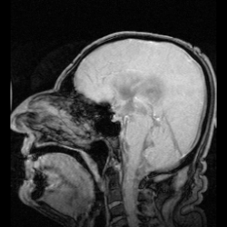
MRI showing pulsations of CSF (normal individual)

Superficial hemosiderosis of the central nervous system is a disease of the brain resulting from chronic iron deposition in neuronal tissues associated with cerebrospinal fluid. This occurs via the deposition of hemosiderin in neuronal tissue, and is associated with neuronal loss, gliosis, and demyelination of neuronal cells. This disease was first discovered in 1908 by R.C. Hamill after performing an autopsy. [1] Detection of this disease was largely post-mortem until the advent of MRI technology, which made diagnosis far easier. Superficial siderosis is largely considered a rare disease, with less than 270 total reported cases in scientific literature as of 2006, [2] and affects people of a wide range of ages with men being approximately three times more frequently affected than women. [3] The number of reported cases of superficial siderosis has increased with advances in MRI technology, but it remains a rare disease.

Contents

Symptoms

Superficial siderosis is characterized by many symptoms resulting from brain damage: [4]

Cause

The most commonly described underlying cause of superficial siderosis is chronic bleeding into the subarachnoid space of the brain, which releases erythrocytes, or blood cells, into the cerebrospinal fluid. The chronic bleeding can come from many sources such as from an arteriovenous malformation or cavernous malformation, myxopapillary ependymoma of the spinal cord, from chronic subdural hematoma, from a ventricular shunt, or from chronic subarachnoid hemorrhage. Chronic bleeding sources can also be a result of past brain surgery or CNS trauma, which may be the most common cause of superficial siderosis, with superficial siderosis showing up many years later. [5] In up to as many as half of all described cases the source of bleeding was never found. [5]

Pathology

Blood cells are not native to the cerebrospinal fluid, and their presence there is problematic. Once they eventually break down, they release the heme containing protein hemoglobin. Hemoglobin breaks down and releases iron-containing heme into the cerebrospinal fluid. In response to this upsurge in heme levels, Bergmann glia and microglia produce heme oxygenase 1. Heme oxygenase 1 breaks down free heme into biliverdin, carbon monoxide, and iron. It is this breakdown of heme that is the source of iron deposition that ultimately causes superficial siderosis. [6] The excess free iron is circulated in the cerebrospinal fluid and deposited in neuronal tissues, where it catalyzes the formation of reactive oxygen species which can damage DNA, lipids and proteins, and is otherwise toxic to the cells. [7]

Iron deposition is prevalent in siderotic brain tissues, with iron concentrations of 1.79 to 8.26 times normal levels. Ferritin, an iron storage protein, is over-produced in response to excess heme by glial cells in order to sequester iron, with production ranging from 20.1 to 27.4 times normal levels. Excess iron, ferritin, and red blood cells may result in xanthochromia of the cerebrospinal fluid. Amongst neuronal cells types, iron deposition appears to be preferential for oligodendroglial cells, which is supported by the belief that they can act as iron sinks in the central nervous system. Schwann cells are also frequently damaged, contributing to demyelination. [6]

Iron deposition is most prevalent in the inferior temporal lobes, the brainstem, the cerebellum, peri-ventricular structures, the spinal cord, and cranial nerve VIII. Iron deposition is also present in cranial nerves I & II, but this damage less frequently presents symptoms when compared to cranial nerve VIII, [3] which can be explained by cranial nerve VIII’s notable segment of glial cells, which are preferentially affected by iron deposition. [8]

The presence of ‘foamy’, ‘spheroid’, or ‘cytoid’ bodies in affected neuronal tissues has been noted but what they are and their origin remains somewhat unclear. They are believed to be swollen axonal bodies, but some evidence exists that they may be astrocytic in origin. [9]

Diagnosis

Early detection of superficial siderosis is usually via MRI. The iron deposition that is characteristic of superficial siderosis shows up as a hypointense band in affected tissues, with a characteristic rim of intensity appearing on the cerebellum; a hyperintense rim is rarely seen. [8] Taking samples of cerebrospinal fluid may also reveal siderosis through xanthochromia, elevated presence of red blood cells, high iron and ferritin concentrations, and elevated levels of the proteins Tau, amyloid beta (Aβ42), neurofilament light chain (NFL), and glial fibrillary acidic protein (GFAP), but the CSF is sometimes normal. [10] Detection is complicated by the fact that superficial siderosis is a rare disease and is not well described in neurological texts, so it may go unnoticed until noticeable symptoms appear. [11]

Treatment

There is no current cure for superficial siderosis, only treatments to help alleviate the current symptoms and to help prevent the development of further symptoms. If a source of bleeding can be identified (sources are frequently not found), then surgical correction of the bleeding source can be performed; this has proved to be effective in halting the development of further symptoms in some cases and has no effect on symptoms that have already presented. [8] Patients with superficial siderosis are often treated with deferiprone (brand name is Ferriprox), a lipid-soluble iron chelator, as this medication has been demonstrated to chelate iron in the central nervous system. [12] Deferiprone use is associated with severe neutropenia, but this risk and associated complications are usually minimised by administering a weekly blood test that measures absolute neutrophil count. [13]

Alleviation of the most common symptom, hearing loss, has been varyingly successful through the use of cochlear implants. Most people do not notice a large improvement after successful implantation, which is most likely due to damage to the vestibulocochlear nerve (cranial nerve VIII) and not the cochlea itself. [14] Some people fare far better, with a return to near-normal hearing, but there is little ability to detect how well a person will respond to this treatment at this time. [15]

Related Research Articles

Cerebrospinal fluid Clear, colorless bodily fluid found in the brain and spinal cord

Cerebrospinal fluid (CSF) is a clear, colorless body fluid found within the tissue that surrounds the brain and spinal cord of all vertebrates. It replaces the body fluid found outside the cells of all bilateral animals.

Nerve Enclosed, cable-like bundle of axons in the peripheral nervous system

A nerve is an enclosed, cable-like bundle of fibers in the peripheral nervous system.

The development of the nervous system, or neural development, or neurodevelopment, refers to the processes that generate, shape, and reshape the nervous system of animals, from the earliest stages of embryonic development to adulthood. The field of neural development draws on both neuroscience and developmental biology to describe and provide insight into the cellular and molecular mechanisms by which complex nervous systems develop, from nematodes and fruit flies to mammals.

Meninges Membranes that envelop the brain and spinal cord

In anatomy, the meninges are the three membranes that envelop the brain and spinal cord. In mammals, the meninges are the dura mater, the arachnoid mater, and the pia mater. Cerebrospinal fluid is located in the subarachnoid space between the arachnoid mater and the pia mater. The primary function of the meninges is to protect the central nervous system.

Pia mater Delicate innermost layer of the meninges, the membranes surrounding the brain and spinal cord

Pia mater, often referred to as simply the pia, is the delicate innermost layer of the meninges, the membranes surrounding the brain and spinal cord. Pia mater is medieval Latin meaning "tender mother". The other two meningeal membranes are the dura mater and the arachnoid mater. Both the pia and arachnoid mater are derivatives of the neural crest while the dura is derived from embryonic mesoderm. The pia mater is a thin fibrous tissue that is permeable to water and small solutes. The pia mater allows blood vessels to pass through and nourish the brain. The perivascular space between blood vessels and pia mater is proposed to be part of a pseudolymphatic system for the brain. When the pia mater becomes irritated and inflamed the result is meningitis.

Choroid plexus

The choroid plexus, or plica choroidea, is a plexus of cells that arises from the tela choroidea in each of the ventricles of the brain. The choroid plexus produces most of the cerebrospinal fluid (CSF) of the central nervous system. CSF is produced and secreted by the regions of the choroid plexus. The choroid plexus consists of modified ependymal cells surrounding a core of capillaries and loose connective tissue.

Iron overload Human disease

Iron overload or haemochromatosis indicates accumulation of iron in the body from any cause. The most important causes are hereditary haemochromatosis (HHC), a genetic disorder, and transfusional iron overload, which can result from repeated blood transfusions.

Dura mater

Dura mater is a thick membrane made of dense irregular connective tissue that surrounds the brain and spinal cord. It is the outermost of the three layers of membrane called the meninges that protect the central nervous system. The other two meningeal layers are the arachnoid mater and the pia mater. The dura surrounds the brain and the spinal cord. It envelops the arachnoid mater, which is responsible for keeping in the cerebrospinal fluid. It is derived primarily from the neural crest cell population, with postnatal contributions of the paraxial mesoderm.

Neurosarcoidosis Medical condition

Neurosarcoidosis refers to a type of sarcoidosis, a condition of unknown cause featuring granulomas in various tissues, in this type involving the central nervous system. Neurosarcoidosis can have many manifestations, but abnormalities of the cranial nerves are the most common. It may develop acutely, subacutely, and chronically. Approximately 5–10 percent of people with sarcoidosis of other organs develop central nervous system involvement. Only 1 percent of people with sarcoidosis will have neurosarcoidosis alone without involvement of any other organs. Diagnosis can be difficult, with no test apart from biopsy achieving a high accuracy rate. Treatment is with immunosuppression. The first case of sarcoidosis involving the nervous system was reported in 1905.

Crown (anatomy) Top of the head

The crown is the top portion of the head behind the vertex. The anatomy of the crown varies between different organisms. The human crown is made of three layers of the scalp above the skull. The crown also covers a range of bone sutures, and contains blood vessels and branches of the trigeminal nerve.

Hemosiderin

Hemosiderin or haemosiderin is an iron-storage complex that is composed of partially digested ferritin and lysosomes. The breakdown of heme gives rise to biliverdin and iron. The body then traps the released iron and stores it as hemosiderin in tissues. Hemosiderin is also generated from the abnormal metabolic pathway of ferritin.

Neuroimmune system

The neuroimmune system is a system of structures and processes involving the biochemical and electrophysiological interactions between the nervous system and immune system which protect neurons from pathogens. It serves to protect neurons against disease by maintaining selectively permeable barriers, mediating neuroinflammation and wound healing in damaged neurons, and mobilizing host defenses against pathogens.

Leptomeningeal cancer Medical condition

Leptomeningeal cancer is a rare complication of cancer in which the disease spreads from the original tumor site to the meninges surrounding the brain and spinal cord. This leads to an inflammatory response, hence the alternative names neoplastic meningitis (NM), malignant meningitis, or carcinomatous meningitis. The term leptomeningeal describes the thin meninges, the arachnoid and the pia mater, between which the cerebrospinal fluid is located. The disorder was originally reported by Eberth in 1870.

Hemosiderosis Iron metabolism disease that has material basis in an accumulation of hemosiderin, an iron-storage complex, resulting in iron overload

Hemosiderosis is a form of iron overload disorder resulting in the accumulation of hemosiderin.

Central nervous system disease Disease of the brain or spinal cord

Central nervous system diseases, also known as central nervous system disorders, are a group of neurological disorders that affect the structure or function of the brain or spinal cord, which collectively form the central nervous system (CNS).

Neuroferritinopathy Medical condition

Neuroferritinopathy is a genetic neurodegenerative disorder characterized by the accumulation of iron in the basal ganglia, cerebellum, and motor cortex of the human brain. Symptoms, which are extrapyramidal in nature, progress slowly and generally do not become apparent until adulthood. These symptoms include chorea, dystonia, and cognitive deficits which worsen with age.

Neurovirology is an interdisciplinary field which represents a melding of clinical neuroscience, virology, immunology, and molecular biology. The main focus of the field is to study viruses capable of infecting the nervous system. In addition to this, the field studies the use of viruses to trace neuroanatomical pathways, for gene therapy, and to eliminate detrimental populations of neural cells.

Neuro-oncology is the study of brain and spinal cord neoplasms, many of which are very dangerous and life-threatening. Among the malignant brain cancers, gliomas of the brainstem and pons, glioblastoma multiforme, and high-grade astrocytoma are among the worst. In these cases, untreated survival usually amounts to only a few months, and survival with current radiation and chemotherapy treatments may extend that time from around a year to a year and a half, possibly two or more, depending on the patient's condition, immune function, treatments used, and the specific type of malignant brain neoplasm. Surgery may in some cases be curative, but, as a general rule, malignant brain cancers tend to regenerate and emerge from remission easily, especially highly malignant cases. In such cases, the goal is to excise as much of the mass and as much of the tumor margin as possible without endangering vital functions or other important cognitive abilities. The Journal of Neuro-Oncology is the longest continuously published journal in the field and serves as a leading reference to those practicing in the area of neuro-oncology.

Neuroinflammation is inflammation of the nervous tissue. It may be initiated in response to a variety of cues, including infection, traumatic brain injury, toxic metabolites, or autoimmunity. In the central nervous system (CNS), including the brain and spinal cord, microglia are the resident innate immune cells that are activated in response to these cues. The CNS is typically an immunologically privileged site because peripheral immune cells are generally blocked by the blood–brain barrier (BBB), a specialized structure composed of astrocytes and endothelial cells. However, circulating peripheral immune cells may surpass a compromised BBB and encounter neurons and glial cells expressing major histocompatibility complex molecules, perpetuating the immune response. Although the response is initiated to protect the central nervous system from the infectious agent, the effect may be toxic and widespread inflammation as well as further migration of leukocytes through the blood–brain barrier.

Neuroangiogenesis is the coordinated growth of nerves and blood vessels. The nervous and blood vessel systems share guidance cues and cell-surface receptors allowing for this synchronised growth. The term neuroangiogenesis only came into use in 2002 and the process was previously known as neurovascular patterning. The combination of neurogenesis and angiogenesis is an essential part of embryonic development and early life. It is thought to have a role in pathologies such as endometriosis, brain tumors, and Alzheimer's disease.

References

  1. Hamill, R.C. (1908) Report of a case of melanosis of the brain, cord, and meninges. J. Nerv. Ment. Dis., 35, 594.
  2. Scheid R, Frisch S, Schroeter ML (October 2009). "Superficial siderosis of the central nervous system - treatment with steroids?". Journal of Clinical Pharmacy and Therapeutics. 34 (5): 603–5. doi:10.1111/j.1365-2710.2009.01030.x. PMID   19753681.
  3. 1 2 3 Fearnley JM, Stevens JM, Rudge P (August 1995). "Superficial siderosis of the central nervous system". Brain. 118 ( Pt 4): 1051–66. doi:10.1093/brain/118.4.1051. PMID   7655881.
  4. Yamawaki T, Sakurai K (July 2013). "[Diagnosis and treatment of superficial siderosis]". Brain Nerve (in Japanese). 65 (7): 843–55. PMID   23832987.
  5. 1 2 McCarron MO, Flynn PA, Owens C, Wallace I, Mirakhur M, Gibson JM, Patterson VH (September 2003). "Superficial siderosis of the central nervous system many years after neurosurgical procedures". Journal of Neurology, Neurosurgery, and Psychiatry. 74 (9): 1326–8. doi:10.1136/jnnp.74.9.1326. PMC   1738683 . PMID   12933948.
  6. 1 2 Koeppen AH, Michael SC, Li D, Chen Z, Cusack MJ, Gibson WM, Petrocine SV, Qian J (October 2008). "The pathology of superficial siderosis of the central nervous system". Acta Neuropathologica. 116 (4): 371–82. doi:10.1007/s00401-008-0421-z. PMID   18696091.
  7. Andersen HH, Johnsen KB, Moos T (May 2014). "Iron deposits in the chronically inflamed central nervous system and contributes to neurodegeneration". Cellular and Molecular Life Sciences. 71 (9): 1607–22. doi:10.1007/s00018-013-1509-8. PMC   3983878 . PMID   24218010.
  8. 1 2 3 Kumar N (April 2007). "Superficial siderosis: associations and therapeutic implications". Archives of Neurology. 64 (4): 491–6. doi: 10.1001/archneur.64.4.491 . PMID   17420310.
  9. Kellermier H, Wang G, Wiley C (April 2009). "Iron localization in superficial siderosis of the central nervous system". Neuropathology. 29 (2): 187–95. doi:10.1111/j.1440-1789.2008.00943.x. PMC   2667447 . PMID   18694445.
  10. Kondziella D, Zetterberg H (October 2008). "Hyperphosphorylation of tau protein in superficial CNS siderosis". Journal of the Neurological Sciences. 273 (1–2): 130–2. doi:10.1016/j.jns.2008.06.009. PMID   18617192.
  11. Simeoni S, Puccetti A, Tinazzi E, Tomelleri G, Corrocher R, Lunardi C (June 2008). "Systemic sclerosis and superficial siderosis of the central nervous system: casuality or causality?". Rheumatology International. 28 (8): 815–8. doi:10.1007/s00296-008-0523-x. PMID   18193230.
  12. Levy M. "Observational Study of Deferiprone (Ferriprox®) in the Treatment of Superficial Siderosis".
  13. Tricta F, Uetrecht J, Galanello R, Connelly J, Rozova A, Spino M, Palmblad J (October 2016). "Deferiprone-induced agranulocytosis: 20 years of clinical observations". American Journal of Hematology. 91 (10): 1026–31. doi:10.1002/ajh.24479. PMC   5129477 . PMID   27415835.
  14. Sydlowski SA, Cevette MJ, Shallop J, Barrs DM (June 2009). "Cochlear implant patients with superficial siderosis". Journal of the American Academy of Audiology. 20 (6): 348–52. doi:10.3766/jaaa.20.6.3. PMID   19594083.
  15. Hathaway B, Hirsch B, Branstetter B (2006). "Successful cochlear implantation in a patient with superficial siderosis". American Journal of Otolaryngology. 27 (4): 255–8. doi:10.1016/j.amjoto.2005.09.020. PMID   16798402.
Classification
D
External resources