TEV protease

Last updated
nuclear-inclusion-a endopeptidase
TEV protease summary.png
TEV protease (white) complexed with peptide substrate (black) with active site triad residues (red). ( PDB: 1lvb )
Identifiers
EC no. 3.4.22.44
CAS no. 139946-51-3
Databases
IntEnz IntEnz view
BRENDA BRENDA entry
ExPASy NiceZyme view
KEGG KEGG entry
MetaCyc metabolic pathway
PRIAM profile
PDB structures RCSB PDB PDBe PDBsum
Search
PMC articles
PubMed articles
NCBI proteins

TEV protease (EC 3.4.22.44, Tobacco Etch Virus nuclear-inclusion-a endopeptidase) is a highly sequence-specific cysteine protease from Tobacco Etch Virus (TEV). [1] It is a member of the PA clan of chymotrypsin-like proteases. [2] Due to its high sequence specificity, TEV protease is frequently used for the controlled cleavage of fusion proteins in vitro and in vivo . [3]

Contents

Origin

The tobacco etch virus encodes its entire genome as a single massive polyprotein (350 kDa). This is cleaved into functional units by the three proteases: P1 protease (1 cleavage site), helper-component protease (1 cleavage site) and TEV protease (7 cleavage sites). [1] The native TEV protease also contains an internal self-cleavage site. This site is slowly cleaved to inactivate the enzyme (the physiological reason for this is unknown).

Structure and function

Structure of TEV protease. The double b-barrels that define the superfamily are highlighted in red. (PDB: 1lvm ) TEV protease beta-barrels.png
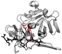
Structure of TEV protease. The double β-barrels that define the superfamily are highlighted in red. ( PDB: 1lvm )

The structure of TEV protease has been solved by X-ray crystallography. [4] It is composed of two β-barrels and a flexible C-terminal tail and displays structural homology to the chymotrypsin superfamily of proteases (PA clan, C4 family by MEROPS classification). [2] Although homologous to cellular serine proteases (such as trypsin, elastase, thrombin etc.), TEV protease uses a cysteine as its catalytic nucleophile [5] (as do many other viral proteases).

Covalent catalysis is performed with an Asp-His-Cys triad, split between the two barrels (Asp on β1 and His and Cys on β2). [6] The substrate is held as a β-sheet, forming an antiparallel interaction with the cleft between the barrels and a parallel interaction with the C-terminal tail. [7] The enzyme therefore forms a binding tunnel around the substrate and side chain interactions control specificity. [4]

Specificity

Surface model of TEV bound to uncleaved substrate (black), also showing the catalytic triad (red). The substrate binds inside an active site tunnel (left). A cutaway (right) shows the complementary shape of the binding tunnel to the substrate. (PDB: 1lvb ) TEV substrate binding tunnel.png
Surface model of TEV bound to uncleaved substrate (black), also showing the catalytic triad (red). The substrate binds inside an active site tunnel (left). A cutaway (right) shows the complementary shape of the binding tunnel to the substrate. ( PDB: 1lvb )

The preferred, native cleavage sequence was first identified by examining the cut sites in the native polyprotein substrate for recurring sequence. The consensus for these native cut sites is ENLYFQ\S where ‘\’ denotes the cleaved peptide bond. [8] Residues of the substrate are labelled P6 to P1 before the cut site and P1’ after the cut site. Early works also measured cleavage of an array of similar substrates to characterise how specific the protease was for the native sequence. [9] [10]

Studies have subsequently used sequencing of cleaved substrates from a pool of randomised sequences to determine preference patterns. [11] [12] Although ENLYFQ\S is the optimal sequence, the protease is active to a greater or lesser extent on a range of substrates (i.e. shows some substrate promiscuity). The highest cleavage is of sequences closest to the consensus EXLYΦQ\φ where X is any residue, Φ is any large or medium hydrophobe and φ is any small hydrophobic or polar residue. Although this sequence is the optimal, sequences with disfavoured residues at some positions can still be cleaved if the rest of the sequence is optimal. [10] [12]

Specificity is endowed by the large contact area between enzyme and substrate. Proteases such as trypsin have specificity for one residue before and after the cleaved bond due to a shallow binding cleft with only one or two pockets that bind the substrate side chains. Conversely, viral proteases such as TEV protease have a long C-terminal tail which completely covers the substrate to create a binding tunnel. This tunnel contains a set of tight binding pockets such that each side chain of the substrate peptide (P6 to P1’) is bound in a complementary site (S6 to S1’). [4]

In particular, peptide side chain P6-Glu contacts a network of three hydrogen bonds; P5-Asn points into the solvent, making no specific interactions (hence the absence of substrate consensus at this position); P4-Leu is buried in a hydrophobic pocket; P3-Tyr is held in a hydrophobic pocket with a short hydrogen bond at the end; P2-Phe is also surrounded by hydrophobes including the face of the triad histidine; P1-Gln forms four hydrogen bonds; and P1’-Ser is only partly enclosed in a shallow hydrophobic groove. [4]

Application as a biochemical tool

One of the main uses of this protein is for removing affinity tags from purified recombinant fusion proteins. The reason for the use of TEV protease as a biochemical tool is its high sequence specificity. This specificity allows for the controlled cleavage of proteins when the preference sequence is inserted into flexible loops. It also makes TEV protease relatively non-toxic in vivo as the recognized sequence scarcely occurs in proteins. [13]

Although rational design has had limited success in changing protease specificity, directed evolution has been used to change the preferred residue either before [14] or after [15] [16] the cleavage site.

However, TEV protease does have limitations as a biochemical tool. It is prone to deactivation by self-cleavage (autolysis), though this can be abolished through a single S219V mutation in the internal cleavage site. [17] The protease expressed alone is also poorly soluble, however several attempts have been made to improve its solubility through directed evolution and computational design. It has also been shown that expression can be improved by fusion to maltose binding protein (MBP) which acts a solubility-enhancing partner. [3]

TEV protease has been reported to show a 10-fold loss of activity at 4 °C. [18] TEV protease shows loss of activity at temperatures above 34 °C. [19] The original TEV protease required the presence of reducing agent for high activity, which could interfere with the function of proteins containing disulfide bonds. After incorporation of various mutations, later "superTEV protease" versions are highly active in the presence or absence of reducing agent. [20] [21] [22]

The molecular weight of this enzyme varies between 25 and 27 kDa depending on the specific construct used.

Related Research Articles

<span class="mw-page-title-main">Proteolysis</span> Breakdown of proteins into smaller polypeptides or amino acids

Proteolysis is the breakdown of proteins into smaller polypeptides or amino acids. Uncatalysed, the hydrolysis of peptide bonds is extremely slow, taking hundreds of years. Proteolysis is typically catalysed by cellular enzymes called proteases, but may also occur by intra-molecular digestion.

<span class="mw-page-title-main">Protease</span> Enzyme that cleaves other proteins into smaller peptides

A protease is an enzyme that catalyzes proteolysis, breaking down proteins into smaller polypeptides or single amino acids, and spurring the formation of new protein products. They do this by cleaving the peptide bonds within proteins by hydrolysis, a reaction where water breaks bonds. Proteases are involved in many biological functions, including digestion of ingested proteins, protein catabolism, and cell signaling.

<span class="mw-page-title-main">Serine protease</span> Class of enzymes

Serine proteases are enzymes that cleave peptide bonds in proteins. Serine serves as the nucleophilic amino acid at the (enzyme's) active site. They are found ubiquitously in both eukaryotes and prokaryotes. Serine proteases fall into two broad categories based on their structure: chymotrypsin-like (trypsin-like) or subtilisin-like.

<span class="mw-page-title-main">Hemagglutinin esterase</span> Glycoprotein present in some enveloped viruses

Hemagglutinin esterase (HEs) is a glycoprotein that certain enveloped viruses possess and use as an invading mechanism. HEs helps in the attachment and destruction of certain sialic acid receptors that are found on the host cell surface. Viruses that possess HEs include influenza C virus, toroviruses, and coronaviruses of the subgenus Embecovirus. HEs is a dimer transmembrane protein consisting of two monomers, each monomer is made of three domains. The three domains are: membrane fusion, esterase, and receptor binding domains.

<span class="mw-page-title-main">Cysteine protease</span> Class of enzymes

Cysteine proteases, also known as thiol proteases, are hydrolase enzymes that degrade proteins. These proteases share a common catalytic mechanism that involves a nucleophilic cysteine thiol in a catalytic triad or dyad.

<i>Potyvirus</i> Genus of positive-strand RNA viruses in the family Potyviridae

Potyvirus is a genus of positive-strand RNA viruses in the family Potyviridae. Plants serve as natural hosts. Like begomoviruses, members of this genus may cause significant losses in agricultural, pastoral, horticultural, and ornamental crops. More than 200 species of aphids spread potyviruses, and most are from the subfamily Aphidinae. The genus contains 190 species and potyviruses account for about thirty percent of all currently known plant viruses.

In molecular biology, the Signal Peptide Peptidase (SPP) is a type of protein that specifically cleaves parts of other proteins. It is an intramembrane aspartyl protease with the conserved active site motifs 'YD' and 'GxGD' in adjacent transmembrane domains (TMDs). Its sequences is highly conserved in different vertebrate species. SPP cleaves remnant signal peptides left behind in membrane by the action of signal peptidase and also plays key roles in immune surveillance and the maturation of certain viral proteins.

Group-specific antigen, or gag, is the polyprotein that contains the core structural proteins of an Ortervirus. It was named as such because scientists used to believe it was antigenic. Now it is known that it makes up the inner shell, not the envelope exposed outside. It makes up all the structural units of viral conformation and provides supportive framework for mature virion.

<span class="mw-page-title-main">HIV-1 protease</span> Enzyme involved with peptide bond hydrolysis in retroviruses

HIV-1 protease (PR) is a retroviral aspartyl protease (retropepsin), an enzyme involved with peptide bond hydrolysis in retroviruses, that is essential for the life-cycle of HIV, the retrovirus that causes AIDS. HIV protease cleaves newly synthesized polyproteins at nine cleavage sites to create the mature protein components of an HIV virion, the infectious form of a virus outside of the host cell. Without effective HIV protease, HIV virions remain uninfectious.

<i>Tobacco etch virus</i> Species of virus

Tobacco etch virus (TEV) is a plant virus in the genus Potyvirus and family Potyviridae. Like other members of the genus Potyvirus, TEV has a monopartite positive-sense, single-stranded RNA genome surrounded by a capsid made from a single viral encoded protein. The virus is a filamentous particle that measures about 730 nm in length. It is transmissible in a non-persistent manner by more than 10 species of aphids including Myzus persicae. It also is easily transmitted by mechanical means but is not known to be transmitted by seeds.

NS2-3 protease is an enzyme responsible for proteolytic cleavage between NS2 and NS3, which are non-structural proteins that form part of the HCV virus particle. NS3 protease of hepatitis C virus, on the other hand, is responsible for the cleavage of non-structural protein downstream. Both of these proteases are directly involved in HCV genome replication, that is, during the viral life-cycle that leads to virus multiplication in the host that has been infected by the virus.

Many major physiological processes depend on regulation of proteolytic enzyme activity and there can be dramatic consequences when equilibrium between an enzyme and its substrates is disturbed. In this prospective, the discovery of small-molecule ligands, like protease inhibitors, that can modulate catalytic activities has an enormous therapeutic effect. Hence, inhibition of the HIV protease is one of the most important approaches for the therapeutic intervention in HIV infection and their development is regarded as major success of structure-based drug design. They are highly effective against HIV and have, since the 1990s, been a key component of anti-retroviral therapies for HIV/AIDS.

<span class="mw-page-title-main">OmpT</span>

OmpT is an aspartyl protease found on the outer membrane of Escherichia coli. OmpT is a subtype of the family of omptin proteases, which are found on some gram-negative species of bacteria.

Pestivirus NS3 polyprotein peptidase is an enzyme. This enzyme catalyses the following chemical reaction

<span class="mw-page-title-main">3C-like protease</span> Class of enzymes

The 3C-like protease (3CLpro) or main protease (Mpro), formally known as C30 endopeptidase or 3-chymotrypsin-like protease, is the main protease found in coronaviruses. It cleaves the coronavirus polyprotein at eleven conserved sites. It is a cysteine protease and a member of the PA clan of proteases. It has a cysteine-histidine catalytic dyad at its active site and cleaves a Gln–(Ser/Ala/Gly) peptide bond.

HIV-2 retropepsin is an enzyme. This enzyme catalyses the following chemical reaction

<span class="mw-page-title-main">PA clan of proteases</span>

The PA clan is the largest group of proteases with common ancestry as identified by structural homology. Members have a chymotrypsin-like fold and similar proteolysis mechanisms but can have identity of <10%. The clan contains both cysteine and serine proteases. PA clan proteases can be found in plants, animals, fungi, eubacteria, archaea and viruses.

<span class="mw-page-title-main">Glutamic protease</span>

Glutamic proteases are a group of proteolytic enzymes containing a glutamic acid residue within the active site. This type of protease was first described in 2004 and became the sixth catalytic type of protease. Members of this group of protease had been previously assumed to be an aspartate protease, but structural determination showed it to belong to a novel protease family. The first structure of this group of protease was scytalidoglutamic peptidase, the active site of which contains a catalytic dyad, glutamic acid (E) and glutamine (Q), which give rise to the name eqolisin. This group of proteases are found primarily in pathogenic fungi affecting plant and human.

<span class="mw-page-title-main">2A self-cleaving peptides</span> Class of peptides

2A self-cleaving peptides, or 2A peptides, is a class of 18–22 aa-long peptides, which can induce ribosomal skipping during translation of a protein in a cell. These peptides share a core sequence motif of DxExNPGP, and are found in a wide range of viral families. 2A peptides are used to help generate polyproteins by causing the ribosome to fail at making a peptide bond.

Rio Negro virus is an alphavirus that was first isolated in Argentina in 1980. The virus was first called Ag80-663 but was renamed to Rio Negro virus in 2005. It is a former member of the Venezuelan equine encephalitis complex (VEEC), which are a group of alphaviruses in the Americas that have the potential to emerge and cause disease. Río Negro virus was recently reclassified as a distinct species. Closely related viruses include Mucambo virus and Everglades virus.

References

  1. 1 2 UniProt: TEV polyprotein: "P04517".
  2. 1 2 Rawlings ND, Barrett AJ, Bateman A (January 2012). "MEROPS: the database of proteolytic enzymes, their substrates and inhibitors". Nucleic Acids Res. 40 (Database issue): D343–50. doi:10.1093/nar/gkr987. PMC   3245014 . PMID   22086950.
  3. 1 2 Kapust RB, Waugh DS (July 2000). "Controlled intracellular processing of fusion proteins by TEV protease". Protein Expr. Purif. 19 (2): 312–8. doi:10.1006/prep.2000.1251. PMID   10873547.
  4. 1 2 3 4 Phan J, Zdanov A, Evdokimov AG, Tropea JE, Peters HK, Kapust RB, Li M, Wlodawer A, Waugh DS (December 2002). "Structural basis for the substrate specificity of tobacco etch virus protease". J. Biol. Chem. 277 (52): 50564–72. doi: 10.1074/jbc.M207224200 . PMID   12377789.
  5. Bazan JF, Fletterick RJ (November 1988). "Viral cysteine proteases are homologous to the trypsin-like family of serine proteases: structural and functional implications". Proc. Natl. Acad. Sci. U.S.A. 85 (21): 7872–6. Bibcode:1988PNAS...85.7872B. doi: 10.1073/pnas.85.21.7872 . PMC   282299 . PMID   3186696.
  6. Dougherty WG, Parks TD, Cary SM, Bazan JF, Fletterick RJ (September 1989). "Characterization of the catalytic residues of the tobacco etch virus 49-kDa proteinase". Virology. 172 (1): 302–10. doi:10.1016/0042-6822(89)90132-3. PMID   2475971.
  7. Tyndall JD, Nall T, Fairlie DP (March 2005). "Proteases universally recognize beta strands in their active sites". Chem. Rev. 105 (3): 973–99. doi:10.1021/cr040669e. PMID   15755082.
  8. Carrington JC, Dougherty WG (May 1988). "A viral cleavage site cassette: identification of amino acid sequences required for tobacco etch virus polyprotein processing". Proc. Natl. Acad. Sci. U.S.A. 85 (10): 3391–5. Bibcode:1988PNAS...85.3391C. doi: 10.1073/pnas.85.10.3391 . PMC   280215 . PMID   3285343.
  9. Dougherty WG, Cary SM, Parks TD (August 1989). "Molecular genetic analysis of a plant virus polyprotein cleavage site: a model". Virology. 171 (2): 356–64. doi:10.1016/0042-6822(89)90603-X. PMID   2669323.
  10. 1 2 Kapust, Rachel B.; Tözsér, József; Copeland, Terry D.; Waugh, David S. (2002-06-28). "The P1' specificity of tobacco etch virus protease". Biochemical and Biophysical Research Communications. 294 (5): 949–955. CiteSeerX   10.1.1.375.4271 . doi:10.1016/S0006-291X(02)00574-0. ISSN   0006-291X. PMID   12074568.
  11. Boulware KT, Jabaiah A, Daugherty PS (June 2010). "Evolutionary optimization of peptide substrates for proteases that exhibit rapid hydrolysis kinetics". Biotechnol. Bioeng. 106 (3): 339–46. doi:10.1002/bit.22693. PMID   20148412. S2CID   205499859.
  12. 1 2 Kostallas G, Löfdahl PÅ, Samuelson P (2011). "Substrate profiling of tobacco etch virus protease using a novel fluorescence-assisted whole-cell assay". PLOS ONE. 6 (1): e16136. Bibcode:2011PLoSO...616136K. doi: 10.1371/journal.pone.0016136 . PMC   3022733 . PMID   21267463.
  13. Parks TD, Leuther KK, Howard ED, Johnston SA, Dougherty WG (February 1994). "Release of proteins and peptides from fusion proteins using a recombinant plant virus proteinase". Anal. Biochem. 216 (2): 413–7. doi:10.1006/abio.1994.1060. PMID   8179197.
  14. Yi L, Gebhard MC, Li Q, Taft JM, Georgiou G, Iverson BL (April 2013). "Engineering of TEV protease variants by yeast ER sequestration screening (YESS) of combinatorial libraries". Proc. Natl. Acad. Sci. U.S.A. 110 (18): 7229–34. Bibcode:2013PNAS..110.7229Y. doi: 10.1073/pnas.1215994110 . PMC   3645551 . PMID   23589865.
  15. Renicke C, Spadaccini R, Taxis C (2013). "A tobacco etch virus protease with increased substrate tolerance at the P1' position". PLOS ONE. 8 (6): e67915. Bibcode:2013PLoSO...867915R. doi: 10.1371/journal.pone.0067915 . PMC   3691164 . PMID   23826349.
  16. Verhoeven KD, Altstadt OC, Savinov SN (March 2012). "Intracellular detection and evolution of site-specific proteases using a genetic selection system". Appl. Biochem. Biotechnol. 166 (5): 1340–54. doi:10.1007/s12010-011-9522-6. PMID   22270548. S2CID   36583382.
  17. Kapust RB, Tözsér J, Fox JD, Anderson DE, Cherry S, Copeland TD, Waugh DS (December 2001). "Tobacco etch virus protease: mechanism of autolysis and rational design of stable mutants with wild-type catalytic proficiency". Protein Eng. 14 (12): 993–1000. doi: 10.1093/protein/14.12.993 . PMID   11809930.
  18. Raran-Kurussi S, Tözsér J, Cherry S, Tropea JE, Waugh DS (15 May 2013). "Differential temperature dependence of tobacco etch virus and rhinovirus 3C proteases". Analytical Biochemistry. 436 (2): 142–144. doi:10.1016/j.ab.2013.01.031. PMC   4196241 . PMID   23395976.
  19. Nallamsetty S, Kapust RB, Tözsér J, Cherry S, Tropea JE, Copeland TD, Waugh DS (November 2004). "Efficient site-specific processing of fusion proteins by tobacco vein mottling virus protease in vivo and in vitro". Protein Expr. Purif. 38 (1): 108–115. doi:10.1016/j.pep.2004.08.016. PMID   15477088.
  20. Cabrita LD, Gilis D, Robertson AL, Dehouck Y, Rooman M, Bottomley SP (2007). "Enhancing the stability and solubility of TEV protease using in silico design". Protein Sci. 16 (11): 2360–7. doi:10.1110/ps.072822507. PMC   2211701 . PMID   17905838.
  21. Correnti CE, Gewe MM, Mehlin C, Bandaranayake AD, Johnsen WA, Rupert PB, Brusniak MY, Clarke M, Burke SE, De Van Der Schueren W, Pilat K, Turnbaugh SM, May D, Watson A, Chan MK, Bahl CD, Olson JM, Strong RK (2018). "Screening, large-scale production and structure-based classification of cystine-dense peptides". Nat Struct Mol Biol. 25 (3): 270–278. doi:10.1038/s41594-018-0033-9. PMC   5840021 . PMID   29483648.
  22. Keeble AH, Turkki P, Stokes S, Khairil Anuar IN, Rahikainen R, Hytönen VP, Howarth M (December 2019). "Approaching infinite affinity through engineering of peptide-protein interaction". Proceedings of the National Academy of Sciences of the United States of America. 116 (52): 26523–26533. Bibcode:2019PNAS..11626523K. doi: 10.1073/pnas.1909653116 . PMC   6936558 . PMID   31822621.