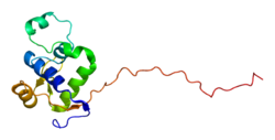
Werner syndrome ATP-dependent helicase, also known as DNA helicase, RecQ-like type 3, is an enzyme that in humans is encoded by the WRN gene. WRN is a member of the RecQ Helicase family. [5] Helicase enzymes generally unwind and separate double-stranded DNA. These activities are necessary before DNA can be copied in preparation for cell division (DNA replication). Helicase enzymes are also critical for making a blueprint of a gene for protein production, a process called transcription. Further evidence suggests that Werner protein plays a critical role in repairing DNA. Overall, this protein helps maintain the structure and integrity of a person's DNA.
The WRN gene is located on the short (p) arm of chromosome 8 between positions 12 and 11.2, from base pair 31,010,319 to base pair 31,150,818.
WRN is a member of the RecQ Helicase family. It is the only RecQ Helicase that contains 3' to 5' exonuclease activity. These exonuclease activities include degradation of recessed 3' ends and initiation of DNA degradation from a gap in dsDNA. WRN is important in repair of double strand breaks by homologous recombination [6] [7] or non-homologous end joining, [8] repair of single nucleotide damages by base excision repair, [9] [10] [5] and is effective in replication arrest recovery. [11] WRN may also be important in telomere maintenance and replication, especially the replication of the G-rich sequences. [12]
WRN is an oligomer that can act as a monomer when unwinding DNA, but as a dimer in solution or a tetramer when complexed with DNA, and has also been observed in hexameric forms. The diffusion of WRN has been measured to 1.62 in nucleoplasm and 0.12 at nucleoli. [13] Orthologs of WRN have been found in a number of other organisms, including Drosophila, Xenopus , and C. elegans. WRN is important to genome stability, and cells with mutations to WRN are more susceptible to DNA damage and DNA breaks. [14]
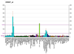
The amino terminus of WRN is involved in both helicase and nuclease activities, while the carboxyl-terminus interacts with p53, an important tumor suppressor. [15] WRN may function as an exonuclease in DNA repair, recombination, or replication, as well as resolution of DNA secondary structures. It is involved in branch migration at Holliday junctions, and it interacts with other DNA replication intermediates. [11] mRNA that codes for WRN has been identified in most human tissues. [15]
Phosphorylation of WRN at serine/threonine inhibits helicase and exonuclease activities which are important to post-replication DNA repair. De-phosphorylation at these sites enhances the catalytic activities of WRN. Phosphorylation may affect other post-translational modifications, including SUMOylation and acetylation. [12] Upon its inhibition by a small molecule in cancer cells harboring a high number of microsatellites (MSI-H), WRN becomes SUMOylated, which leads to is ubiquitylation and subsequent degradation. [16]
Methylation of WRN causes the gene to turn off. This suppresses the production of the WRN protein and its functions in DNA repair. [17]
Werner syndrome is caused by mutations in the WRN gene. [15] More than 20 mutations in the WRN gene are known to cause Werner syndrome. Many of these mutations result in an abnormally shortened Werner protein. Evidence suggests that the altered protein is not transported into the cell nucleus, where it normally interacts with DNA. [18] This shortened protein may also be broken down too quickly, leading to a loss of Werner protein in the cell. Without normal Werner protein in the nucleus, cells cannot perform the tasks of DNA replication, repair, and transcription. [19] Researchers are still determining how these mutations cause the appearance of premature aging seen in Werner syndrome.
Recently, WRN has been identified as a synthetic lethality target in cancers containing a high number of microsatellites. [20] These microsatellite-high (MSI-H) cancers have defects in their mismatch repair machinery (dMMR), which leads to the expansion of (TA)n dinucleotide repeats in the genome. These expanded (TA) dinucleotide microsatellites lead to the formation of secondary DNA structures (e.g. G-quadruplex) and rely on WRN to repair these bulky lesions. [21] Because of this therapeutic hypothesis, inhibition of WRN has become an area of high interest for targeted therapies of MSI-H cancers, especially those that do not respond to immune checkpoint inhibition or chemotherapy. [22]
Cells expressing limiting amounts of WRN have elevated mutation frequencies compared with wildtype cells. [23] Increased mutation may give rise to cancer. Patients with Werner Syndrome, with homozygous mutations in the WRN gene, have an increased incidence of cancers, including soft tissue sarcomas, osteosarcoma, thyroid cancer and melanoma. [24]
Mutations in WRN are rare in the general population. The rate of heterozygous loss-of-function mutation in WRN is approximately one per million. In a Japanese population the rate is 6 per 1,000, which is higher, but still infrequent. [25]
Mutational defects in the WRN gene are relatively rare in cancer cells compared to the frequency of epigenetic alterations in WRN that reduce WRN expression and could contribute to carcinogenesis. The situation is similar to other DNA repair genes whose expression is reduced in cancers due to mainly epigenetic alterations rather than mutations (see Frequencies of epimutations in DNA repair genes).[ citation needed ]
The table shows results of analysis of 630 human primary tumors for WRN CpG island hypermethylation. [26] This hypermethylation caused reduced protein expression of WRN, a common event in tumorigenesis. [26]
Cancer | Frequency of reduction in cancer [26] |
---|---|
Colorectal cancer | 37.9% |
Non-small cell lung cancer | 37.5% |
Gastric cancer | 25% |
Prostate cancer | 20% |
Breast cancer | 17.2% |
Thyroid cancer | 12.5% |
Non-Hodgkin lymphoma | 23.7% |
Acute myeloblastic leukemia | 4.8% |
Chondrosarcomas | 33.3% |
Osteosarcomas | 11.1% |
WRN is active in homologous recombination. Cells defective in the WRN gene have a 23-fold reduction in spontaneous mitotic recombination, with especial deficiency in conversion-type events. [27] WRN defective cells, when exposed to x-rays, have more chromosome breaks and micronuclei than cells with wild-type WRN. [28] Cells defective in the WRN gene are not more sensitive than wild-type cells to gamma-irradiation, UV light, 4 – 6 cyclobutane pyrimidines, or mitomycin C, but are sensitive to type I and type II topoisomerase inhibitors. [29] These findings suggested that the WRN protein takes part in homologous recombinational repair and in the processing of stalled replication forks. [30]
WRN has an important role in non-homologous end joining (NHEJ) DNA repair. As shown by Shamanna et al., [8] WRN is recruited to double-strand breaks (DSBs) and participates in NHEJ with its enzymatic and non-enzymatic functions. At DSBs, in association with Ku (protein), it promotes standard or canonical NHEJ (c-NHEJ), repairing double-strand breaks in DNA with its enzymatic functions and with a fair degree of accuracy. WRN inhibits an alternative form of NHEJ, called alt-NHEJ or microhomology-mediated end joining (MMEJ). MMEJ is an inaccurate mode of repair for double-strand breaks.
WRN has a role in base excision repair (BER) of DNA. As shown by Das et al., [9] WRN associates with NEIL1 in the early damage-sensing step of BER. WRN stimulates NEIL1 in excision of oxidative lesions. NEIL1 is a DNA glycosylase that initiates the first step in BER by cleaving bases damaged by reactive oxygen species (ROS) and introducing a DNA strand break via NEIL1's associated lyase activity. [31] NEIL1 recognizes (targets) and removes certain ROS-damaged bases and then incises the abasic site via β,δ elimination, leaving 3′ and 5′ phosphate ends. NEIL1 recognizes oxidized pyrimidines, formamidopyrimidines, thymine residues oxidized at the methyl group, and both stereoisomers of thymine glycol. [32]
WRN also participates in BER through its interaction with Polλ. [10] WRN binds to the catalytic domain of Polλ and specifically stimulates DNA gap filling by Polλ over 8-oxo-G followed by strand displacement synthesis. This allows WRN to promote long-patch DNA repair synthesis by Polλ during MUTYH-initiated repair of 8-oxo-G:A mispairs.
WRN is also involved in replication arrest recovery. If WRN is defective, replication arrest results in accumulation of DSBs and enhanced chromosome fragmentation. [33] As shown by Pichierri et al., [33] WRN interacts with the RAD9-RAD1-HUS1 (9.1.1) complex, one of the central factors of the replication checkpoint. This interaction is mediated by the binding of the RAD1 subunit to the N-terminal region of WRN and is instrumental for WRN relocalization to nuclear foci and its phosphorylation in response to replication arrest. (In the absence of DNA damage or replication fork stalling, WRN protein remains localized to the nucleoli. [34] ) The interaction of WRN with the 9.1.1 complex results in prevention of DSB formation at stalled replication forks. [33]
The p53 protein and WRN helicase engage in direct protein-protein interaction. [35] Increased cellular WRN levels elicit increased cellular p53 levels and also potentiate p53-mediated apoptosis. [35] This finding suggests that WRN helicase participates in the activation of p53 in response to certain types of DNA damage. [35] p53-mediated apoptosis is attenuated in cells from patients with Werner syndrome. [36]
Both repair of DNA damage and apoptosis are enzymatic processes necessary for maintaining integrity of the genome in humans. Cells with insufficient DNA repair tend to accumulate DNA damages, and when such cells are also defective in apoptosis they tend to survive even though excessive DNA damages are present. [37] Replication of DNA in such deficient cells tends to lead to mutations and such mutations may cause cancer. Thus Werner syndrome helicase appears to have two roles related to the prevention of cancer, where the first role is to promote repair of specific types of damage and the second role is to induce apoptosis if the level of such DNA damage is beyond the cell’s repair capability [37]
Merging with "Clinical significance" section
Werner syndrome ATP-dependent helicase has been shown to interact with:
Werner syndrome (WS) or Werner's syndrome, also known as "adult progeria", is a rare, autosomal recessive disorder which is characterized by the appearance of premature aging.
Helicases are a class of enzymes thought to be vital to all organisms. Their main function is to unpack an organism's genetic material. Helicases are motor proteins that move directionally along a nucleic acid phosphodiester backbone, separating two hybridized nucleic acid strands, using energy from ATP hydrolysis. There are many helicases, representing the great variety of processes in which strand separation must be catalyzed. Approximately 1% of eukaryotic genes code for helicases.
RecQ helicase is a family of helicase enzymes initially found in Escherichia coli that has been shown to be important in genome maintenance. They function through catalyzing the reaction ATP + H2O → ADP + P and thus driving the unwinding of paired DNA and translocating in the 3' to 5' direction. These enzymes can also drive the reaction NTP + H2O → NDP + P to drive the unwinding of either DNA or RNA.
Bloom syndrome is a rare autosomal recessive genetic disorder characterized by short stature, predisposition to the development of cancer, and genomic instability. BS is caused by mutations in the BLM gene which is a member of the RecQ DNA helicase family. Mutations in genes encoding other members of this family, namely WRN and RECQL4, are associated with the clinical entities Werner syndrome and Rothmund–Thomson syndrome, respectively. More broadly, Bloom syndrome is a member of a class of clinical entities that are characterized by chromosomal instability, genomic instability, or both and by cancer predisposition.
Base excision repair (BER) is a cellular mechanism, studied in the fields of biochemistry and genetics, that repairs damaged DNA throughout the cell cycle. It is responsible primarily for removing small, non-helix-distorting base lesions from the genome. The related nucleotide excision repair pathway repairs bulky helix-distorting lesions. BER is important for removing damaged bases that could otherwise cause mutations by mispairing or lead to breaks in DNA during replication. BER is initiated by DNA glycosylases, which recognize and remove specific damaged or inappropriate bases, forming AP sites. These are then cleaved by an AP endonuclease. The resulting single-strand break can then be processed by either short-patch or long-patch BER.
A DNA repair-deficiency disorder is a medical condition due to reduced functionality of DNA repair.
DNA mismatch repair protein Mlh1 or MutL protein homolog 1 is a protein that in humans is encoded by the MLH1 gene located on chromosome 3. The gene is commonly associated with hereditary nonpolyposis colorectal cancer. Orthologs of human MLH1 have also been studied in other organisms including mouse and the budding yeast Saccharomyces cerevisiae.
Ku70 is a protein that, in humans, is encoded by the XRCC6 gene.
Ku80 is a protein that, in humans, is encoded by the XRCC5 gene. Together, Ku70 and Ku80 make up the Ku heterodimer, which binds to DNA double-strand break ends and is required for the non-homologous end joining (NHEJ) pathway of DNA repair. It is also required for V(D)J recombination, which utilizes the NHEJ pathway to promote antigen diversity in the mammalian immune system.
Replication protein A 70 kDa DNA-binding subunit is a protein that in humans is encoded by the RPA1 gene.
Bloom syndrome protein is a protein that in humans is encoded by the BLM gene and is not expressed in Bloom syndrome.
Flap endonuclease 1 is an enzyme that in humans is encoded by the FEN1 gene.
DNA excision repair protein ERCC-6 is a protein that in humans is encoded by the ERCC6 gene. The ERCC6 gene is located on the long arm of chromosome 10 at position 11.23.
Exonuclease 1 is an enzyme that in humans is encoded by the EXO1 gene.
DNA polymerase delta catalytic subunit(DPOD1) is an enzyme that is encoded in the human by the POLD1 gene, in the DNA polymerase delta complex. DPOD1 is responsible for synthesizing the lagging strand of DNA, and has also been implicated in some activities at the leading strand. The DPOD1 subunit encodes both DNA polymerizing and exonuclease domains, which provide the protein an important second function in proofreading to ensure replication accuracy during DNA synthesis, and in a number of types of replication-linked DNA repair following DNA damage.
Fanconi anemia group J protein is a protein that in humans is encoded by the BRCA1-interacting protein 1 (BRIP1) gene.
ATP-dependent DNA helicase Q4 is an enzyme that in humans is encoded by the RECQL4 gene.
ATPase WRNIP1 is an enzyme that is encoded by the WRNIP1 gene in humans. The protein is a member of AAA ATPase family.
Progeroid syndromes (PS) are a group of rare genetic disorders that mimic physiological aging, making affected individuals appear to be older than they are. The term progeroid syndrome does not necessarily imply progeria, which is a specific type of progeroid syndrome.
A hereditary cancer syndrome is a genetic disorder in which inherited genetic mutations in one or more genes predispose the affected individuals to the development of cancer and may also cause early onset of these cancers. Hereditary cancer syndromes often show not only a high lifetime risk of developing cancer, but also the development of multiple independent primary tumors.