RecBCD

Last updated
Exodeoxyribonuclease V
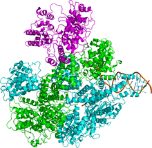
RecBCD 1W36.png
Figure 1 The crystallographic structure of the RecBCD enzyme ( PDB: 1W36 ). [1] The RecB, RecC, and RecD subunits of the enzyme are colored cyan, green, and magenta respectively while the partially unwound DNA helix to which the enzyme is bound is colored brown.
Identifiers
EC no. 3.1.11.5
CAS no. 37350-26-8
Databases
IntEnz IntEnz view
BRENDA BRENDA entry
ExPASy NiceZyme view
KEGG KEGG entry
MetaCyc metabolic pathway
PRIAM profile
PDB structures RCSB PDB PDBe PDBsum
Search
PMC articles
PubMed articles
NCBI proteins

Exodeoxyribonuclease V (EC 3.1.11.5, RecBCD, Exonuclease V, Escherichia coli exonuclease V, E. coli exonuclease V, gene recBC endoenzyme, RecBC deoxyribonuclease, gene recBC DNase, gene recBCD enzymes) is an enzyme of E. coli that initiates recombinational repair from potentially lethal double strand breaks in DNA which may result from ionizing radiation, replication errors, endonucleases, oxidative damage, and a host of other factors. [2] [3] The RecBCD enzyme is both a helicase that unwinds, or separates the strands of DNA, and a nuclease that makes single-stranded nicks in DNA. [1] It catalyses exonucleolytic cleavage (in the presence of ATP) in either 5′- to 3′- or 3′- to 5′-direction to yield 5′-phosphooligonucleotides.

Contents

Structure

The enzyme complex is composed of three different subunits called RecB, RecC, and RecD and hence the complex is named RecBCD (Figure 1). Before the discovery of the recD gene, [4] the enzyme was known as “RecBC.” Each subunit is encoded by a separate gene:

genechainproteinfunction
RecBβ P08394 3'-5' helicase, nuclease
RecCγ P07648 Recognizes Chi (crossover hotspot instigator)
RecDα P04993 5'-3' helicase

Function

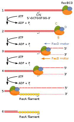
Figure 2 RecBCD pathway of homologous recombination where ATP is in excess. Chi Recombination Model for Wikipedia.tif
Figure 2 RecBCD pathway of homologous recombination where ATP is in excess.

Both the RecD and RecB subunits are helicases, i.e., energy-dependent molecular motors that unwind DNA (or RNA in the case of other proteins). The RecB subunit in addition has a nuclease function. [5] Finally, RecBCD enzyme (perhaps the RecC subunit) recognizes a specific sequence in DNA, 5'-GCTGGTGG-3', known as Chi (sometimes designated with the Greek letter χ).

RecBCD is unusual amongst helicases because it has two helicases that travel with different rates [6] and because it can recognize and be altered by the Chi DNA sequence. [7] [8] RecBCD avidly binds an end of linear double-stranded (ds) DNA. The RecD helicase travels on the strand with a 5' end at which the enzyme initiates unwinding, and RecB on the strand with a 3' end. RecB is slower than RecD, so that a single-stranded (ss) DNA loop accumulates ahead of RecB (Figure 2). This produces DNA structures with two ss tails (a shorter 3’ ended tail and a longer 5’ ended tail) and one ss loop (on the 3' ended strand) observed by electron microscopy. [9] The ss tails can anneal to produce a second ss loop complementary to the first one; such twin-loop structures were initially referred to as “rabbit ears.”

Mechanism of action

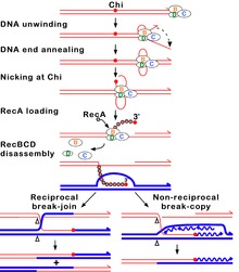
Figure 3 Beginning of the RecBCD pathway of homologous recombination where Mg is in excess. HR RecBCD RecA.svg
Figure 3 Beginning of the RecBCD pathway of homologous recombination where Mg is in excess.

During unwinding the nuclease in RecB can act in different ways depending on the reaction conditions, notably the ratio of the concentrations of Mg2+ ions and ATP. (1) If ATP is in excess, the enzyme simply nicks the strand with Chi (the strand with the initial 3' end) (Figure 2). [10] [11] Unwinding continues and produces a 3' ss tail with Chi near its terminus. This tail can be bound by RecA protein, which promotes strand exchange with an intact homologous DNA duplex. [12] When RecBCD reaches the end of the DNA, all three subunits disassemble and the enzyme remains inactive for an hour or more; [13] a RecBCD molecule that acted at Chi does not attack another DNA molecule. (2) If Mg2+ ions are in excess, RecBCD cleaves both DNA strands endonucleolytically, although the 5' tail is cleaved less often (Figure 3). [14] When RecBCD encounters a Chi site on the 3' ended strand, unwinding pauses and digestion of the 3' tail is reduced. [15] When RecBCD resumes unwinding, it now cleaves the opposite strand (i.e., the 5' tail) [16] [17] and loads RecA protein onto the 3’-ended strand. [12] After completing reaction on one DNA molecule, the enzyme quickly attacks a second DNA, on which the same reactions occur as on the first DNA.

Although neither reaction has been verified by analysis of intracellular DNA, due to the transient nature of reaction intermediates, genetic evidence indicates that the first reaction more nearly mimics that in cells. [2] For example, the activity of Chi is influenced by nucleotides to its 3' side, both in cells and in reactions with ATP in excess but not with Mg2+ in excess [PMIDs 27401752, 27330137]. RecBCD mutants lacking detectable exonuclease activity retain high Chi hotspot activity in cells and nicking at Chi outside cells. [18] A Chi site on one DNA molecule in cells reduces or eliminates Chi activity on another DNA, perhaps reflecting the Chi-dependent disassembly of RecBCD observed in vitro under conditions of excess ATP and nicking of DNA at Chi. [19] [20]

Under both reaction conditions, the 3' strand remains intact downstream of Chi. The RecA protein is then actively loaded onto the 3' tail by RecBCD. [12] At some undetermined point RecBCD dissociates from the DNA, although RecBCD can unwind at least 60 kb of DNA without falling off. RecA initiates exchange of the DNA strand to which it is bound with the identical, or nearly identical, strand in an intact DNA duplex; this strand exchange generates a joint DNA molecule, such as a D-loop (Figure 2). The joint DNA molecule is thought to be resolved either by replication primed by the invading 3’ ended strand containing Chi or by cleavage of the D-loop and formation of a Holliday junction. The Holliday junction can be resolved into linear DNA by the RuvABC complex or dissociated by the RecG protein. Each of these events can generate intact DNA with new combinations of genetic markers by which the parental DNAs may differ. This process, homologous recombination, completes the repair of the double-stranded DNA break.

RecD1 and RecD2

RecD enzymes are divided into two groups RecD1 (known as RecD) and RecD2. [21] Many organisms have a recD gene even though the other members of a recBCD complex, i. e. rec B and recC, are not present. [22] For instance, the bacterium Deinococcus radiodurans , that has an extraordinary DNA repair capability, is an example of an organism that does not possess a recB or recC gene, and yet does have a recD gene. [22] In the bacterium Escherichia coli , RecD protein is part of the well studied RecBCD complex that is necessary for recombinational DNA repair (as described above). In the bacterium Bacillus subtilis , RecD2 protein has a role as a modulator of replication restart and also a modulator of the RecA recombinase. [21] RecD2 may inhibit unwanted recombination events when replication forks are stalled, and also may have a role in displacing RecA protein from recombination intermediates in order to permit advance of the replication fork. [21]

Applications

RecBCD is a model enzyme for the use of single molecule fluorescence as an experimental technique used to better understand the function of protein-DNA interactions. [23] The enzyme is also useful in removing linear DNA, either single- or double-stranded, from preparations of circular double-stranded DNA, since it requires a DNA end for activity.

Related Research Articles

<span class="mw-page-title-main">DNA replication</span> Biological process

In molecular biology, DNA replication is the biological process of producing two identical replicas of DNA from one original DNA molecule. DNA replication occurs in all living organisms acting as the most essential part of biological inheritance. This is essential for cell division during growth and repair of damaged tissues, while it also ensures that each of the new cells receives its own copy of the DNA. The cell possesses the distinctive property of division, which makes replication of DNA essential.

<span class="mw-page-title-main">DNA polymerase</span> Form of DNA replication

A DNA polymerase is a member of a family of enzymes that catalyze the synthesis of DNA molecules from nucleoside triphosphates, the molecular precursors of DNA. These enzymes are essential for DNA replication and usually work in groups to create two identical DNA duplexes from a single original DNA duplex. During this process, DNA polymerase "reads" the existing DNA strands to create two new strands that match the existing ones. These enzymes catalyze the chemical reaction

DNA primase is an enzyme involved in the replication of DNA and is a type of RNA polymerase. Primase catalyzes the synthesis of a short RNA segment called a primer complementary to a ssDNA template. After this elongation, the RNA piece is removed by a 5' to 3' exonuclease and refilled with DNA.

<span class="mw-page-title-main">Helicase</span> Class of enzymes to unpack an organisms genes

Helicases are a class of enzymes thought to be vital to all organisms. Their main function is to unpack an organism's genetic material. Helicases are motor proteins that move directionally along a nucleic acid phosphodiester backbone, separating two hybridized nucleic acid strands, using energy from ATP hydrolysis. There are many helicases, representing the great variety of processes in which strand separation must be catalyzed. Approximately 1% of eukaryotic genes code for helicases.

<span class="mw-page-title-main">Nuclease</span> Class of enzymes which cleave nucleic acids

In biochemistry, a nuclease is an enzyme capable of cleaving the phosphodiester bonds that link nucleotides together to form nucleic acids. Nucleases variously affect single and double stranded breaks in their target molecules. In living organisms, they are essential machinery for many aspects of DNA repair. Defects in certain nucleases can cause genetic instability or immunodeficiency. Nucleases are also extensively used in molecular cloning.

<span class="mw-page-title-main">RuvABC</span> Protein complex

RuvABC is a complex of three proteins that mediate branch migration and resolve the Holliday junction created during homologous recombination in bacteria. As such, RuvABC is critical to bacterial DNA repair.

DNA gyrase, or simply gyrase, is an enzyme within the class of topoisomerase and is a subclass of Type II topoisomerases that reduces topological strain in an ATP dependent manner while double-stranded DNA is being unwound by elongating RNA-polymerase or by helicase in front of the progressing replication fork. It is the only known enzyme to actively contribute negative supercoiling to DNA, while it also is capable of relaxing positive supercoils. It does so by looping the template to form a crossing, then cutting one of the double helices and passing the other through it before releasing the break, changing the linking number by two in each enzymatic step. This process occurs in bacteria, whose single circular DNA is cut by DNA gyrase and the two ends are then twisted around each other to form supercoils. Gyrase is also found in eukaryotic plastids: it has been found in the apicoplast of the malarial parasite Plasmodium falciparum and in chloroplasts of several plants. Bacterial DNA gyrase is the target of many antibiotics, including nalidixic acid, novobiocin, albicidin, and ciprofloxacin.

RecQ helicase is a family of helicase enzymes initially found in Escherichia coli that has been shown to be important in genome maintenance. They function through catalyzing the reaction ATP + H2O → ADP + P and thus driving the unwinding of paired DNA and translocating in the 3' to 5' direction. These enzymes can also drive the reaction NTP + H2O → NDP + P to drive the unwinding of either DNA or RNA.

<span class="mw-page-title-main">Exonuclease</span> Class of enzymes; type of nuclease

Exonucleases are enzymes that work by cleaving nucleotides one at a time from the end (exo) of a polynucleotide chain. A hydrolyzing reaction that breaks phosphodiester bonds at either the 3′ or the 5′ end occurs. Its close relative is the endonuclease, which cleaves phosphodiester bonds in the middle (endo) of a polynucleotide chain. Eukaryotes and prokaryotes have three types of exonucleases involved in the normal turnover of mRNA: 5′ to 3′ exonuclease (Xrn1), which is a dependent decapping protein; 3′ to 5′ exonuclease, an independent protein; and poly(A)-specific 3′ to 5′ exonuclease.
<span class="mw-page-title-main">Homologous recombination</span> Genetic recombination between identical or highly similar strands of genetic material

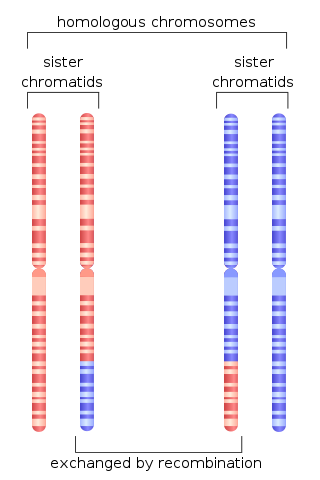
Homologous recombination is a type of genetic recombination in which genetic information is exchanged between two similar or identical molecules of double-stranded or single-stranded nucleic acids.

<span class="mw-page-title-main">Werner syndrome helicase</span> Enzyme found in humans

Werner syndrome ATP-dependent helicase, also known as DNA helicase, RecQ-like type 3, is an enzyme that in humans is encoded by the WRN gene. WRN is a member of the RecQ Helicase family. Helicase enzymes generally unwind and separate double-stranded DNA. These activities are necessary before DNA can be copied in preparation for cell division. Helicase enzymes are also critical for making a blueprint of a gene for protein production, a process called transcription. Further evidence suggests that Werner protein plays a critical role in repairing DNA. Overall, this protein helps maintain the structure and integrity of a person's DNA.

Deoxyribonuclease IV (phage-T4-induced) is catalyzes the degradation nucleotides in DsDNA by attacking the 5'-terminal end.

<span class="mw-page-title-main">Exonuclease 1</span> Protein-coding gene in the species Homo sapiens

Exonuclease 1 is an enzyme that in humans is encoded by the EXO1 gene.

<span class="mw-page-title-main">RECQL</span> Protein-coding gene in the species Homo sapiens

ATP-dependent DNA helicase Q1 is an enzyme that in humans is encoded by the RECQL gene.

<span class="mw-page-title-main">T7 DNA polymerase</span> Enzyme

T7 DNA polymerase is an enzyme used during the DNA replication of the T7 bacteriophage. During this process, the DNA polymerase “reads” existing DNA strands and creates two new strands that match the existing ones. The T7 DNA polymerase requires a host factor, E. coli thioredoxin, in order to carry out its function. This helps stabilize the binding of the necessary protein to the primer-template to improve processivity by more than 100-fold, which is a feature unique to this enzyme. It is a member of the Family A DNA polymerases, which include E. coli DNA polymerase I and Taq DNA polymerase.

A Chi site or Chi sequence is a short stretch of DNA in the genome of a bacterium near which homologous recombination is more likely to occur than on average across the genome. Chi sites serve as stimulators of DNA double-strand break repair in bacteria, which can arise from radiation or chemical treatments, or result from replication fork breakage during DNA replication. The sequence of the Chi site is unique to each group of closely related organisms; in E. coli and other enteric bacteria, such as Salmonella, the core sequence is 5'-GCTGGTGG-3' plus important nucleotides about 4 to 7 nucleotides to the 3' side of the core sequence. The existence of Chi sites was originally discovered in the genome of bacteriophage lambda, a virus that infects E. coli, but is now known to occur about 1000 times in the E. coli genome.

<span class="mw-page-title-main">Circular chromosome</span> Type of chromosome

A circular chromosome is a chromosome in bacteria, archaea, mitochondria, and chloroplasts, in the form of a molecule of circular DNA, unlike the linear chromosome of most eukaryotes.

The RecF pathway, also called the RecFOR pathway, is a pathway of homologous recombination that repairs DNA in bacteria. It repairs breaks that occur on only one of DNA's two strands, known as single-strand gaps. The RecF pathway can also repair double-strand breaks in DNA when the RecBCD pathway, another pathway of homologous recombination in bacteria, is inactivated by mutations. Like the RecBCD pathway, the RecF pathway requires RecA for strand invasion. The two pathways are also similar in their phases of branch migration, in which the Holliday junction slides in one direction, and resolution, in which the Holliday junctions are cleaved apart by enzymes.

DNA Polymerase V is a polymerase enzyme involved in DNA repair mechanisms in bacteria, such as Escherichia coli. It is composed of a UmuD' homodimer and a UmuC monomer, forming the UmuD'2C protein complex. It is part of the Y-family of DNA Polymerases, which are capable of performing DNA translesion synthesis (TLS). Translesion polymerases bypass DNA damage lesions during DNA replication - if a lesion is not repaired or bypassed the replication fork can stall and lead to cell death. However, Y polymerases have low sequence fidelity during replication. When the UmuC and UmuD' proteins were initially discovered in E. coli, they were thought to be agents that inhibit faithful DNA replication and caused DNA synthesis to have high mutation rates after exposure to UV-light. The polymerase function of Pol V was not discovered until the late 1990s when UmuC was successfully extracted, consequent experiments unequivocally proved UmuD'2C is a polymerase. This finding lead to the detection of many Pol V orthologs and the discovery of the Y-family of polymerases.

Stephen Charles Kowalczykowski is a Distinguished Professor of Microbiology and Molecular Genetics at the University of California at Davis. His research focuses on the biochemistry and molecular biology of DNA repair and homologous recombination. His lab combines fluorescence microscopy, optical trapping and microfluidics to manipulate and visualize single molecules of DNA and the enzymes involved in processing and repairing DNA. He calls this scientific approach, "visual biochemistry". Stephen Kowalczykowski was elected to the American Society for Arts and Science in 2005, the National Academy of Sciences in 2007 and was a Harvey Society Lecturer at Rockefeller University in 2012.
References

  1. 1 2 Singleton MR, Dillingham MS, Gaudier M, Kowalczykowski SC, Wigley DB (November 2004). "Crystal structure of RecBCD enzyme reveals a machine for processing DNA breaks". Nature. 432 (7014): 187–93. Bibcode:2004Natur.432..187S. doi:10.1038/nature02988. PMID   15538360. S2CID   2916995.
  2. 1 2 Smith, GR (June 2012). "How RecBCD Enzyme and Chi Promote DNA Break Repair and Recombination: a Molecular Biologist's View". Microbiol Mol Biol Rev. 76 (2): 217–28. doi:10.1128/MMBR.05026-11. PMC   3372252 . PMID   22688812.
  3. Spies M, Kowalczykowski SC (2003). "Homologous recombination by RecBCD and RecF pathways". In Higgins P (ed.). Bacterial Chromosomes. Washington, D.C.: ASM Press. pp. 389–403. ISBN   1-55581-232-5.
  4. Amundsen SK, Taylor AF, Chaudhury AM, Smith GR (August 1986). "recD: the gene for an essential third subunit of exonuclease V." Proc Natl Acad Sci U S A. 83 (15): 5558–62. Bibcode:1986PNAS...83.5558A. doi: 10.1073/pnas.83.15.5558 . PMC   386327 . PMID   3526335.
  5. Yu M, Souaya J, Julin DA (February 1998). "The 30-kDa C-terminal domain of the RecB protein is critical for the nuclease activity, but not the helicase activity, of the RecBCD enzyme from Escherichia coli". Proc Natl Acad Sci U S A. 95 (3): 981–6. Bibcode:1998PNAS...95..981Y. doi: 10.1073/pnas.95.3.981 . PMC   18645 . PMID   9448271.
  6. Taylor AF, Smith GR (June 2003). "RecBCD enzyme is a DNA helicase with fast and slow motors of opposite polarity". Nature. 423 (6942): 889–93. Bibcode:2003Natur.423..889T. doi:10.1038/nature01674. PMID   12815437. S2CID   4302346.
  7. Taylor AF, Smith GR (June 1992). "RecBCD enzyme is altered upon cutting DNA at a Chi recombination hotspot". Proc Natl Acad Sci U S A. 89 (12): 5226–30. Bibcode:1992PNAS...89.5226T. doi: 10.1073/pnas.89.12.5226 . PMC   49264 . PMID   1535156.
  8. Amundsen SK, Taylor AF, Reddy M, Smith GR (December 2007). "Intersubunit signaling in RecBCD enzyme, a complex protein machine regulated by Chi hot spots". Genes Dev. 21 (24): 3296–307. doi:10.1101/gad.1605807. PMC   2113030 . PMID   18079176.
  9. Taylor A, Smith GR (November 1980). "Unwinding and rewinding of DNA by the RecBC enzyme". Cell. 22 (2 Pt 2): 447–57. doi:10.1016/0092-8674(80)90355-4. PMID   6256081. S2CID   22123298.
  10. Ponticelli, A. S.; Schultz, D. W.; Taylor, A. F.; Smith, G. R. (May 1985). "Chi-dependent DNA strand cleavage by RecBC enzyme". Cell. 41 (1): 145–151. doi:10.1016/0092-8674(85)90069-8. PMID   3888404. S2CID   38361329.
  11. Taylor AF, Schultz DW, Ponticelli AS, Smith GR (May 1985). "RecBC enzyme nicking at Chi sites during DNA unwinding: location and orientation-dependence of the cutting". Cell. 41 (1): 153–63. doi:10.1016/0092-8674(85)90070-4. PMID   3888405. S2CID   8551482.
  12. 1 2 3 Anderson DG, Kowalczykowski SC (July 1997). "The translocating RecBCD enzyme stimulates recombination by directing RecA protein onto ssDNA in a Chi-regulated manner". Cell. 90 (1): 77–86. doi: 10.1016/S0092-8674(00)80315-3 . PMID   9230304.
  13. Taylor AF, Smith GR (April 1999). "Regulation of homologous recombination: Chi inactivates RecBCD enzyme by disassembly of the three subunits". Genes Dev. 13 (7): 890–900. doi:10.1101/gad.13.7.890. PMC   316601 . PMID   10197988.
  14. Dixon DA, Kowalczykowski SC (April 1993). "The recombination hotspot Chi is a regulatory sequence that acts by attenuating the nuclease activity of the E. coli RecBCD enzyme". Cell. 73 (1): 87–96. doi:10.1016/0092-8674(93)90162-J. PMID   8384931. S2CID   19718817.
  15. Spies M, Amitani I, Baskin RJ, Kowalczykowski SC (November 2007). "RecBCD enzyme switches lead motor subunits in response to Chi recognition". Cell. 131 (4): 694–705. doi:10.1016/j.cell.2007.09.023. PMC   2151923 . PMID   18022364.
  16. Taylor AF, Smith GR (October 1995). "Strand specificity of nicking of DNA at Chi sites by RecBCD enzyme. Modulation by ATP and magnesium levels". J Biol Chem. 270 (41): 24459–67. doi: 10.1074/jbc.270.41.24459 . PMID   7592661.
  17. Anderson DG, Kowalczykowski SC (March 1997). "The recombination hot spot chi is a regulatory element that switches the polarity of DNA degradation by the RecBCD enzyme". Genes Dev. 11 (5): 571–81. doi: 10.1101/gad.11.5.571 . PMID   9119222.
  18. Amundsen SK, Smith GR (January 2007). "Chi hotspot activity in Escherichia coli without RecBCD exonuclease activity: implications for the mechanism of recombination". Genetics. 175 (1): 41–54. doi:10.1534/genetics.106.065524. PMC   1774988 . PMID   17110484.
  19. Köppen A, Krobitsch S, Thoms B, Wackernagel W (July 1995). "Interaction with the recombination hot spot Chi in vivo converts the RecBCD enzyme of Escherichia coli into a Chi-independent recombinase by inactivation of the RecD subunit". Proc Natl Acad Sci U S A. 92 (14): 6249–53. Bibcode:1995PNAS...92.6249K. doi: 10.1073/pnas.92.14.6249 . PMC   41495 . PMID   7541534.
  20. Myers RS, Kuzminov A, Stahl FW (July 1995). "The recombination hot spot Chi activates RecBCD recombination by converting Escherichia coli to a recD mutant phenocopy". Proc Natl Acad Sci U S A. 92 (14): 6244–8. Bibcode:1995PNAS...92.6244M. doi: 10.1073/pnas.92.14.6244 . PMC   41494 . PMID   7603978.
  21. 1 2 3 Ramos C, Hernández-Tamayo R, López-Sanz M, Carrasco B, Serrano E, Alonso JC, Graumann PL, Ayora S (April 2022). "The RecD2 helicase balances RecA activities". Nucleic Acids Res. 50 (6): 3432–3444. doi:10.1093/nar/gkac131. PMC   8989531 . PMID   35234892.
  22. 1 2 Montague M, Barnes C, Smith HO, Chuang RY, Vashee S (October 2009). "The evolution of RecD outside of the RecBCD complex". J Mol Evol. 69 (4): 360–71. doi:10.1007/s00239-009-9290-x. PMID   19841849.
  23. Bianco PR, Brewer LR, Corzett M, Balhorn R, Yeh Y, Kowalczykowski SC, Baskin RJ (January 2001). "Processive translocation and DNA unwinding by individual RecBCD enzyme molecules". Nature. 409 (6818): 374–8. Bibcode:2001Natur.409..374B. doi:10.1038/35053131. PMID   11201750. S2CID   4399125.