Pectinesterase

Last updated
pectinesterase
2ntb.png
Pectin methylesterase from Dickeya dadantii in complex with hexasaccharide. PDB 2ntb [1]
Identifiers
EC no. 3.1.1.11
CAS no. 9025-98-3
Databases
IntEnz IntEnz view
BRENDA BRENDA entry
ExPASy NiceZyme view
KEGG KEGG entry
MetaCyc metabolic pathway
PRIAM profile
PDB structures RCSB PDB PDBe PDBsum
Gene Ontology AmiGO / QuickGO
Search
PMC articles
PubMed articles
NCBI proteins

Pectinesterase (EC 3.1.1.11; systematic name pectin pectylhydrolase) is a ubiquitous cell-wall-associated enzyme that presents several isoforms that facilitate plant cell wall modification and subsequent breakdown. It catalyzes the following reaction:

Contents

pectin + n H2O = n methanol + pectate

It is found in all higher plants as well as in some bacteria and fungi. Pectinesterase functions primarily by altering the localised pH of the cell wall resulting in alterations in cell wall integrity.

Pectinesterase catalyses the de-esterification of pectin into pectate and methanol. Pectin is one of the main components of the plant cell wall. In plants, pectinesterase plays an important role in cell wall metabolism during fruit ripening. In plant bacterial pathogens such as Erwinia carotovora and in fungal pathogens such as Aspergillus niger , pectinesterase is involved in maceration and soft-rotting of plant tissue. Plant pectinesterases are regulated by pectinesterase inhibitors, which are ineffective against microbial enzymes. [2]

Function

Recent studies[ citation needed ] have shown that the manipulation of pectinesterase expression can influence numerous physiological processes. In plants, pectinesterase plays a role in the modulation of cell wall mechanical stability during fruit ripening, cell wall extension during pollen germination and pollen tube growth, abscission, stem elongation, tuber yield and root development. Pectinesterase has also been shown to play a role in a plants response to pathogen attack. A cell wall-associated pectinesterase of Nicotiana tabacum is involved in host cell receptor recognition for the tobacco mosaic virus movement protein and it has been shown that this interaction is required for cell-to-cell translocation of the virus.

Pectinesterase action on the components of the plant cell wall can produce two diametrically opposite effects. The first being a contribution to the stiffening of the cell wall by producing blocks of unesterified carboxyl groups that can interact with calcium ions forming a pectate gel. The other being that proton release may stimulate the activity of cell wall hydrolases contributing to cell wall loosening.

Esterification of pectin

Pectins form approximately 35% of the dry weight of dicot cell walls. They are polymerised in the cis Golgi, methylesterified in the medial Golgi and substituted with side chains in the trans Golgi cisternae. Pectin biochemistry can be rather complicated but put simply, the pectin backbone comprises 3 types of polymer: homogalacturonan (HGA); rhamnogalacturonan I (RGI); rhamnogalacturonan II (RGII).

Homogalacturonan is highly methyl-esterified when exported into cell walls and is subsequently de-esterified by the action of pectinesterase and other pectic enzymes. Pectinesterase catalyses the de-esterification of methyl-esterified D-galactosiduronic acid units in pectic compounds yielding substrates for depolymerising enzymes, particularly acidic pectins and methanol.

Most of the purified plant pectinesterases have neutral or alkaline isoelectric points and are bound to the cell wall via electrostatic interactions. Pectinesterases can however display acidic isoelectric points as detected in soluble fractions of plant tissues. Until recently, it was generally assumed that plant pectinesterases remove methyl esters in a progressive block-wise fashion, giving rise to long contiguous stretches of un-esterified GalA residues in homogalacturonan domains of pectin. Alternatively it was thought that fungal pectinesterases had a random activity resulting in the de-esterification of single GalA residues per enzyme/substrate interactions. It has now been shown that some plant pectinesterase isoforms may exhibit both mechanisms and that such mechanisms are driven by alterations in pH. The optimal pH of higher plants is usually between pH 7 and pH 8 although the pH of pectinesterase from fungi and bacteria is usually much lower than this.

Molecular biology and biochemistry

PE proteins are synthesised as pre-proteins of 540–580 amino acids possessing a signal sequence and a large amino-terminal extension of around 22 kDa. This terminal extension is eventually removed to yield a mature protein of 34-37 kDa. Most PEs lack consensus sequences for N-glycosylation in the mature protein, although at least one site is present in the amino-terminal extension region.

Spatial and temporal regulation of pectinesterase activity during plant development is based on a large family of isoforms. Recently, the systematic sequencing of the Arabidopsis thaliana genome has led to the identification of 66 open reading frames that are annotated as pectinesterases, most of which are encoded as large pre-proproteins. The signal peptide pre-region is required for targeting the enzyme to the endoplasmic reticulum and consists of about 25 amino acid residues. These N-terminal regions contain several glycosylation sites and it is thought that these sites also play a role in targeting.

Pectinesterase is thought to be secreted to the apoplasm with highly methylated pectin although at some point along this secretory pathway the N-terminal pro-peptide is cleaved off. Currently, the role of the pro-region is unknown although it has been hypothesised that it may act as an intramolecular chaperone, ensuring correct folding or deactivating activity until PE insertion in the cell wall is complete.

Recently, particular attention has been devoted to molecular studies of pectinesterase leading to the characterisation of several related isoforms in various higher plant species. Some of these pectinesterases were shown to be ubiquitously expressed, whereas others are specifically expressed during fruit ripening, germination of the pollen grain, or stem elongation. Such data suggests that pectinesterases are encoded by a family of genes that are differentially regulated in cell type in response to specific developmental or environmental cues.

Plant isoforms

Several pectinesterase isoforms differing in molecular weight, isoelectric point and biochemical activity have been identified in dicotyledonous plants. Pectinesterase isoforms are encoded by a family of genes, some of which are constitutively expressed throughout the plant, whereas others are differentially expressed in specific tissues and at different developmental stages. Isoforms of pectinesterase differ in various biochemical parameters such as relative molecular mass, isoelectric point, optimum pH, substrate affinity, ion-requirement and location.

Structure

Pectinesterase, catalytic
Identifiers
SymbolPectinesterase_cat
Pfam PF01095
InterPro IPR000070
PROSITE PDOC00413
Available protein structures:
Pfam   structures / ECOD  
PDB RCSB PDB; PDBe; PDBj
PDBsum structure summary
PDB 1gq8 , 1qjv 1xg2 , 2nsp , 2nst , 2nt6 , 2nt9 , 2ntb , 2ntp , 2ntq

The N-terminal pro-peptides of pectinesterase are variable in size and sequence and show a low level of amino acid identity. Alternatively the C-terminal catalytic region is highly conserved and constitutes the mature enzyme. The first three-dimensional structure solved for a plant pectinesterase was for an isoform from carrot (Daucus carota) root and consists of a right-handed parallel β-helix as seen in all the carbohydrate esterase family CE-8, a transmembrane domain and a pectin binding cleft. [3] Similarly several pectinesterase structures have been elucidated in fungi and E.coli and share most of the structural motifs seen in plants.

Prokaryotic and eukaryotic pectinesterases share a few regions of sequence similarity. The crystal structure of pectinesterase from Erwinia chrysanthemi revealed a beta-helix structure similar to that found in pectinolytic enzymes, though it is different from most structures of esterases. [4] The putative catalytic residues are in a similar location to those of the active site and substrate-binding cleft of pectate lyase.

Related Research Articles

<span class="mw-page-title-main">Protein</span> Biomolecule consisting of chains of amino acid residues

Proteins are large biomolecules and macromolecules that comprise one or more long chains of amino acid residues. Proteins perform a vast array of functions within organisms, including catalysing metabolic reactions, DNA replication, responding to stimuli, providing structure to cells and organisms, and transporting molecules from one location to another. Proteins differ from one another primarily in their sequence of amino acids, which is dictated by the nucleotide sequence of their genes, and which usually results in protein folding into a specific 3D structure that determines its activity.

<span class="mw-page-title-main">Pectin</span> Structural carbohydrate in the cell walls of land plants and some algae

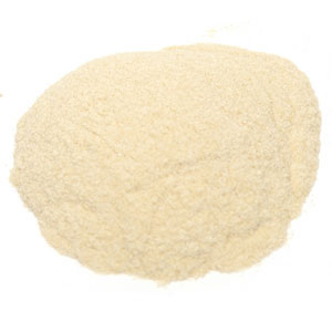
Pectin is a heteropolysaccharide, a structural acid contained in the primary lamella, in the middle lamella, and in the cell walls of terrestrial plants. The principal chemical component of pectin is galacturonic acid which was isolated and described by Henri Braconnot in 1825. Commercially produced pectin is a white-to-light-brown powder, produced from citrus fruits for use as an edible gelling agent, especially in jams and jellies, dessert fillings, medications, and sweets; and as a food stabiliser in fruit juices and milk drinks, and as a source of dietary fiber.

Pectinases are a group of enzymes that breaks down pectin, a polysaccharide found in plant cell walls, through hydrolysis, transelimination and deesterification reactions. Commonly referred to as pectic enzymes, they include pectolyase, pectozyme, and polygalacturonase, one of the most studied and widely used commercial pectinases. It is useful because pectin is the jelly-like matrix which helps cement plant cells together and in which other cell wall components, such as cellulose fibrils, are embedded. Therefore, pectinase enzymes are commonly used in processes involving the degradation of plant materials, such as speeding up the extraction of fruit juice from fruit, including apples and sapota. Pectinases have also been used in wine production since the 1960s. The function of pectinase in brewing is twofold, first it helps break down the plant material and so helps the extraction of flavors from the mash. Secondly the presence of pectin in finished wine causes a haze or slight cloudiness. Pectinase is used to break this down and so clear the wine.

<span class="mw-page-title-main">Hemagglutinin esterase</span> Glycoprotein present in some enveloped viruses

Hemagglutinin esterase (HEs) is a glycoprotein that certain enveloped viruses possess and use as an invading mechanism. HEs helps in the attachment and destruction of certain sialic acid receptors that are found on the host cell surface. Viruses that possess HEs include influenza C virus, toroviruses, and coronaviruses of the subgenus Embecovirus. HEs is a dimer transmembrane protein consisting of two monomers, each monomer is made of three domains. The three domains are: membrane fusion, esterase, and receptor binding domains.

<span class="mw-page-title-main">Catalytic triad</span> Set of three coordinated amino acids

A catalytic triad is a set of three coordinated amino acids that can be found in the active site of some enzymes. Catalytic triads are most commonly found in hydrolase and transferase enzymes. An acid-base-nucleophile triad is a common motif for generating a nucleophilic residue for covalent catalysis. The residues form a charge-relay network to polarise and activate the nucleophile, which attacks the substrate, forming a covalent intermediate which is then hydrolysed to release the product and regenerate free enzyme. The nucleophile is most commonly a serine or cysteine amino acid, but occasionally threonine or even selenocysteine. The 3D structure of the enzyme brings together the triad residues in a precise orientation, even though they may be far apart in the sequence.

<span class="mw-page-title-main">Lipoxygenase</span>

Lipoxygenases (LOX) are a family of (non-heme) iron-containing enzymes, more specifically oxidative enzymes, most of which catalyze the dioxygenation of polyunsaturated fatty acids in lipids containing a cis,cis-1,4-pentadiene into cell signaling agents that serve diverse roles as autocrine signals that regulate the function of their parent cells, paracrine signals that regulate the function of nearby cells, and endocrine signals that regulate the function of distant cells.

<i>Dickeya dadantii</i> Disease-causing Gram Negative Bacillus

Dickeya dadantii is a gram-negative bacillus that belongs to the family Pectobacteriaceae. It was formerly known as Erwinia chrysanthemi but was reassigned as Dickeya dadantii in 2005. Members of this family are facultative anaerobes, able to ferment sugars to lactic acid, have nitrate reductase, but lack oxidases. Even though many clinical pathogens are part of the order Enterobacterales, most members of this family are plant pathogens. D. dadantii is a motile, nonsporing, straight rod-shaped cell with rounded ends, much like the other members of the genus, Dickeya. Cells range in size from 0.8 to 3.2 μm by 0.5 to 0.8 μm and are surrounded by numerous flagella (peritrichous).

The pelB leader sequence is a sequence of amino acids which, when attached to a protein, directs the protein to the bacterial periplasm, where the sequence is removed by a signal peptidase. Specifically, pelB refers to pectate lyase B of Erwinia carotovora CE. The leader sequence consists of the 22 N-terminal amino acid residues. This leader sequence can be attached to any other protein resulting in a transfer of such a fused protein to the periplasmic space of Gram-negative bacteria, such as Escherichia coli, often used in genetic engineering. Protein secretion can increase the stability of cloned gene products. For instance it was shown that the half-life of the recombinant proinsulin is increased 10-fold when the protein is secreted to the periplasmic space.

Phospholipase D (EC 3.1.4.4, lipophosphodiesterase II, lecithinase D, choline phosphatase, PLD; systematic name phosphatidylcholine phosphatidohydrolase) is an enzyme of the phospholipase superfamily that catalyses the following reaction

<span class="mw-page-title-main">Phosphodiesterase 3</span> Class of enzymes

PDE3 is a phosphodiesterase. The PDEs belong to at least eleven related gene families, which are different in their primary structure, substrate affinity, responses to effectors, and regulation mechanism. Most of the PDE families are composed of more than one gene. PDE3 is clinically significant because of its role in regulating heart muscle, vascular smooth muscle and platelet aggregation. PDE3 inhibitors have been developed as pharmaceuticals, but their use is limited by arrhythmic effects and they can increase mortality in some applications.

<span class="mw-page-title-main">Sucrase-isomaltase</span>

Sucrase-isomaltase is a bifunctional glucosidase located on the brush border of the small intestine, encoded by the human gene SI. It is a dual-function enzyme with two GH31 domains, one serving as the isomaltase, the other as a sucrose alpha-glucosidase. It has preferential expression in the apical membranes of enterocytes. The enzyme’s purpose is to digest dietary carbohydrates such as starch, sucrose and isomaltose. By further processing the broken-down products, energy in the form of ATP can be generated.

<span class="mw-page-title-main">Chlorophyllase</span> Enzyme in chlorophyll metabolism

Chlorophyllase is an essential enzyme in chlorophyll metabolism. It is a membrane proteins commonly known as chlase (EC 3.1.1.14, CLH) with systematic name chlorophyll chlorophyllidohydrolase. It catalyzes the reaction

<span class="mw-page-title-main">Branched-chain amino acid aminotransferase</span> Aminotransferase enzyme

Branched-chain amino acid aminotransferase (BCAT), also known as branched-chain amino acid transaminase, is an aminotransferase enzyme (EC 2.6.1.42) which acts upon branched-chain amino acids (BCAAs). It is encoded by the BCAT2 gene in humans. The BCAT enzyme catalyzes the conversion of BCAAs and α-ketoglutarate into branched chain α-keto acids and glutamate.

<span class="mw-page-title-main">Tyrosylprotein sulfotransferase</span> Enzyme

Tyrosylprotein sulfotransferase is an enzyme that catalyzes tyrosine sulfation.

<span class="mw-page-title-main">Wall-associated kinase</span>

Wall-associated kinases (WAKs) are one of many classes of plant proteins known to serve as a medium between the extracellular matrix (ECM) and cytoplasm of cell walls. They are serine-threonine kinases that contain epidermal growth factor (EGF) repeats, a cytoplasmic kinase and are located in the cell walls. They provide a linkage between the inner and outer surroundings of cell walls. WAKs are under a group of receptor-like kinases (RLK) that are actively involved in sensory and signal transduction pathways especially in response to foreign attacks by pathogens and in cell development. On the other hand, pectins are an abundant group of complex carbohydrates present in the primary cell wall that play roles in cell growth and development, protection, plant structure and water holding capacity.

Pectate lyase is an enzyme involved in the maceration and soft rotting of plant tissue. Pectate lyase is responsible for the eliminative cleavage of pectate, yielding oligosaccharides with 4-deoxy-α-D-mann-4-enuronosyl groups at their non-reducing ends. The protein is maximally expressed late in pollen development. It has been suggested that the pollen expression of pectate lyase genes might relate to a requirement for pectin degradation during pollen tube growth.

Pectin lyase is a polysaccharide enzyme with a complex structure that is present in plant cell walls. It has a significant role in pectin degradation and different biotechnological and industrial applications. It can be found in different organisms.

The enzyme tannase (EC 3.1.1.20) catalyzes the following reaction:

Reticulons are a group of evolutionary conservative proteins residing predominantly in endoplasmic reticulum, primarily playing a role in promoting membrane curvature. In addition, reticulons may play a role in nuclear pore complex formation, vesicle formation, and other processes yet to be defined. They have also been linked to oligodendrocyte roles in inhibition of neurite outgrowth. Some studies link RTNs with Alzheimer's disease and amyotrophic lateral sclerosis.

<span class="mw-page-title-main">Polygalacturonase</span>

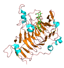
Endo-polygalacturonase (EC 3.2.1.15, pectin depolymerase, pectolase, pectin hydrolase, and poly-α-1,4-galacturonide glycanohydrolase; systematic name (1→4)-α-D-galacturonan glycanohydrolase (endo-cleaving)) is an enzyme that hydrolyzes the α-1,4 glycosidic bonds between galacturonic acid residues:

References

  1. Fries, M.; Ihrig, J.; Brocklehurst, K.; Shevchik, V. E.; Pickersgill, R. W. (2007). "Molecular basis of the activity of the phytopathogen pectin methylesterase". The EMBO Journal. 26 (17): 3879–3887. doi:10.1038/sj.emboj.7601816. PMC   2000356 . PMID   17717531.
  2. Giovane A, Tsernoglou D, Camardella L, Di Matteo A, Raiola A, Bonivento D, De Lorenzo G, Cervone F, Bellincampi D (2005). "Structural basis for the interaction between pectin methylesterase and a specific inhibitor protein". Plant Cell. 17 (3): 849–858. doi:10.1105/tpc.104.028886. PMC   1069703 . PMID   15722470.
  3. PDB: 1GQ8 ; Johansson K, El-Ahmad M, Friemann R, Jörnvall H, Markovic O, Eklund H (March 2002). "Crystal structure of plant pectin methylesterase". FEBS Lett. 514 (2–3): 243–9. doi: 10.1016/S0014-5793(02)02372-4 . PMID   11943159.
  4. PDB: 1QJV ; Pickersgill RW, Smith D, Jenkins J, Mayans O, Worboys K (2001). "Three-dimensional structure of Erwinia chrysanthemi pectin methylesterase reveals a novel esterase active site". J. Mol. Biol. 305 (4): 951–960. doi:10.1006/jmbi.2000.4324. PMID   11162105.