Biliverdin reductase B

Last updated
BLVRB
Available structures
PDB Ortholog search: PDBe RCSB
Identifiers
Aliases BLVRB , BVRB, FLR, HEL-S-10, SDR43U1, Biliverdin reductase B
External IDs OMIM: 600941 MGI: 2385271 HomoloGene: 573 GeneCards: BLVRB
EC number 1.3.1.24
Orthologs
SpeciesHumanMouse
Entrez
Ensembl
UniProt
RefSeq (mRNA)

NM_000713

NM_001290525
NM_144923

RefSeq (protein)

NP_000704

NP_001277454
NP_659172

Location (UCSC) Chr 19: 40.45 – 40.47 Mb Chr 7: 27.15 – 27.17 Mb
PubMed search [3] [4]
Wikidata
View/Edit Human View/Edit Mouse

Biliverdin reductase B is a protein that in humans is encoded by the BLVRB gene. [5]

Contents

Structure

The BLVRB gene was localized to chromosome 19, the specific region being 19q13.13 to q13.2; this was done using fluorescence in situ hybridization. [6]

BLVRB encodes a protein that is a 206-residue monomeric enzyme. [7] The structure of BVR-B has a single-domain architecture consisting of a central parallel beta-sheet with alpha-helices on either side. This characteristic dinucleotide binding fold comprises, in this case, a seven-stranded parallel beta-sheet further extended by an antiparallel strand. In addition to the seven long strands of the main pleated sheet, a short parallel beta-sheet (strands 6a and 6c) is formed within the loop joining strand 6 and alpha-helix F. The central beta-sheet and the two groups of helices are held together mainly through hydrophobic interactions. One group of helices is made up of alpha-helices C, D, and E. The second group is composed of alpha-helices A and F and includes a short 310-helix between strands beta2 and beta3, in contrast to typical dinucleotide binding proteins in which a regular alpha-helix flanks these beta-strands. The most flexible loop in the structure corresponds to loop 120, between strand 5 and alpha-helix E, which contains residues with the highest main chain B-factors, with the exception of the N-terminal region. [8]

Function

The final step in heme metabolism in mammals is catalyzed by the cytosolic biliverdin reductase enzymes A and B (EC 1.3.1.24). [5] From a functional standpoint, it has been hypothesized that BLRVB is identical to flavin reductase (FR), an enzyme that catalyzes the NADPH-dependent reduction of FMN and Methylene Blue and, in the presence of redox couplers, the reduction of methaemoglobin. [9] [10]

There have been two isoforms of BLVRB, I and II, that have been isolated and characterized. The purified enzymes were monomers with a molecular weight of about 21,000, and they used NADPH and NADH as electron donors for the reduction of biliverdin. The identified Km values of isozymes I and II for NADPH are 35.9 and 13.1 μM, respectively, whereas those for NADH are 5.6 and 8.2, indicating that NADPH rather than NADH acts as the physiological electron donor in reaction. The NADPH-dependent enzyme activities are inhibited by substrate concentrations in excess of 3-4 μM. The optimum pH of the reaction with NADPH for isozymes I and II is 8.2. [11] Flavin reductase/biliverdin-IXbeta reductase has also been shown to exhibit ferric reductase activity, with an apparent K(m) of 2.5 μM for the ferric iron. The ferric reductase reaction requires NAD(P)H and FMN. This activity is intriguing, as haem cleavage in the foetus produces non-alpha isomers of biliverdin and ferric iron, both of which are substrates for flavin reductase/biliverdin-IXbeta reductase. [12]

Clinical significance

As BLVRB is a promiscuous enzyme catalysing the pyridine-nucleotide-dependent reduction of a variety of flavins, biliverdins, PQQ (pyrroloquinoline quinone), and ferric ion. Mechanistically it is a good model for BVR-A (biliverdin-IXalpha reductase), a potential pharmacological target for neonatal jaundice, and also a potential target for adjunct therapy to maintain protective levels of biliverdin-IXalpha during organ transplantation. [13]

Interactions

BLVRB binds to human heme oxygenase-1 (hHO-1) in conjunction with cytochrome p450 reductase to catalyzes the NADPH-cytochrome P450 reductase-dependent oxidation of heme to biliverdin, CO, and free iron. [14]

Related Research Articles

<span class="mw-page-title-main">Respiratory complex I</span> Protein complex involved in cellular respiration

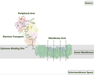
Respiratory complex I, EC 7.1.1.2 is the first large protein complex of the respiratory chains of many organisms from bacteria to humans. It catalyzes the transfer of electrons from NADH to coenzyme Q10 (CoQ10) and translocates protons across the inner mitochondrial membrane in eukaryotes or the plasma membrane of bacteria.

<span class="mw-page-title-main">Rossmann fold</span>

The Rossmann fold is a tertiary fold found in proteins that bind nucleotides, such as enzyme cofactors FAD, NAD+, and NADP+. This fold is composed of alternating beta strands and alpha helical segments where the beta strands are hydrogen bonded to each other forming an extended beta sheet and the alpha helices surround both faces of the sheet to produce a three-layered sandwich. The classical Rossmann fold contains six beta strands whereas Rossmann-like folds, sometimes referred to as Rossmannoid folds, contain only five strands. The initial beta-alpha-beta (bab) fold is the most conserved segment of the Rossmann fold. The motif is named after Michael Rossmann who first noticed this structural motif in the enzyme lactate dehydrogenase in 1970 and who later observed that this was a frequently occurring motif in nucleotide binding proteins.

<span class="mw-page-title-main">Flavin adenine dinucleotide</span> Redox-active coenzyme

In biochemistry, flavin adenine dinucleotide (FAD) is a redox-active coenzyme associated with various proteins, which is involved with several enzymatic reactions in metabolism. A flavoprotein is a protein that contains a flavin group, which may be in the form of FAD or flavin mononucleotide (FMN). Many flavoproteins are known: components of the succinate dehydrogenase complex, α-ketoglutarate dehydrogenase, and a component of the pyruvate dehydrogenase complex.

<span class="mw-page-title-main">Flavoprotein</span> Protein family

Flavoproteins are proteins that contain a nucleic acid derivative of riboflavin. These proteins are involved in a wide array of biological processes, including removal of radicals contributing to oxidative stress, photosynthesis, and DNA repair. The flavoproteins are some of the most-studied families of enzymes.

<span class="mw-page-title-main">Thromboxane-A synthase</span> Mammalian protein found in Homo sapiens

Thromboxane A synthase 1 , also known as TBXAS1, is a cytochrome P450 enzyme that, in humans, is encoded by the TBXAS1 gene.

<span class="mw-page-title-main">Heme oxygenase</span> Class of enzymes

Heme oxygenase, or haem oxygenase, is an enzyme that catalyzes the degradation of heme to produce biliverdin, ferrous ion, and carbon monoxide.

<span class="mw-page-title-main">Protein contact map</span>

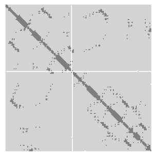
A protein contact map represents the distance between all possible amino acid residue pairs of a three-dimensional protein structure using a binary two-dimensional matrix. For two residues and , the element of the matrix is 1 if the two residues are closer than a predetermined threshold, and 0 otherwise. Various contact definitions have been proposed: The distance between the Cα-Cα atom with threshold 6-12 Å; distance between Cβ-Cβ atoms with threshold 6-12 Å ; and distance between the side-chain centers of mass.

<span class="mw-page-title-main">Biliverdin reductase</span> Class of enzymes

Biliverdin reductase (BVR) is an enzyme found in all tissues under normal conditions, but especially in reticulo-macrophages of the liver and spleen. BVR facilitates the conversion of biliverdin to bilirubin via the reduction of a double bond between the second and third pyrrole ring into a single bond.

<span class="mw-page-title-main">Lanosterol 14 alpha-demethylase</span> Protein-coding gene in the species Homo sapiens

Lanosterol 14α-demethylase (CYP51A1) is the animal version of a cytochrome P450 enzyme that is involved in the conversion of lanosterol to 4,4-dimethylcholesta-8(9),14,24-trien-3β-ol. The cytochrome P450 isoenzymes are a conserved group of proteins that serve as key players in the metabolism of organic substances and the biosynthesis of important steroids, lipids, and vitamins in eukaryotes. As a member of this family, lanosterol 14α-demethylase is responsible for an essential step in the biosynthesis of sterols. In particular, this protein catalyzes the removal of the C-14α-methyl group from lanosterol. This demethylation step is regarded as the initial checkpoint in the transformation of lanosterol to other sterols that are widely used within the cell.

<span class="mw-page-title-main">Shikimate dehydrogenase</span> Enzyme involved in amino acid biosynthesis

In enzymology, a shikimate dehydrogenase (EC 1.1.1.25) is an enzyme that catalyzes the chemical reaction

In enzymology, a cholestanetriol 26-monooxygenase (EC 1.14.13.15) is an enzyme that catalyzes the chemical reaction

In enzymology, a ferric-chelate reductase (EC 1.16.1.7) is an enzyme that catalyzes the chemical reaction

Flavin reductase a class of enzymes. There are a variety of flavin reductases, which bind free flavins and through hydrogen bonding, catalyze the reduction of these molecules to a reduced flavin. Riboflavin, or vitamin B, and flavin mononucleotide are two of the most well known flavins in the body and are used in a variety of processes which include metabolism of fat and ketones and the reduction of methemoglobin in erythrocytes. Flavin reductases are similar and often confused for ferric reductases because of their similar catalytic mechanism and structures.

In enzymology, a leghemoglobin reductase (EC 1.6.2.6) is an enzyme that catalyzes the chemical reaction

<span class="mw-page-title-main">NADPH—hemoprotein reductase</span> Enzyme

In enzymology, a NADPH—hemoprotein reductase is an enzyme that catalyzes the chemical reaction

<span class="mw-page-title-main">AKR1C3</span> Protein-coding gene in the species Homo sapiens

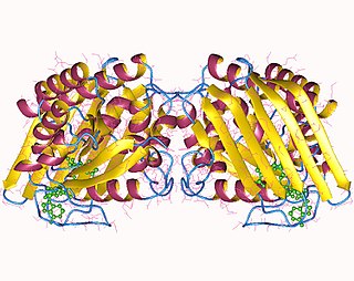
Aldo-keto reductase family 1 member C3 (AKR1C3), also known as 17β-hydroxysteroid dehydrogenase type 5 is a key steroidogenic enzyme that in humans is encoded by the AKR1C3 gene.

<span class="mw-page-title-main">AKR1C1</span> Protein-coding gene in the species Homo sapiens

Aldo-keto reductase family 1 member C1 also known as 20α-hydroxysteroid dehydrogenase, 3α-hydroxysteroid dehydrogenase, and dihydrodiol dehydrogenase 1/2 is an enzyme that in humans is encoded by the AKR1C1 gene.

<span class="mw-page-title-main">3α-Hydroxysteroid dehydrogenase</span> Protein-coding gene in the species Homo sapiens

3α-Hydroxysteroid dehydrogenase is an enzyme that in humans is encoded by the AKR1C4 gene. It is known to be necessary for the synthesis of the endogenous neurosteroids allopregnanolone, THDOC, and 3α-androstanediol. It is also known to catalyze the reversible conversion of 3α-androstanediol (5α-androstane-3α,17β-diol) to dihydrotestosterone and vice versa.

Nitric oxide reductase (NAD(P), nitrous oxide-forming) (EC 1.7.1.14, fungal nitric oxide reductase, cytochrome P450nor, NOR (ambiguous)) is an enzyme with systematic name nitrous oxide:NAD(P) oxidoreductase. This enzyme catalyses the following chemical reaction

<span class="mw-page-title-main">Biliverdin reductase A</span> Protein-coding gene in the species Homo sapiens

Biliverdin reductase A is a protein that in humans is encoded by the BLVRA gene.

References

  1. 1 2 3 GRCh38: Ensembl release 89: ENSG00000090013 - Ensembl, May 2017
  2. 1 2 3 GRCm38: Ensembl release 89: ENSMUSG00000040466 - Ensembl, May 2017
  3. "Human PubMed Reference:". National Center for Biotechnology Information, U.S. National Library of Medicine.
  4. "Mouse PubMed Reference:". National Center for Biotechnology Information, U.S. National Library of Medicine.
  5. 1 2 "Entrez Gene: Biliverdin reductase B".
  6. Saito F, Yamaguchi T, Komuro A, Tobe T, Ikeuchi T, Tomita M, Nakajima H (1995). "Mapping of the newly identified biliverdin-IX beta reductase gene (BLVRB) to human chromosome 19q13.13-->q13.2 by fluorescence in situ hybridization". Cytogenetics and Cell Genetics. 71 (2): 179–81. doi:10.1159/000134102. PMID   7656592.
  7. Yamaguchi T, Komuro A, Nakano Y, Tomita M, Nakajima H (Dec 1993). "Complete amino acid sequence of biliverdin-IX beta reductase from human liver". Biochemical and Biophysical Research Communications. 197 (3): 1518–23. doi:10.1006/bbrc.1993.2649. PMID   8280170.
  8. Pereira PJ, Macedo-Ribeiro S, Párraga A, Pérez-Luque R, Cunningham O, Darcy K, Mantle TJ, Coll M (Mar 2001). "Structure of human biliverdin IXbeta reductase, an early fetal bilirubin IXbeta producing enzyme". Nature Structural Biology. 8 (3): 215–20. doi:10.1038/84948. PMID   11224564. S2CID   8276076.
  9. Shalloe F, Elliott G, Ennis O, Mantle TJ (Jun 1996). "Evidence that biliverdin-IX beta reductase and flavin reductase are identical". The Biochemical Journal. 316 (2): 385–7. doi:10.1042/bj3160385. PMC   1217361 . PMID   8687377.
  10. Komuro A, Tobe T, Hashimoto K, Nakano Y, Yamaguchi T, Nakajima H, Tomita M (Jun 1996). "Molecular cloning and expression of human liver biliverdin-IX beta reductase". Biological & Pharmaceutical Bulletin. 19 (6): 796–804. doi: 10.1248/bpb.19.796 . PMID   8799475.
  11. Yamaguchi T, Komoda Y, Nakajima H (Sep 1994). "Biliverdin-IX alpha reductase and biliverdin-IX beta reductase from human liver. Purification and characterization". The Journal of Biological Chemistry. 269 (39): 24343–8. doi: 10.1016/S0021-9258(19)51088-2 . PMID   7929092.
  12. Cunningham O, Gore MG, Mantle TJ (Jan 2000). "Initial-rate kinetics of the flavin reductase reaction catalysed by human biliverdin-IXbeta reductase (BVR-B)". The Biochemical Journal. 345 (2): 393–9. doi:10.1042/bj3450393. PMC   1220769 . PMID   10620517.
  13. Smith LJ, Browne S, Mulholland AJ, Mantle TJ (May 2008). "Computational and experimental studies on the catalytic mechanism of biliverdin-IXbeta reductase" (PDF). The Biochemical Journal. 411 (3): 475–84. doi:10.1042/BJ20071495. PMID   18241201.
  14. Wang J, de Montellano PR (May 2003). "The binding sites on human heme oxygenase-1 for cytochrome p450 reductase and biliverdin reductase". The Journal of Biological Chemistry. 278 (22): 20069–76. doi: 10.1074/jbc.M300989200 . PMID   12626517.

This article incorporates text from the United States National Library of Medicine, which is in the public domain.