In anatomy, the atlas (C1) is the most superior (first) cervical vertebra of the spine and is located in the neck. It is named for Atlas of Greek mythology because, just as Atlas supported the globe, it supports the entire head.

Cholesteatoma is a destructive and expanding growth consisting of keratinizing squamous epithelium in the middle ear and/or mastoid process. Cholesteatomas are not cancerous as the name may suggest, but can cause significant problems because of their erosive and expansile properties. This can result in the destruction of the bones of the middle ear (ossicles), as well as growth through the base of the skull into the brain. They often become infected and can result in chronically draining ears. Treatment almost always consists of surgical removal.
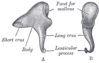
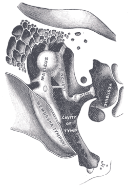
The incus or anvil is a bone in the middle ear. The anvil-shaped small bone is one of three ossicles in the middle ear. The incus receives vibrations from the malleus, to which it is connected laterally, and transmits these to the stapes medially. The incus is so-called because of its resemblance to an anvil.
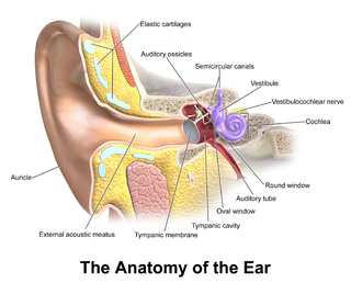
In the anatomy of humans and various other tetrapods, the eardrum, also called the tympanic membrane or myringa, is a thin, cone-shaped membrane that separates the external ear from the middle ear. Its function is to transmit sound from the air to the ossicles inside the middle ear, and then to the oval window in the fluid-filled cochlea. Hence, it ultimately converts and amplifies vibration in air to vibration in cochlear fluid. The malleus bone bridges the gap between the eardrum and the other ossicles.

In anatomy, the temporomandibular joints (TMJ) are the two joints connecting the jawbone to the skull. It is a bilateral synovial articulation between the temporal bone of the skull above and the mandible below; it is from these bones that its name is derived. This joint is unique in that it is a bilateral joint that functions as one unit. Since the TMJ is connected to the mandible, the right and left joints must function together and therefore are not independent of each other.
Articles related to anatomy include:

The temporal bones are situated at the sides and base of the skull, and lateral to the temporal lobes of the cerebral cortex.

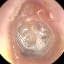
The tympanic cavity is a small cavity surrounding the bones of the middle ear. Within it sit the ossicles, three small bones that transmit vibrations used in the detection of sound.

The squamous part of temporal bone, or temporal squama, forms the front and upper part of the temporal bone, and is scale-like, thin, and translucent.

The tympanic part of the temporal bone is a curved plate of bone lying below the squamous part of the temporal bone, in front of the mastoid process, and surrounding the external part of the ear canal.
In anatomy, arterial tree is used to refer to all arteries and/or the branching pattern of the arteries. This article regards the human arterial tree. Starting from the aorta:

The carotid triangle is a portion of the anterior triangle of the neck.

Anton Freiherr von Tröltsch was a German otologist who was a native of Schwabach.
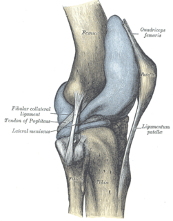
The articular capsule of the knee joint is the wide and lax joint capsule of the knee. It is thin in front and at the side, and contains the patella, ligaments, menisci, and bursae of the knee. The capsule consists of an inner synovial membrane, and an outer fibrous membrane separated by fatty deposits anteriorly and posteriorly.

The following outline is provided as an overview of and topical guide to human anatomy:
Inferior alveolar nerve block is a nerve block technique which induces anesthesia (numbness) in the areas of the mouth and face innervated by one of the inferior alveolar nerves which are paired on the left and right side. These areas are the skin and mucous membranes of the lower lip, the skin of the chin, the lower teeth and the labial gingiva of the anterior teeth, all unilaterally to the midline of the side on which the block is administered. However, depending on technique, the long buccal nerve may not be anesthetized by an IANB and therefore an area of buccal gingiva adjacent to the lower posterior teeth will retain normal sensation unless that nerve is anesthetized separately, via a (long) buccal nerve block. The inferior alveolar nerve is a branch of the mandibular nerve, the third division of the trigeminal nerve. This procedure attempts to anaesthetise the inferior alveolar nerve prior to it entering the mandibular foramen on the medial surface of the mandibular ramus.
Tympanic membrane retraction describes a condition in which a part of the eardrum lies deeper within the ear than its normal position.
The Notch of Rivinus is a small defect in the posterior edge of the bony annular tympanic ring. The defect is located just superior to the tympano-mastoid suture line in the posterior ear canal. Following identification of the spine of Henle it is possible to follow the tympano-mastoid suture line medially towards the annular ring. At this location the Chorda Tympani Nerve is often identified. Just superior to this the Notch of Rivinus can be seen and the neck of the malleus occupies the notch and often is the superior limit of a tympanomeatal flap.

The ligaments of malleus are three ligaments that attach the malleus in the middle ear. They are the anterior, lateral and superior ligaments.

The malleus or hammer is a hammer-shaped small bone or ossicle of the middle ear which connects with the incus and is attached to the inner surface of the eardrum. The word is Latin for hammer or mallet. It transmits the sound vibrations from the eardrum to the incus.