Tethered spinal cord syndrome

Last updated
Tethered spinal cord syndrome
Other namesOccult spinal dysraphism sequence
Specialty Neurosurgery

Tethered cord syndrome (TCS) refers to a group of neurological disorders that relate to malformations of the spinal cord. [1] Various forms include tight filum terminale, lipomeningomyelocele, split cord malformations (diastematomyelia), occult, dermal sinus tracts, and dermoids. All forms involve the pulling of the spinal cord at the base of the spinal canal, literally a tethered cord. [1] The spinal cord normally hangs loose in the canal, free to move up and down with growth, and with bending and stretching. A tethered cord, however, is held taut at the end or at some point in the spinal canal. In children, a tethered cord can force the spinal cord to stretch as they grow. In adults the spinal cord stretches in the course of normal activity, usually leading to progressive spinal cord damage if untreated. [1] TCS is often associated with the closure of a spina bifida. It can be congenital, such as in tight filum terminale, or the result of injury later in life. [2]

Contents

Signs and symptoms

In children, symptoms may include:

Skin manifestation typical of tethered cord syndrome Skin manifestation that is classically seen in tethered cord syndrome.jpg
Skin manifestation typical of tethered cord syndrome

Tethered spinal cord syndrome may go undiagnosed until adulthood, when sensory, motor, bowel, and bladder control issues emerge. This delayed presentation of symptoms relates to the degree of strain on the spinal cord over time. [4]

Tethering may also develop after spinal cord injury. Scar tissue can block the flow of fluids around the spinal cord. Fluid pressure may cause cysts to form in the spinal cord, a condition called syringomyelia. This can lead to additional loss of movement or feeling, or the onset of pain or autonomic nervous system symptoms. [5]

In adults, onset of symptoms typically include:

Neurological symptoms can include a mixed picture of upper and lower motor neuron findings, such as amyotrophy, hyperreflexia or hyporeflexia, a positive stretch sign, and pathologic plantar response, occurring in the same limb. Profound sensory changes, such as loss of pain, temperature, and proprioceptive sensations, are common. [6] Last, progressive symptoms of a neuropathic bladder are noted on over 70% of adult patients, versus only 20% to 30% of children. These symptoms include urinary frequency and urgency, feeling of incomplete voiding, poor voluntary control, nocturia, and urge and stress incontinence. Chronic recurrent infections are common and occasionally lead to nephrolithiasis (kidney stones), kidney failure, or kidney transplantation. Female patients also give a history of ineffective labor and postpartum rectal prolapse, presumably due to an atonic pelvic floor. [7] [8]

Cause

Tethered spinal cord can be caused by various conditions but the main cause is when tissue attachments limit the movement of the spinal cord in the spinal column which causes abnormal stretching of the cord. The tethered spinal cord syndrome is correlated with having the causes: [9]

Tethered spinal cord is a disorder and not a mechanism so it does not spread to other people and there are no measures that can be done to prevent it beforehand. The only preventative measure that is successful is to surgically untether the spinal cord though there might already be irreversible damage. [11]

Spina bifida

In tethered spinal cord cases spina bifida can be accompanied by tethering of the spinal cord but in rare cases with Spina bifida occulta. Tethering of the spinal cord tends to occur in the cases of Spina bifida with mylomeningocele. In most people the spine grows faster than the spinal cord during development which causes the end of the spinal cord to appear to rise relative to the bony spine next to it. By the time of birth the spinal cord is located between L1 and L2. In a baby with Spina bifida the spinal cord is still attached to the skin around it preventing it from rising properly. [12] This occurs because the spinal cord in a child with Spina bifida is low lying and tethered at the bottom. At the time of birth the mylomeningocele is separated from the skin but the spinal cord is still stuck in the same place. As the child begins to grow the spinal cord remains in the same place becoming stretched out causing the tight cord and the tethering at the end. With this type of tethering there is an interference with the blood supply to the nerves and body which can then cause the deterioration of the body causing orthopedic, neurological, and urological problems. With milder forms of Spina bifida such as Occulta, may be related to the degree of strain on the cord which can become worse with physical activity, injury, pregnancy, bone spurs, or spinal stenosis. The tethered cord in this case might not be diagnosed until adulthood when it worsens and can still cause neurological, orthopedic, and urological dysfunctions. [11]

Mechanism

Tethered spinal cord syndrome is a clinical entity which is manifested by progressive motor and sensory changes in:

In order to understand the pathophysiology that is involved in a tethered spinal cord, the reduction/oxidation ratio has to be used in vivo of cytochrome alpha and alpha 3 to signal the oxidative metabolic functioning in humans. Studies have found that marked metabolic and electrophysiological susceptibility to hypoxic stress to the lumbar and sacral portion of the spinal cord under traction with various weights. [13] Similar effects were found in redox behavior of tethered spinal cord during the surgical procedures to repair it. This can be due to impairment of mitochondrial oxidative metabolism under constant or intermittent stretching. The act of prolonged stretching can lead to structural damage to the neural perikarya and eventually the axons in neurons. The untethering process can improve the oxidative metabolism and can help to repair injured neurons. [14]

Diagnosis

For children younger than eight weeks of age (and possibly in utero), a tethered cord may be observed using ultrasonography. Ultrasonography may still be useful through age 5 in limited circumstances. [15]

MRI imaging appears to be the gold standard for diagnosing a tethered cord. [16] [17]

A tethered cord is often diagnosed as a "low conus". The conus medullaris (or lower termination of the spinal cord) normally terminates at or above the L1-2 disk space (where L1 is the first, or topmost lumbar vertebra). After about 3 months of age, a conus below the L1-2 disk space may indicate a tethered cord and termination below L3-4 is unmistakably tethered. "Cord tethering is often assumed when the conus is below the normal L2-3 level.

TCS, however, is a clinical diagnosis that should be based on "neurological and musculoskeletal signs and symptoms. Imaging features are in general obtained to support rather than make the diagnosis." [18] This is especially true for cases of occult tethered cord, where the patient has the symptoms of tethered cord syndrome but MRI reveals the conus to be above the L2 level. [7] [8] Clinical evaluation may include a simple rectal examination and may also include invasive or non-invasive urological examination. [19] [20] "Bladder dysfunction occurs in ~40% of patients affected by tethered cord syndrome. ... [I]t may be the earliest sign of the syndrome." [21]

Treatment

Because neurological deficits are generally irreversible, early surgery is recommended when symptoms begin to worsen. [22] [23] In children, early surgery is recommended [24] to prevent further neurological deterioration, including but not limited to chronic urinary incontinence.

In adults, surgery to detether (free) the spinal cord can reduce the size and further development of cysts in the cord and may restore some function or alleviate other symptoms. Although detethering is the common surgical approach to TCS, [25] another surgical option for adults is a spine-shortening vertebral osteotomy. [25] [26] A vertebral osteotomy aims to indirectly relieve the excess tension on the spinal cord by removing a portion of the spine, shortening it. This procedure offers a unique benefit in that the spinal cord remains fixated to the spine, preventing retethering and spinal cord injury as possible surgical complications. However, its complexity and limited “track record” presently keeps vertebral osteotomies reserved as an option for patients who have failed in preventing retethering after detethering procedure(s). [27]

Other treatment is symptomatic and supportive. Medications such as NSAIDs, opiates, synthetic opiates, COX-2 inhibitors, and off-label applications of tricyclic antidepressants combined with anti-seizure compounds have yet to prove they are of value in treatment of this affliction's pain manifestations. There is anecdotal evidence that TENS units may benefit some patients.

Treatment may be needed in adults who, while previously asymptomatic, begin to experience pain, lower back degeneration, scoliosis, neck and upper back problems and bladder control issues. Surgery on adults with minimal symptoms is somewhat controversial. For example, a website from the Columbia University Department of Neurosurgery says, "For the child that has reached adult height with minimal if any symptoms, some neurosurgeons would advocate careful observation only." [1] However, surgery for those who have worsening symptoms is less controversial. If the only abnormality is a thickened, shortened filum, then a limited lumbosacral laminectomy with division of the filum may be sufficient to relieve the symptoms. [28]

This syndrome was first noticed in the late 19th century. While information has been available for years, little widespread blind research has been done. More research has been called for, and doctors have conducted many studies with good results. There is a low morbidity rate, and no complications have been documented other than those typical of any type of back surgery. The association of this condition with others has been noticed, and needs further research to understand such relationships. TCS is causally linked to Chiari malformation and any affirmative diagnosis of TCS must be followed by screening for Chiari's several degrees. TCS may also be related to Ehlers–Danlos syndrome, or Klippel–Feil syndrome, which should also be screened for upon a positive TCS diagnosis. Spinal compression and the resulting relief is a known issue with this disorder. Like with the early-onset form, this disease form is linked to the Arnold–Chiari malformation, in which the brain is pulled or lowers into the top of the spine. [29]

Prognosis

The disorder progresses with age, but the aforementioned treatments can help prevent or sometimes relieve symptoms. With treatment, individuals with tethered spinal cord syndrome have a normal life expectancy. Studies have shown surgery can help improve low back pain, urinary symptoms leg weakness and walking distance. [30] [8] [31] However, most neurological and motor impairments are irreversible. [32] [33]

Related Research Articles

<span class="mw-page-title-main">Foix–Alajouanine syndrome</span> Medical condition

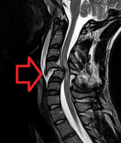
Foix–Alajouanine syndrome, also called subacute ascending necrotizing myelitis, is a disease caused by an arteriovenous malformation of the spinal cord. In particular, most cases involve dural arteriovenous malformations that present in the lower thoracic or lumbar spinal cord. The condition is named after Charles Foix and Théophile Alajouanine who first described the condition in 1926.

<span class="mw-page-title-main">Syringomyelia</span> Disorder in which a cyst forms in the spinal cord

Syringomyelia is a generic term referring to a disorder in which a cyst or cavity forms within the spinal cord. Often, syringomyelia is used as a generic term before an etiology is determined. This cyst, called a syrinx, can expand and elongate over time, destroying the spinal cord. The damage may result in loss of feeling, paralysis, weakness, and stiffness in the back, shoulders, and extremities. Syringomyelia may also cause a loss of the ability to feel extremes of hot or cold, especially in the hands. It may also lead to a cape-like bilateral loss of pain and temperature sensation along the upper chest and arms. The combination of symptoms varies from one patient to another depending on the location of the syrinx within the spinal cord, as well as its extent.

<span class="mw-page-title-main">Spina bifida</span> Birth defect of the spinal cord

Spina bifida /ˌspaɪnə ˈbɪfɪdə/ is a birth defect in which there is incomplete closing of the spine and the membranes around the spinal cord during early development in pregnancy. There are three main types: spina bifida occulta, meningocele and myelomeningocele. Meningocele and myelomeningocele may be grouped as spina bifida cystica. The most common location is the lower back, but in rare cases it may be in the middle back or neck.

<span class="mw-page-title-main">Chiari malformation</span> Medical condition

Chiari malformation (CM) is a structural defect in the cerebellum, characterized by a downward displacement of one or both cerebellar tonsils through the foramen magnum. CMs can cause headaches, difficulty swallowing, vomiting, dizziness, neck pain, unsteady gait, poor hand coordination, numbness and tingling of the hands and feet, and speech problems. Less often, people may experience ringing or buzzing in the ears, weakness, slow heart rhythm, or fast heart rhythm, curvature of the spine (scoliosis) related to spinal cord impairment, abnormal breathing, such as central sleep apnea, characterized by periods of breathing cessation during sleep, and, in severe cases, paralysis.

<span class="mw-page-title-main">Spinal cord injury</span> Injury to the main nerve bundle in the back of humans

A spinal cord injury (SCI) is damage to the spinal cord that causes temporary or permanent changes in its function. Symptoms may include loss of muscle function, sensation, or autonomic function in the parts of the body served by the spinal cord below the level of the injury. Injury can occur at any level of the spinal cord and can be complete, with a total loss of sensation and muscle function at lower sacral segments, or incomplete, meaning some nervous signals are able to travel past the injured area of the cord up to the Sacral S4-5 spinal cord segments. Depending on the location and severity of damage, the symptoms vary, from numbness to paralysis, including bowel or bladder incontinence. Long term outcomes also range widely, from full recovery to permanent tetraplegia or paraplegia. Complications can include muscle atrophy, loss of voluntary motor control, spasticity, pressure sores, infections, and breathing problems.

<span class="mw-page-title-main">Arachnoiditis</span> Inflammation of the arachnoid mater

Arachnoiditis is an inflammatory condition of the arachnoid mater or 'arachnoid', one of the membranes known as meninges that surround and protect the central nervous system. The outermost layer of the meninges is the dura mater and adheres to inner surface of the skull and vertebrae. The arachnoid is under or "deep" to the dura and is a thin membrane that adheres directly to the surface of the brain and spinal cord.

<span class="mw-page-title-main">Cauda equina syndrome</span> Nerve damage at the end of the spinal cord

Cauda equina syndrome (CES) is a condition that occurs when the bundle of nerves below the end of the spinal cord known as the cauda equina is damaged. Signs and symptoms include low back pain, pain that radiates down the leg, numbness around the anus, and loss of bowel or bladder control. Onset may be rapid or gradual.

<span class="mw-page-title-main">Conus medullaris</span> Lower part of the spinal cord

The conus medullaris or conus terminalis is the tapered, lower end of the spinal cord. It occurs near lumbar vertebral levels 1 (L1) and 2 (L2), occasionally lower. The upper end of the conus medullaris is usually not well defined, however, its corresponding spinal cord segments are usually S1–S5.

<span class="mw-page-title-main">Central canal</span> Cerebrospinal fluid-filled space around the spinal cord

The central canal is the cerebrospinal fluid-filled space that runs through the spinal cord. The central canal lies below and is connected to the ventricular system of the brain, from which it receives cerebrospinal fluid, and shares the same ependymal lining. The central canal helps to transport nutrients to the spinal cord as well as protect it by cushioning the impact of a force when the spine is affected.

<span class="mw-page-title-main">Kyphoscoliosis</span> Medical condition

Kyphoscoliosis describes an abnormal curvature of the spine in both the coronal and sagittal planes. It is a combination of kyphosis and scoliosis. This musculoskeletal disorder often leads to other issues in patients, such as under-ventilation of lungs, pulmonary hypertension, difficulty in performing day-to-day activities, psychological issues emanating from anxiety about acceptance among peers, especially in young patients. It can also be seen in syringomyelia, Friedreich's ataxia, spina bifida, kyphoscoliotic Ehlers–Danlos syndrome (kEDS), and Duchenne muscular dystrophy due to asymmetric weakening of the paraspinal muscles.

Neurogenic bladder dysfunction, often called by the shortened term neurogenic bladder, refers to urinary bladder problems due to disease or injury of the central nervous system or peripheral nerves involved in the control of urination. There are multiple types of neurogenic bladder depending on the underlying cause and the symptoms. Symptoms include overactive bladder, urinary urgency, frequency, incontinence or difficulty passing urine. A range of diseases or conditions can cause neurogenic bladder including spinal cord injury, multiple sclerosis, stroke, brain injury, spina bifida, peripheral nerve damage, Parkinson's disease, or other neurodegenerative diseases. Neurogenic bladder can be diagnosed through a history and physical as well as imaging and more specialized testing. In addition to symptomatic treatment, treatment depends on the nature of the underlying disease and can be managed with behavioral changes, medications, surgeries, or other procedures. The symptoms of neurogenic bladder, especially incontinence, can severely degrade a person's quality of life.

<span class="mw-page-title-main">Central cord syndrome</span> Human spinal cord disorder

Central cord syndrome (CCS) is the most common form of cervical spinal cord injury. It is characterized by loss of power and sensation in arms and hands. It usually results from trauma which causes damage to the neck, leading to major injury to the central corticospinal tract of the spinal cord. CCS most frequently occurs among older persons with cervical spondylosis, however, it also may occur in younger individuals.

<span class="mw-page-title-main">Diastematomyelia</span> Medical condition

Diastematomyelia is a congenital disorder in which a part of the spinal cord is split, usually at the level of the upper lumbar vertebra in the longitudinal (sagittal) direction. Females are affected much more commonly than males. This condition occurs in the presence of an osseous, cartilaginous or fibrous septum in the central portion of the spinal canal which then produces a complete or incomplete sagittal division of the spinal cord into two hemicords. When the split does not reunite distally to the spur, the condition is referred to as diplomyelia, which is true duplication of the spinal cord.

<span class="mw-page-title-main">Rachischisis</span> Neural tube defect in which the spinal cord is exposed

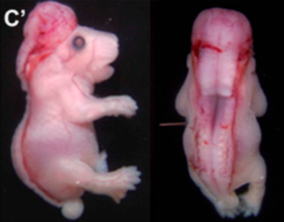
Rachischisis is a developmental birth defect involving the neural tube. This anomaly occurs in utero, when the posterior neuropore of the neural tube fails to close by the 27th intrauterine day. As a consequence the vertebrae overlying the open portion of the spinal cord do not fully form and remain unfused and open, leaving the spinal cord exposed. Patients with rachischisis have motor and sensory deficits, chronic infections, and disturbances in bladder function. This defect often occurs with anencephaly.

<span class="mw-page-title-main">Spinal disease</span> Diseases involving the vertebral column

Spinal disease refers to a condition impairing the backbone. These include various diseases of the back or spine ("dorso-"), such as kyphosis. Dorsalgia refers to back pain. Some other spinal diseases include spinal muscular atrophy, ankylosing spondylitis, lumbar spinal stenosis, spina bifida, spinal tumors, osteoporosis and cauda equina syndrome.

Karin Marie Muraszko is an American pediatric neurosurgeon.

<span class="mw-page-title-main">Nicholas Theodore</span> American neurosurgeon

Nicholas Theodore is an American neurosurgeon and researcher at Johns Hopkins University School of Medicine. He is known for his work in spinal trauma, minimally invasive surgery, robotics, and personalized medicine. He is Director of the Neurosurgical Spine Program at Johns Hopkins and Co-Director of the Carnegie Center for Surgical Innovation at Johns Hopkins.

<span class="mw-page-title-main">Cervicocranial syndrome</span> Medical condition

Cervicocranial syndrome or is a neurological illness. It is a combination of symptoms that are caused by an abnormality in the neck. The bones of the neck that are affected are cervical vertebrae (C1-C7). This syndrome can be identified by confirming cervical bone shifts, collapsed cervical bones or misalignment of the cervical bone leading to improper functioning of cervical spinal nerves. Cervicocranial syndrome is either congenital or acquired. Some examples of diseases that could result in cervicocranial syndrome are Chiari disease, Klippel-Feil malformation osteoarthritis, and trauma. Treatment options include neck braces, pain medication and surgery. The quality of life for individuals suffering from CCJ syndrome can improve through surgery.

Caudal duplication, is a rare congenital disorder in which various structures of the caudal region, embryonic cloaca, and neural tube exhibit a spectrum of abnormalities such as duplication and malformations. The exact causes of the condition is unknown, though there are several theories implicating abnormal embryological development as a cause for the condition. Diagnosis is often made during prenatal development of the second trimester through anomaly scans or immediately after birth. However, rare cases of adulthood diagnosis has also been observed. Treatment is often required to correct such abnormalities according to the range of symptoms present, whilst treatment options vary from conservative expectant management to resection of caudal tissue to restore normal function or appearance. As a rare congenital disorder, the prevalence at birth is less than 1 per 100,000 with less than 100 cases reported worldwide.

A lipomyelomeningocele is a type of closed neural tube defect that affects around 3 to 6 babies out of 100,000 births. It is an example of a spinal lipoma, which is a collection of adipose tissue, or fat, that is located at or around the spinal cord. Essentially, it is a tumor of fat located on the spinal cord. A lipomyelomeningocele defect is present at birth and can present with physical defect of the spine and back, and it can also cause various neurological symptoms such as weakness and bladder and bowel incontinence. The general treatment for this spinal defect is surgical detachment of the lipoma from the spine.

References

  1. 1 2 3 4 "Tethered Spinal Cord", Columbia University Department of Neurosurgery.
  2. "Tethered Cord Syndrome - an overview". ScienceDirect Topics. Archived from the original on 2022-11-28. Retrieved 2022-11-28.
  3. 1 2 3 4 5 6 Sanford Schneider (2009). "Neurological Assessment of Tethered Spinal Cord". In Yamada, Shokei (ed.). Tethered Cord Syndrome in Children and Adults (Rev. ed.). Thieme Medical Publishers, Inc. 44. ISBN   978-1-60406-241-0.
  4. "Tethered Spinal Cord Syndrome | National Institute of Neurological Disorders and Stroke". www.ninds.nih.gov. Retrieved 2022-11-28.
  5. "Syringomyelia Fact Sheet | National Institute of Neurological Disorders and Stroke". www.ninds.nih.gov. Retrieved 2022-11-28.
  6. Emos, Marc Christopher; Rosner, Julie (2022), "Neuroanatomy, Upper Motor Nerve Signs", StatPearls, Treasure Island (FL): StatPearls Publishing, PMID   31082126 , retrieved 2022-11-28
  7. 1 2 "Recurrence of symptoms post-surgery". Chiari Online Support Group. 6 August 2013. Retrieved 2017-12-12.
  8. 1 2 3 Zingman, Alissa; Tuchman, Kelly; Henderson, Fraser; Francomano, Clair A. (2022). "Patient-Reported Outcomes Following Sectioning of the Filum Terminale for Treatment of Tethered Cord Syndrome Associated With Ehlers-Danlos Syndrome". Cureus. 14 (5): e24679. doi: 10.7759/cureus.24679 . PMC   9160501 . PMID   35663696.
  9. Agarwal, Nitin; Hansberry, David R.; Goldstein, Ira M. (2017-08-04). "Tethered Cord as a Complication of Chronic Cerebral Spinal Fluid Diversion". International Journal of Spine Surgery. 11 (4): 26. doi: 10.14444/4026 . ISSN   2211-4599. PMC   5779270 . PMID   29372130.
  10. Zalatimo, Omar. "Tethered Spinal Cord Syndrome". AANS. Retrieved 2017-11-07.
  11. 1 2 "Spinal Cord Tethering A common cause of deterioration in Spina Bilda" (PDF). Spina Bifida Association. Archived from the original (PDF) on Aug 4, 2016.
  12. "Spina Bifida - an overview". ScienceDirect Topics. Archived from the original on 2022-11-28. Retrieved 2022-11-28.
  13. Yamada, S.; Zinke, D. E.; Sanders, D. (1981). "Pathophysiology of "tethered cord syndrome"". Journal of Neurosurgery. 54 (4): 494–503. doi:10.3171/jns.1981.54.4.0494. ISSN   0022-3085. PMID   6259301.
  14. Yamada, S.; Zinke, D. E.; Sanders, D. (April 1981). "Pathophysiology of "tethered cord syndrome"". Journal of Neurosurgery. 54 (4): 494–503. doi:10.3171/jns.1981.54.4.0494. ISSN   0022-3085. PMID   6259301.
  15. Nelson, Jr., Marvin D. (2010). "Ultrasonic Evaluation of Tethered Cord Syndrome". In Yamada, Shokei (ed.). Tethered Cord Syndrome in Children and Adults (Rev. ed.). Thieme Medical Publishers, Inc. p. 72. ISBN   978-1-60406-241-0. After the patient reaches 5 years of age, it becomes increasingly difficult to image the thoracic spinal cord to adequately evaluate pulsations.
  16. Nelson, Jr., Stephen L. (6 October 2013). "Clinical Summary: Tethered Spinal Cord". Medlink.
  17. Hinshaw Jr., David B. (2010). "Imaging of Tethered Spinal Cord". In Yamada, Shokei (ed.). Tethered Cord Syndrome in Children and Adults (Rev. ed.). Thieme Medical Publishers, Inc. p. 51. ISBN   978-1-60406-241-0. In the mid- and late 1980s, magnetic resonance imaging (MRI) developed to a degree that allowed visualization of the spinal anatomy without the use of x-radiation or subarachnoid injection of contrast agents. It has emerged as the most useful noninvasive modality, providing excellent detail of anatomy and characterization of soft tissue anomalies. ... MRI has become the primary imaging modality for tethered cords and has both facilitated earlier diagnosis and tailored treatment of these disorders.
  18. Khoury, Antoine E. (2010). "Clinical Experience in Urological Involvement with Tethered Cord Syndrome". In Yamada, Shokei (ed.). Tethered Cord Syndrome in Children and Adults (Rev. ed.). Thieme Medical Publishers, Inc. p. 90. ISBN   978-1-60406-241-0. The diagnosis of TCS is primarily based on neurological and musculoskeletal signs and symptoms. Imaging features are in general obtained to supportrather than make the diagnosis. MRI has revolutionized the noninvasive evaluation of spinal cord lesions. The major obstacle to obtaining an MRI in children is the need for sedation or general anesthesia. Patients with tethered cord have displacement of the conus and elongation of the cord, with a sometimes thickened or fatty density in the filum.
  19. Kitchens DM, Herndon CD, Joseph DB (2007). "Pediatric urodynamics: basic concepts for the neurosurgeon". Neurosurg Focus. 23 (2): E8. doi: 10.3171/FOC-07/08/E8 . PMID   17961012.
  20. Hadley, H. Roger (2010). "Lower Urinary Tract Dysfunction in Tethered Cord Syndrome". In Yamada, Shokei (ed.). Tethered Cord Syndrome in Children and Adults (Rev. ed.). Thieme Medical Publishers, Inc. p. 77. ISBN   978-1-60406-241-0. Urodynamic testing has emerged as the quintessential evaluation to explicitly identify, document, and quantify the effects of neurological dysfunction on the urinary system.
  21. Hadley, H. Roger (2010). "Lower Urinary Tract Dysfunction in Tethered Cord Syndrome". In Yamada, Shokei (ed.). Tethered Cord Syndrome in Children and Adults (Rev. ed.). Thieme Medical Publishers, Inc. p. 47. ISBN   978-1-60406-241-0.
  22. "TETHERED CORD SYNDROME".
  23. Iskandar BJ, Fulmer BB, Hadley MN, Oakes WJ (2001). "Congenital tethered spinal cord syndrome in adults". Neurosurgical Focus. 10 (1): 1–5. doi: 10.3171/foc.2001.10.1.8 . PMID   16749759.
  24. "Tethered spinal cord syndrome". National Institutes of Health. Retrieved 2008-08-28.
  25. 1 2 Kokubun, S.; Ozawa, H.; Aizawa, T.; Ly, N. M.; Tanaka, Y (July 2011). "Spine-shortening osteotomy for patients with tethered cord syndrome caused by lipomyelomeningocele". Journal of Neurosurgery: Spine. 15 (1): 21–27. doi:10.3171/2011.2.SPINE10114. PMID   21495816.
  26. Miyakoshi, N.; Abe, E.; Suzuki, T.; Kido, T.; Chiba, M.; Shimada, Y. (2009). "Spine-shortening vertebral osteotomy for tethered cord syndrome: report of three cases". Spine. 34 (22): E823–E825. doi:10.1097/BRS.0b013e3181af2607. PMID   19829247. S2CID   40180033.
  27. Shih, P.; Halpin, R.; Ganju, A.; Liu, J.; Koski, T. (2010). "Management of Recurrent Adult Tethered Cord Syndrome". Neurosurgical Focus. 29 (1): E5. doi: 10.3171/2010.3.FOCUS1073 . PMID   20594003. S2CID   6452934.
  28. "Adult Tethered Cord". UCLA. Retrieved 2 July 2013.
  29. Wehby MC, O'Hollaren PS, Abtin K, Hume JL, Richards BJ (2004). "Occult tight filum terminale syndrome: results of surgical untethering". Pediatric Neurosurgery. 40 (2): 51–7, discussion 58. doi:10.1159/000078908. PMID   15292632. S2CID   12094020.
  30. Warder, D. E.; Oakes, W. J. (1993). "Tethered cord syndrome and the conus in a normal position". Neurosurgery. 33 (3): 374–378. doi:10.1227/00006123-199309000-00004. PMID   8413866 . Retrieved 2022-06-09.
  31. Wehby, M. C.; O'Hollaren, P. S.; Abtin, K.; Hume, J. L.; Richards, B. J. (2004). "Occult Tight Filum terminale Syndrome: Results of Surgical Untethering". Pediatric Neurosurgery. 40 (2): 51–7, discussion 58. doi:10.1159/000078908. PMID   15292632. S2CID   12094020 . Retrieved 2022-06-09.
  32. Fitzgerald, Kevin. "Tethered spinal cord syndrome". Answers.com .[ unreliable medical source? ]
  33. "Facts on Tethered Cord Syndrome". Spinabifida. Retrieved 2022-05-02.