Mir-184

Last updated
mir-184
MiR-184 secondary structure.png
miR-184 microRNA secondary structure and sequence conservation
Identifiers
Symbolmir-184
Rfam RF00657
miRBase family MIPF0000059
NCBI Gene 406960
HGNC 31555
OMIM 613146
Other data
RNA type microRNA
Domain(s) Eukaryota; Chordata
PDB structures PDBe

In molecular biology, miR-184 microRNA is a short non-coding RNA molecule. MicroRNAs (miRNAs) function as posttranscriptional regulators of expression levels of other genes by several mechanisms. [1] Several targets for miR-184 have been described, including that of mediators of neurological development, apoptosis and it has been suggested that miR-184 plays an essential role in development. [2]

Contents

MicroRNAs can bind to the three prime untranslated region (3'UTR) of the target messenger RNA (mRNA). [3] Binding of the miRNA can hinder translation of mRNA by promoting degradation or inducing deadenylation. [4]

Genomic location

miR-184 is a single copy gene and evolutionarily conserved at the nucleotide level from flies to humans. [5] In humans, miR-184 is located within region 25.1 on the q-arm of chromosome 15, and its corresponding transcript is comparatively small (84bp) which is not encoded near other clustered miRNAs. [6] In the mouse genome, miR-184 is located in an imprinted locus on mouse chromosome 9, and it is 55 kb away from the nearest coding gene. [7]

The genomic region immediately surrounding miR-184 does not contain a classic CpG island, but does contain several CpG-rich sequences that are suitable for MBD1 binding. [8]

Expression

miR-184 displays a tissue- and developmental-specific expression pattern. In mammals, mature miR-184 is particularly enriched in the brain and testis, [7] along with the corneal epithelium. [9] Depolarization of cortical neurons results in pri-miR-184 expression in an allele specific manner. [7] High expression is observed in suprabasal cells of the corneal epithelium in the mouse model, along with expression in mouse testis and brain tissue. [7] [9] In Zebrafish, it is expressed in lens, hatching gland and epidermis (shown by Northern blot). [10] miR-184 is expressed ubiquitously in Drosophila embryos, larvae and adults, and its expression pattern displays dynamic changes during the development of embryo, especially in the central nervous system. [2] [5] However, the temporal and spatial expression pattern of miR-184 is still being debated.

Role in neuronal cells

C. Liu et al. showed that Methyl-CpG binding protein 1 (MBD1) regulates the expression of several miRNAs in adult neural stem/progenitor cells (aNSCs) and, specifically, that miR-184 is directly repressed by MBD1. High levels of miR-184 promotes cell proliferation but inhibits differentiation of aNSCs, whereas inhibition of miR-184 rescued phenotypes associated with MBD1 deficiency. [11]

Numblike (Numbl) is known to be important in embryonic neural stem cell function and cortical brain development and has been identified as a downstream target of miR-184. [12] [13] It has been found that exogenously expressed Numbl could rescue aNSC proliferation and differentiation deficits resulting from either elevated miR-184 or MBD1 deficiency. [11]

Other Targets

An analysis of the primary transcript of miR-184 (pri-mir-184) in several mouse tissues revealed specific expression in the brain and testis. Its expression is repressed by the binding of methyl-CpG binding protein 2 (MeCP2) to its promoter, but is upregulated by the release of MeCP2 after depolarization, suggesting a link between miRNAs and DNA methylation pathways . [7] J. Yu et al. demonstrated that the lipid phosphatase SH2-containing phosphoinositide 5'phosphatase 2 (SHIP2) is a target of miRNA-205 (miR-205) in epithelial cells, and that the corneal epithelial-specific miR-184 can interfere with the ability of miR-205 to suppress SHIP2 levels. The mechanism by which miR-184 negatively regulates miR-205 appears to be unique, and is the first example of a miRNA negatively regulating another to maintain levels of a target protein. miR-184 does not directly affect SHIP2 translation, but instead prevents miR-205 from interacting with SHIP2 mRNA. Interfering with miR-205 function by using a synthetic antagomir, or by the ectopic expression of miR-184, is thought to lead to a coordinated damping of the Akt signaling pathway via SHIP2 induction. [14]

R. Weitzel et al. showed that miR-184 mediates NFAT1 translational regulation in umbilical cord blood (UCB) graft CD4+ T-cells leading to blunted allogenic responses. [15]

J. Roberts et al. found that miR-184 repressed the expression of Argonaute 2 in epidermal keratinocytes. [16] Similarly, Tattikota et al. showed miR-184 reduced Argonaute 2 levels in the MIN6 mouse pancreatic beta islet cell line. [17]

Furthermore, miR-184 has multiple roles in Drosophila female germline development. [18]

Finally, a recent study identified miR-184 as essential for embryonic corneal commitment of pluripotent stem cells. [19]

Disease relevance

• A single base mutation in the seed region of miR-184 causes EDICT syndrome, a hereditary eye disease. [20]
• A mutation altering the miR-184 seed region causes familial keratoconus with cataract. [21]
Rett syndrome. [7]
• Several forms of cancer (see below) including elevation of miR-184 levels in squamous cell carcinoma of the tongue. [22] All-trans-retinoic acid induces miR-184 expression in neuroblastoma cell line and ectopic miR-184 causes apoptosis. [23]
• miR-184 has been implicated in ischemia-induced retinal neovascularization. [24]

Angiogenesis and cancer

Dysregulation of miRNA expression is thought to play a part in abnormal gene expression in cancer cells, and miR-184 has been implicated in several forms of cancer. [22] [25] MYCN has been found to contribute to tumorigenesis, in part, by repressing miR-184, leading to increased levels of the serine/threonine kinase, AKT2. AKT2 is a major effector of the phosphatidylinositol 3-kinase (PI3K) pathways, one of the most potent survival pathways in cancer, and is a direct target of miR-184. It has been suggested that MYCN provides a tumourigenic effect, in part, by protecting AKT2 mRNA from degradation by miR-184, permitting the PI3K pathway to remain functional. [26]

miR-184 has been found to be significantly increased in the tumor cells in comparison with the normal epithelial cells of the tongue. High miR-184 levels were not only detected in the tumor tissues, but also in the plasma of patients with tongue squamous cell carcinoma (SCC). Decreased plasma levels of miR-184 were observed in patients after surgical removal of the primary tumor, suggesting that it is a potential oncogenic miRNA in tongue SCC. Inhibiting miR-184 promotes apoptosis as well as hindering cell proliferation in cultured tongue SCC cells. [27] Furthermore, over expression of miR-184 in neuroblastoma cell lines results in apoptosis. [23] SND1 expression is regulated by miR-184 in gliomas. [28]

See also

Related Research Articles

<span class="mw-page-title-main">Regulation of gene expression</span> Modifying mechanisms used by cells to increase or decrease the production of specific gene products

Regulation of gene expression, or gene regulation, includes a wide range of mechanisms that are used by cells to increase or decrease the production of specific gene products. Sophisticated programs of gene expression are widely observed in biology, for example to trigger developmental pathways, respond to environmental stimuli, or adapt to new food sources. Virtually any step of gene expression can be modulated, from transcriptional initiation, to RNA processing, and to the post-translational modification of a protein. Often, one gene regulator controls another, and so on, in a gene regulatory network.

<span class="mw-page-title-main">Piwi</span>

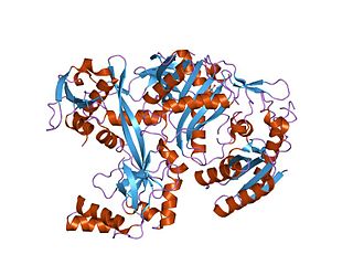
Piwi genes were identified as regulatory proteins responsible for stem cell and germ cell differentiation. Piwi is an abbreviation of P-elementInduced WImpy testis in Drosophila. Piwi proteins are highly conserved RNA-binding proteins and are present in both plants and animals. Piwi proteins belong to the Argonaute/Piwi family and have been classified as nuclear proteins. Studies on Drosophila have also indicated that Piwi proteins have no slicer activity conferred by the presence of the Piwi domain. In addition, Piwi associates with heterochromatin protein 1, an epigenetic modifier, and piRNA-complementary sequences. These are indications of the role Piwi plays in epigenetic regulation. Piwi proteins are also thought to control the biogenesis of piRNA as many Piwi-like proteins contain slicer activity which would allow Piwi proteins to process precursor piRNA into mature piRNA.

The Let-7 microRNA precursor was identified from a study of developmental timing in C. elegans, and was later shown to be part of a much larger class of non-coding RNAs termed microRNAs. miR-98 microRNA precursor from human is a let-7 family member. Let-7 miRNAs have now been predicted or experimentally confirmed in a wide range of species (MIPF0000002). miRNAs are initially transcribed in long transcripts called primary miRNAs (pri-miRNAs), which are processed in the nucleus by Drosha and Pasha to hairpin structures of about 70 nucleotide. These precursors (pre-miRNAs) are exported to the cytoplasm by exportin5, where they are subsequently processed by the enzyme Dicer to a ~22 nucleotide mature miRNA. The involvement of Dicer in miRNA processing demonstrates a relationship with the phenomenon of RNA interference.

mir-9/mir-79 microRNA precursor family

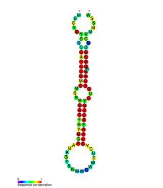
The miR-9 microRNA, is a short non-coding RNA gene involved in gene regulation. The mature ~21nt miRNAs are processed from hairpin precursor sequences by the Dicer enzyme. The dominant mature miRNA sequence is processed from the 5' arm of the mir-9 precursor, and from the 3' arm of the mir-79 precursor. The mature products are thought to have regulatory roles through complementarity to mRNA. In vertebrates, miR-9 is highly expressed in the brain, and is suggested to regulate neuronal differentiation. A number of specific targets of miR-9 have been proposed, including the transcription factor REST and its partner CoREST.

mir-10 microRNA precursor family

The mir-10 microRNA precursor is a short non-coding RNA gene involved in gene regulation. It is part of an RNA gene family which contains mir-10, mir-51, mir-57, mir-99 and mir-100. mir-10, mir-99 and mir-100 have now been predicted or experimentally confirmed in a wide range of species. miR-51 and miR-57 have currently only been identified in the nematode Caenorhabditis elegans.

mir-129 microRNA precursor family

The miR-129 microRNA precursor is a small non-coding RNA molecule that regulates gene expression. This microRNA was first experimentally characterised in mouse and homologues have since been discovered in several other species, such as humans, rats and zebrafish. The mature sequence is excised by the Dicer enzyme from the 5' arm of the hairpin. It was elucidated by Calin et al. that miR-129-1 is located in a fragile site region of the human genome near a specific site, FRA7H in chromosome 7q32, which is a site commonly deleted in many cancers. miR-129-2 is located in 11p11.2.

mir-130 microRNA precursor family

In molecular biology, miR-130 microRNA precursor is a small non-coding RNA that regulates gene expression. This microRNA has been identified in mouse, and in human. miR-130 appears to be vertebrate-specific miRNA and has now been predicted or experimentally confirmed in a range of vertebrate species. Mature microRNAs are processed from the precursor stem-loop by the Dicer enzyme. In this case, the mature sequence is excised from the 3' arm of the hairpin. It has been found that miR-130 is upregulated in a type of cancer called hepatocellular carcinoma. It has been shown that miR-130a is expressed in the hematopoietic stem/progenitor cell compartment but not in mature blood cells.

mir-19 microRNA precursor family

There are 89 known sequences today in the microRNA 19 (miR-19) family but it will change quickly. They are found in a large number of vertebrate species. The miR-19 microRNA precursor is a small non-coding RNA molecule that regulates gene expression. Within the human and mouse genome there are three copies of this microRNA that are processed from multiple predicted precursor hairpins:

mir-1 microRNA precursor family

The miR-1 microRNA precursor is a small micro RNA that regulates its target protein's expression in the cell. microRNAs are transcribed as ~70 nucleotide precursors and subsequently processed by the Dicer enzyme to give products at ~22 nucleotides. In this case the mature sequence comes from the 3' arm of the precursor. The mature products are thought to have regulatory roles through complementarity to mRNA. In humans there are two distinct microRNAs that share an identical mature sequence, and these are called miR-1-1 and miR-1-2.

The miR-34 microRNA precursor family are non-coding RNA molecules that, in mammals, give rise to three major mature miRNAs. The miR-34 family members were discovered computationally and later verified experimentally. The precursor miRNA stem-loop is processed in the cytoplasm of the cell, with the predominant miR-34 mature sequence excised from the 5' arm of the hairpin.

<span class="mw-page-title-main">MBD3</span> Protein-coding gene in the species Homo sapiens

Methyl-CpG-binding domain protein 3 is a protein that in humans is encoded by the MBD3 gene.

miR-137

In molecular biology, miR-137 is a short non-coding RNA molecule that functions to regulate the expression levels of other genes by various mechanisms. miR-137 is located on human chromosome 1p22 and has been implicated to act as a tumor suppressor in several cancer types including colorectal cancer, squamous cell carcinoma and melanoma via cell cycle control.

mir-200

In molecular biology, the miR-200 microRNA is a short RNA molecule. MicroRNAs function to regulate the expression levels of other genes by binding and cleaving mRNAs or inhibiting translation. The miR-200 family contains miR-200a, miR-200b, miR-200c, miR-141, and miR-429. There is growing evidence to suggest that miR-200 microRNAs are involved in cancer metastasis.

miR-203

In molecular biology miR-203 is a short non-coding RNA molecule. MicroRNAs function to regulate the expression levels of other genes by several mechanisms, such as translational repression and Argonaute-catalyzed messenger RNA cleavage. miR-203 has been identified as a skin-specific microRNA, and it forms an expression gradient that defines the boundary between proliferative epidermal basal progenitors and terminally differentiating suprabasal cells. It has also been found upregulated in psoriasis and differentially expressed in some types of cancer.

mir-205 Micro RNA involved in the regulation of multiple genes

In molecular biology miR-205 microRNA is a short RNA molecule. MicroRNAs function to regulate the expression levels of other genes by several mechanisms. They are involved in numerous cellular processes, including development, proliferation, and apoptosis. Currently, it is believed that miRNAs elicit their effect by silencing the expression of target genes.

High-throughput sequencing of RNA isolated by crosslinking immunoprecipitation (HITS-CLIP) is a variant of CLIP for genome-wide mapping protein–RNA binding sites or RNA modification sites in vivo. HITS-CLIP was originally used to generate genome-wide protein-RNA interaction maps for the neuron-specific RNA-binding protein and splicing factor NOVA1 and NOVA2; since then a number of other splicing factor maps have been generated, including those for PTB, RbFox2, SFRS1, hnRNP C, and even N6-Methyladenosine (m6A) mRNA modifications.

miR-138

miR-138 is a family of microRNA precursors found in animals, including humans. MicroRNAs are typically transcribed as ~70 nucleotide precursors and subsequently processed by the Dicer enzyme to give a ~22 nucleotide product. The excised region or, mature product, of the miR-138 precursor is the microRNA mir-138.

In molecular biology, competing endogenous RNAs regulate other RNA transcripts by competing for shared microRNAs (miRNAs). Models for ceRNA regulation describe how changes in the expression of one or multiple miRNA targets alter the number of unbound miRNAs and lead to observable changes in miRNA activity - i.e., the abundance of other miRNA targets. Models of ceRNA regulation differ greatly. Some describe the kinetics of target-miRNA-target interactions, where changes in the expression of one target species sequester one miRNA species and lead to changes in the dysregulation of the other target species. Others attempt to model more realistic cellular scenarios, where multiple RNA targets are affecting multiple miRNAs and where each target pair is co-regulated by multiple miRNA species. Some models focus on mRNA 3' UTRs as targets, and others consider long non-coding RNA targets as well. It's evident that our molecular-biochemical understanding of ceRNA regulation remains incomplete.

Epigenetic regulation of neurogenesis is the role that epigenetics plays in the regulation of neurogenesis.

References

  1. Cullen BR (December 2004). "Transcription and processing of human microRNA precursors". Mol. Cell. 16 (6): 861–5. doi: 10.1016/j.molcel.2004.12.002 . PMID   15610730.
  2. 1 2 Li P, Peng J, Hu J, Xu Z, Xie W, Yuan L (March 2010). "Localized expression pattern of miR-184 in Drosophila". Mol Biol Rep. 38 (1): 355–8. doi:10.1007/s11033-010-0115-1. PMID   20339929. S2CID   2939105.
  3. Hutvágner G, Zamore PD (April 2002). "RNAi: nature abhors a double-strand". Curr. Opin. Genet. Dev. 12 (2): 225–32. doi:10.1016/S0959-437X(02)00290-3. PMID   11893497.
  4. Hutvágner G, Zamore PD (September 2002). "A microRNA in a multiple-turnover RNAi enzyme complex". Science. 297 (5589): 2056–60. Bibcode:2002Sci...297.2056H. doi:10.1126/science.1073827. PMID   12154197. S2CID   16969059.
  5. 1 2 Aboobaker AA, Tomancak P, Patel N, Rubin GM, Lai EC (December 2005). "Drosophila microRNAs exhibit diverse spatial expression patterns during embryonic development". Proc. Natl. Acad. Sci. U.S.A. 102 (50): 18017–22. Bibcode:2005PNAS..10218017A. doi: 10.1073/pnas.0508823102 . PMC   1306796 . PMID   16330759.
  6. Weitzel RP, Lesniewski ML, Greco NJ, Laughlin MJ (October 2010). "Reduced methyl-CpG protein binding contributing to miR-184 expression in umbilical cord blood CD4(+) T-cells". Leukemia. 25 (1): 169–72. doi: 10.1038/leu.2010.227 . PMID   20927133.
  7. 1 2 3 4 5 6 Nomura T, Kimura M, Horii T, et al. (April 2008). "MeCP2-dependent repression of an imprinted miR-184 released by depolarization". Hum. Mol. Genet. 17 (8): 1192–9. doi:10.1093/hmg/ddn011. PMID   18203756.
  8. Jørgensen HF, Ben-Porath I, Bird AP (April 2004). "Mbd1 is recruited to both methylated and nonmethylated CpGs via distinct DNA binding domains". Mol. Cell. Biol. 24 (8): 3387–95. doi:10.1128/mcb.24.8.3387-3395.2004. PMC   381685 . PMID   15060159.
  9. 1 2 Ryan DG, Oliveira-Fernandes M, Lavker RM (2006). "MicroRNAs of the mammalian eye display distinct and overlapping tissue specificity". Mol. Vis. 12: 1175–84. PMID   17102797.
  10. Wienholds E, Kloosterman WP, Miska E, et al. (July 2005). "MicroRNA expression in zebrafish embryonic development". Science. 309 (5732): 310–1. Bibcode:2005Sci...309..310W. doi:10.1126/science.1114519. PMID   15919954. S2CID   38939571.
  11. 1 2 Liu C, Teng ZQ, Santistevan NJ, et al. (May 2010). "Epigenetic regulation of miR-184 by MBD1 governs neural stem cell proliferation and differentiation". Cell Stem Cell. 6 (5): 433–44. doi:10.1016/j.stem.2010.02.017. PMC   2867837 . PMID   20452318.
  12. Li HS, Wang D, Shen Q, et al. (December 2003). "Inactivation of Numb and Numblike in embryonic dorsal forebrain impairs neurogenesis and disrupts cortical morphogenesis". Neuron. 40 (6): 1105–18. doi: 10.1016/S0896-6273(03)00755-4 . PMID   14687546.
  13. Petersen PH, Zou K, Hwang JK, Jan YN, Zhong W (October 2002). "Progenitor cell maintenance requires numb and numblike during mouse neurogenesis". Nature. 419 (6910): 929–34. Bibcode:2002Natur.419..929P. doi:10.1038/nature01124. PMID   12410312. S2CID   4412567.
  14. Yu J, Ryan DG, Getsios S, Oliveira-Fernandes M, Fatima A, Lavker RM (December 2008). "MicroRNA-184 antagonizes microRNA-205 to maintain SHIP2 levels in epithelia". Proc. Natl. Acad. Sci. U.S.A. 105 (49): 19300–5. Bibcode:2008PNAS..10519300Y. doi: 10.1073/pnas.0803992105 . PMC   2587229 . PMID   19033458.
  15. Weitzel RP, Lesniewski ML, Haviernik P, et al. (June 2009). "microRNA 184 regulates expression of NFAT1 in umbilical cord blood CD4+ T cells". Blood . 113 (26): 6648–57. doi:10.1182/blood-2008-09-181156. PMC   2710921 . PMID   19286996.
  16. Roberts JC, Warren RB, Griffiths CE, Ross K (2013). "Expression of microRNA-184 in keratinocytes represses argonaute 2". J. Cell. Physiol. 228 (12): 2314–23. doi:10.1002/jcp.24401. PMID   23696368. S2CID   206052733.
  17. Tattikota SG, Rathjen T, McAnulty SJ, Wessels HH, Akerman I, van de Bunt M, Hausser J, Esguerra JL, Musahl A, Pandey AK, You X, Chen W, Herrera PL, Johnson PR, O'Carroll D, Eliasson L, Zavolan M, Gloyn AL, Ferrer J, Shalom-Feuerstein R, Aberdam D, Poy MN (2014). "Argonaute2 mediates compensatory expansion of the pancreatic β cell". Cell Metab. 19 (1): 122–34. doi:10.1016/j.cmet.2013.11.015. PMC   3945818 . PMID   24361012.
  18. Iovino N, Pane A, Gaul U (July 2009). "miR-184 has multiple roles in Drosophila female germline development". Dev. Cell . 17 (1): 123–33. doi:10.1016/j.devcel.2009.06.008. hdl: 21.11116/0000-0007-F64E-D . PMID   19619497.
  19. Shalom-Feuerstein R, Serror L, De La Forest Divonne S, Petit I, Aberdam E, Camargo L, Damour O, Vigouroux C, Solomon A, Gaggioli C, Itskovitz-Eldor J, Ahmad S, Aberdam D (May 2012). "Pluripotent stem cell model reveals essential roles for miR-450b-5p and miR-184 in embryonic corneal lineage specification". Stem Cells. 30 (5): 898–909. doi: 10.1002/stem.1068 . PMID   22367714.
  20. Iliff BW, Riazuddin SA, Gottsch JD (January 2012), "A Single-Base Substitution in the Seed Region of miR-184 Causes EDICT Syndrome", Invest. Ophthalmol. Vis. Sci. , 53 (1): 348–53, doi:10.1167/iovs.11-8783, PMC   3292370 , PMID   22131394
  21. Hughes AE, Bradley DT, Campbell M, Lechner J, Dash DP, Simpson DA, Willoughby CE (2011). "Mutation Altering the miR-184 Seed Region Causes Familial Keratoconus with Cataract". American Journal of Human Genetics. 89 (5): 628–33. doi:10.1016/j.ajhg.2011.09.014. PMC   3213395 . PMID   21996275.
  22. 1 2 Wong TS, Liu XB, Wong BY, Ng RW, Yuen AP, Wei WI (May 2008). "Mature miR-184 as Potential Oncogenic microRNA of Squamous Cell Carcinoma of Tongue". Clin. Cancer Res. 14 (9): 2588–92. doi: 10.1158/1078-0432.CCR-07-0666 . PMID   18451220.
  23. 1 2 Chen Y, Stallings RL (February 2007). "Differential patterns of microRNA expression in neuroblastoma are correlated with prognosis, differentiation, and apoptosis". Cancer Res. 67 (3): 976–83. doi: 10.1158/0008-5472.CAN-06-3667 . PMID   17283129.
  24. Shen J, Yang X, Xie B, et al. (July 2008). "MicroRNAs regulate ocular neovascularization". Mol. Ther. 16 (7): 1208–16. doi:10.1038/mt.2008.104. PMC   3033219 . PMID   18500251.
  25. Hayashita Y, Osada H, Tatematsu Y, et al. (November 2005). "A polycistronic microRNA cluster, miR-17-92, is overexpressed in human lung cancers and enhances cell proliferation". Cancer Res. 65 (21): 9628–32. doi: 10.1158/0008-5472.CAN-05-2352 . PMID   16266980.
  26. Foley NH, Bray IM, Tivnan A, et al. (2010). "MicroRNA-184 inhibits neuroblastoma cell survival through targeting the serine/threonine kinase AKT2". Mol. Cancer . 9: 83. doi: 10.1186/1476-4598-9-83 . PMC   2864218 . PMID   20409325.
  27. Wong TS, Ho WK, Chan JY, Ng RW, Wei WI (2009). "Mature miR-184 and squamous cell carcinoma of the tongue". ScientificWorldJournal . 9: 130–2. doi: 10.1100/tsw.2009.12 . PMC   5823126 . PMID   19219377.
  28. Emdad L, Janjic A, Alzubi MA, Hu B, Santhekadur PK, Menezes ME, Shen XN, Das SK, Sarkar D, Fisher PB (2015). "Suppression of miR-184 in malignant gliomas upregulates SND1 and promotes tumor aggressiveness". Neuro-Oncology. 17 (3): 419–29. doi:10.1093/neuonc/nou220. PMC   4483100 . PMID   25216670.