Axenfeld syndrome

Last updated
Axenfeld syndrome
Other namesAxenfeld-Rieger syndrome or Hagedoom syndrome
Axenfeld syndrome.jpg
a) Microdontia and hypodontia. b) Slit pupil and iris atrophy right eye. c) Corectopia with iris atrophy left eye. d) Posterior embryotoxon right eye. e) Posterior embryotoxon left eye. f) Broad peripheral anterior synechiae right eye. [1]
Specialty Medical genetics   OOjs UI icon edit-ltr-progressive.svg

Axenfeld syndrome is a rare autosomal dominant [2] disorder, which affects the development of the teeth, eyes, and abdominal region. [3]

Dominance (genetics) relationship between alleles of a gene, in which the phenotypic effect of one allele masks the phenotypic effect (phenotype) of another allele at the same locus

Dominance, in genetics, is the phenomenon of one variant (allele) of a gene on a chromosome masking or overriding the effect of a different variant of the same gene on the other copy of the chromosome. The first variant is termed dominant and the second recessive. This state of having two different variants of the same gene on each chromosome is originally caused by a mutation in one of the genes, either new or inherited. The terms autosomal dominant or autosomal recessive are used to describe gene variants on non-sex chromosomes (autosomes) and their associated traits, while those on sex chromosomes (allosomes) are termed X-linked dominant, X-linked recessive or Y-linked, and these show a very different inheritance and presentation pattern to autosomal traits which depends on the sex of the individual. Additionally, there are other forms of dominance such as incomplete dominance, in which a gene variant has a partial effect compared to when it is present on both chromosomes, and co-dominance, in which different variants on each chromosome both show their associated traits.

Contents

Pathophysiology

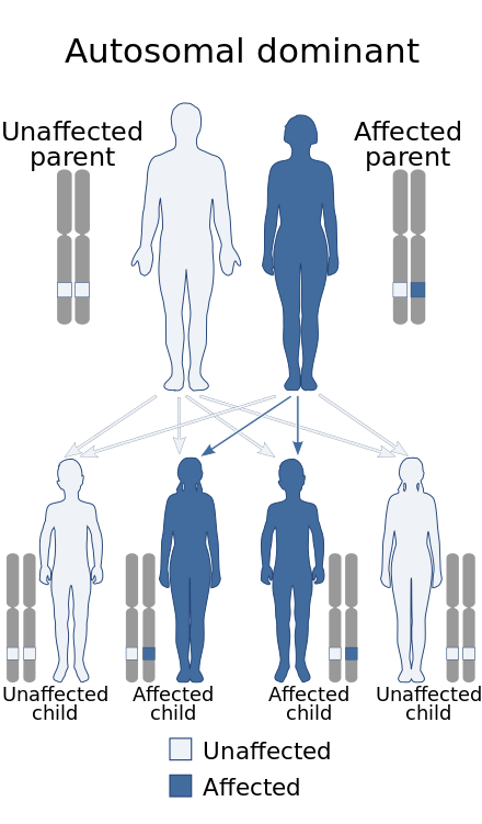
Axenfeld syndrome has an autosomal dominant pattern of inheritance. Autosomal dominant - en.svg
Axenfeld syndrome has an autosomal dominant pattern of inheritance.

The molecular genetics of Axenfeld syndrome are poorly understood, but center on three genes identified by cloning of chromosomal breakpoints from patients. [5] [6]

Molecular genetics is the field of biology that studies the structure and function of genes at a molecular level and thus employs methods of both molecular biology and genetics. The study of chromosomes and gene expression of an organism can give insight into heredity, genetic variation, and mutations. This is useful in the study of developmental biology and in understanding and treating genetic diseases.

This disorder is inheritable as an autosomal dominant trait, [4] which means the defective gene is located on an autosome, and only one copy of the gene is sufficient to cause the disorder when inherited from a parent who has the disorder. As shown in the diagram, this gives a 50/50 chance of offspring inheriting the condition from an affected parent.[ citation needed ]

Autosome Any chromosome other than a sex chromosome.

An autosome is a chromosome that is not an allosome. The members of an autosome pair in a diploid cell have the same morphology, unlike those in allosome pairs which may have different structures. The DNA in autosomes is collectively known as atDNA or auDNA.

Diagnosis

Although most recognized for its correlation with the onset of glaucoma, the malformation is not limited to the eye, as Axenfeld syndrome when associated with the PITX2 genetic mutation usually presents congenital malformations of the face, teeth, and skeletal system. [7]

Glaucoma eye disease that is characterized by an unstable or a sustained increase in the intraocular pressure which the eye cannot withstand without damage to its structure or impairment of its function

Glaucoma is a group of eye diseases which result in damage to the optic nerve and cause vision loss. The most common type is open-angle glaucoma with less common types including closed-angle glaucoma and normal-tension glaucoma. Open-angle glaucoma develops slowly over time and there is no pain. Peripheral vision may begin to decrease followed by central vision resulting in blindness if not treated. Closed-angle glaucoma can present gradually or suddenly. The sudden presentation may involve severe eye pain, blurred vision, mid-dilated pupil, redness of the eye, and nausea. Vision loss from glaucoma, once it has occurred, is permanent.

PITX2 protein-coding gene in the species Homo sapiens

Paired-like homeodomain transcription factor 2 also known as pituitary homeobox 2 is a protein that in humans is encoded by the PITX2 gene.

The most characteristic feature affecting the eye is a distinct corneal posterior arcuate ring, known as an "embryotoxon". [8] The iris is commonly adherent to the Schwalbe's line (posterior surface of the cornea).[ citation needed ]

Schwalbes line

Schwalbe's line is the anatomical line found on the interior surface of the eye's cornea, and delineates the outer limit of the corneal endothelium layer. Specifically, it represents the termination of Descemet's membrane. In many cases it can be seen via gonioscopy.

Diagnosis One of the three known genetic mutations which cause Rieger Syndrome can be identified through genetic samples analysis. About 40% of Axenfeld-Rieger sufferers have displayed mutations in genes PITX2, [7] FOXC1, and PAX6. [9] [10] The difference between Type 1, 2, and 3 Axenfeld Syndrome is the genetic cause, all three types display the same symptoms and abnormalities. [11]

Forkhead box C1 protein-coding gene in the species Homo sapiens

Forkhead box C1, also known as FOXC1, is a protein which in humans is encoded by the FOXC1 gene.

PAX6 protein-coding gene in the species Homo sapiens

Paired box protein Pax-6, also known as aniridia type II protein (AN2) or oculorhombin, is a protein that in humans is encoded by the PAX6 gene.

The OMIM classification is as follows:

Type OMIM Gene
Type 1 180500 PITX2
Type 2 601499 possibly FOXO1A [12]
Type 3 602482 FOXC1
DeHauwere syndrome 109120 Unknown [9]

Detection of any of these mutations can give patients a clear diagnosis and prenatal procedures such as preimplantation genetic diagnosis, chorionic villus sampling and amniocentesis can be offered to patients and prospective parents. [13] [ failed verification ]

Management

Eponym

It is named after the German ophthalmologist Theodor Axenfeld [14] [8] who studied anterior segment disorders, especially those such as Rieger Syndrome and the Axenfeld Anomaly.[ citation needed ]

Axenfeld-Rieger syndrome is characterized by abnormalities of the eyes, teeth, and facial structure. [11] Rieger Syndrome, by medical definition, is determined by the presence of malformed teeth, underdeveloped anterior segment of the eyes, and cardiac problems associated with the Axenfeld anomaly. [12] The term "Rieger syndrome" is sometimes used to indicate an association with glaucoma. [7] Glaucoma occurs in up to 50% of patients with Rieger Syndrome. Glaucoma develops during adolescence or late childhood, but often occurs in infancy. [8] [9] In addition, a prominent Schwalbe's line, an opaque ring around the cornea known as posterior embryotoxon, may arise with hypoplasia of the iris. [5] Below average height and stature, stunted development of the mid-facial features and mental deficiencies may also be observed in patients. [5]

See also

Related Research Articles

Kabuki syndrome rare disease

Kabuki syndrome is a pediatric congenital disorder of genetic origin. It affects multiple parts of the body with varying symptoms and severity, although the most common is the characteristic facial appearance. It is quite rare, affecting roughly one in 32,000 births. It was first identified and described in 1981 by two Japanese groups, led by scientists Norio Niikawa and Yoshikazu Kuroki. It is named Kabuki syndrome because of the facial resemblance of affected individuals to stage makeup used in kabuki, a Japanese traditional theatrical form.
CHARGE syndrome syndrome that is characterized by a pattern of congenital anomalies including choanal atresia and malformations of the heart, inner ear, and retina

CHARGE syndrome is a rare syndrome caused by a genetic disorder. First described in 1979, the acronym "CHARGE" came into use for newborn children with the congenital features of coloboma of the eye, heart defects, atresia of the nasal choanae, retardation of growth and/or development, genital and/or urinary abnormalities, and ear abnormalities and deafness. These features are no longer used in making a diagnosis of CHARGE syndrome, but the name remains. About two thirds of cases are due to a CHD7 mutation. CHARGE syndrome occurs only in 0.1–1.2 per 10,000 live births; as of 2009 it was the leading cause of congenital deafblindness in the US.

Fraser syndrome Fraser syndrome is a rare clinical entity including as main characteristics cryptophthalmos and syndactyly

Fraser syndrome is an autosomal recessive congenital disorder. Fraser syndrome is named for the geneticist George R. Fraser, who first described the syndrome in 1962.

Simpson–Golabi–Behmel syndrome X-linked disease characterized by pre- and postnatal overgrowth and craniofacial, skeletal, cardiac and renal abnormalities

Simpson–Golabi–Behmel syndrome (SGBS), is a rare inherited congenital disorder that can cause craniofacial, skeletal, cardiac, and renal abnormalities. The syndrome is inherited in an X-linked recessive fashion, where males express the phenotype and females usually do not. Females that possess one copy of the mutation are considered to be carriers of the syndrome and may express varying degrees of the phenotype.

22q13 deletion syndrome Rare genetic syndrome

22q13 deletion syndrome, also known as Phelan–McDermid syndrome (PMS), is a genetic disorder caused by deletions or rearrangements on the q terminal end of chromosome 22. Any abnormal genetic variation in the q13 region that presents with significant manifestations (phenotype) typical of a terminal deletion may be diagnosed as 22q13 deletion syndrome. There is disagreement among researchers as to the exact definition of 22q13 deletion syndrome. The Developmental Synaptopathies Consortium defines PMS as being caused by SHANK3 mutations, a definition that appears to exclude terminal deletions. The requirement to include SHANK3 in the definition is supported by many but not by those who first described 22q13 deletion syndrome.

Chromosome abnormality abnormal number or structure of chromosomes

A chromosomal disorder, anomaly, aberration, or mutation is a missing, extra, or irregular portion of chromosomal DNA. It can be from a typical number of chromosomes or a structural abnormality in one or more chromosomes. Chromosome mutation was formerly used in a strict sense to mean a change in a chromosomal segment, involving more than one gene. The term "karyotype" refers to the full set of chromosomes from an individual; this can be compared to a "normal" karyotype for the species via genetic testing. A chromosome anomaly may be detected or confirmed in this manner. Chromosome anomalies usually occur when there is an error in cell division following meiosis or mitosis. There are many types of chromosome anomalies. They can be organized into two basic groups, numerical and structural anomalies.

Papillorenal syndrome autosomal dominant disease characterized by optic nerve coloboma and renal disease that has material basis in heterozygous mutation in the PAX2 gene on chromosome 10q24

Papillorenal syndrome, is an autosomal dominant genetic disorder marked by underdevelopment (hypoplasia) of the kidney and colobomas of the optic nerve.
Frank–ter Haar syndrome

Frank ter Haar-syndrome (FTHS), also known as Ter Haar-syndrome, is a rare disease characterized by abnormalities that affect bone, heart, and eye development. Children born with the disease usually die very young.

Angelman syndrome Human disease

Angelman syndrome (AS) is a genetic disorder that mainly affects the nervous system. Symptoms include a small head and a specific facial appearance, severe intellectual disability, developmental disability, speaking problems, balance and movement problems, seizures, and sleep problems. Children usually have a happy personality and have a particular interest in water. The symptoms generally become noticeable by one year of age.

Congenital generalized lipodystrophy is an extremely rare autosomal recessive condition, characterized by an extreme scarcity of fat in the subcutaneous tissues. It is a type of lipodystophy disorder where the magnitude of fat loss determines the severity of metabolic complications. Only 250 cases of the condition have been reported, and it is estimated that it occurs in 1 in 10 million people worldwide.

A Finnish heritage disease is a genetic disease or disorder that is significantly more common in people whose ancestors were ethnic Finns, natives of Finland and Sweden (Meänmaa) and Russia. 36 rare diseases are regarded as Finnish heritage diseases. The diseases are not restricted to Finns; they are genetic diseases with far wider distribution in the world, but due to founder effects and genetic isolation they are more common in Finns.

Lujan–Fryns syndrome

Lujan–Fryns syndrome (LFS) is an X-linked genetic disorder that causes mild to moderate intellectual disability and features described as Marfanoid habitus, referring to a group of physical characteristics similar to those found in Marfan syndrome. These features include a tall, thin stature and long, slender limbs. LFS is also associated with psychopathology and behavioral abnormalities, and it exhibits a number of malformations affecting the brain and heart. The disorder is inherited in an X-linked dominant manner, and is attributed to a missense mutation in the MED12 gene. There is currently no treatment or therapy for the underlying MED12 malfunction, and the exact cause of the disorder remains unclear.

Malpuech facial clefting syndrome, also called Malpuech syndrome or Gypsy type facial clefting syndrome, is a rare congenital syndrome. It is characterized by facial clefting, a caudal appendage, growth deficiency, intellectual and developmental disability, and abnormalities of the renal system (kidneys) and the male genitalia. Abnormalities of the heart, and other skeletal malformations may also be present. The syndrome was initially described by Georges Malpuech and associates in 1983. It is thought to be genetically related to Juberg-Hayward syndrome. Malpuech syndrome has also been considered as part of a spectrum of congenital genetic disorders associated with similar facial, urogenital and skeletal anomalies. Termed "3MC syndrome", this proposed spectrum includes Malpuech, Michels and Mingarelli-Carnevale (OSA) syndromes. Mutations in the COLLEC11 and MASP1 genes are believed to be a cause of these syndromes. The incidence of Malpuech syndrome is unknown. The pattern of inheritance is autosomal recessive, which means a defective (mutated) gene associated with the syndrome is located on an autosome, and the syndrome occurs when two copies of this defective gene are inherited.

Nasodigitoacoustic syndrome, also called Keipert syndrome, is a rare congenital syndrome first described by J.A. Keipert and colleagues in 1973. The syndrome is characterized by a misshaped nose, broad thumbs and halluces, brachydactyly, sensorineural hearing loss, facial features such as hypertelorism, and developmental delay. It is believed to be inherited in an X-linked recessive manner, which means a genetic mutation causing the disorder is located on the X chromosome, and while two copies of the mutated gene must be inherited for a female to be born with the disorder, just one copy is sufficient to cause a male to be born with the disorder. Nasodigitoacoustic syndrome is likely caused by a mutated gene located on the X chromosome between positions Xq22.2–q28. The incidence of the syndrome has not been determined, but it is considered to affect less than 200,000 people in the United States, and no greater than 1 per 2,000 in Europe. It is similar to Keutel, Muenke, Rubinstein and Teunissen-Cremers syndrome.

Floating–Harbor syndrome Floating-Harbor syndrome is a genetic developmental disorder characterized by facial dysmorphism, short stature with delayed bone age, and expressive language delay

Floating–Harbor syndrome, also known as Pelletier–Leisti syndrome, is a rare disease with fewer than 50 cases described in the literature. It is usually diagnosed in early childhood and is characterized by the triad of proportionate short stature with delayed bone age, characteristic facial appearance, and delayed speech development. Although its cause is unknown, it is thought to result from genetic mutation, and diagnosis is established by the presence of a heterozygous SRCAP mutation in those with clinical findings of FHS.

Liebenberg Syndrome is a rare autosomal genetic disease that involves a deletion mutation upstream of the PITX1 gene, which is one that's responsible for the body's organization, specifically in forming lower limbs. In animal studies, when this deletion was introduced to developing birds, their wing buds were noted to take on limb-like structures.

Primary juvenile glaucoma is glaucoma that develops due to ocular hypertension and is evident either at birth or within the first few years of life. It is caused due to abnormalities in the anterior chamber angle development that obstruct aqueous outflow in the absence of systemic anomalies or other ocular malformation.

Barx homeobox 1 protein-coding gene in the species Homo sapiens

BARX homeobox 1 is a protein that in humans is encoded by the BARX1 gene.

References

  1. Dhir, L; Frimpong-Ansah, K; Habib, Nabil E (2008). "Missed case of Axenfeld-Rieger syndrome: a case report". Cases Journal. 1 (1): 299. doi:10.1186/1757-1626-1-299. PMC   2585579 . PMID   18990239.
  2. Vieira, Véronique; David, Gabriel; Roche, Olivier; de la Houssaye, Guillaume; Boutboul, Sandrine; Arbogast, Laurence; Kobetz, Alexandra; Orssaud, Christophe; Camand, Olivier; Schorderet, Daniel F.; Munier, Francis; Rossi, Annick; Delezoide, Anne Lise; Marsac, Cécile; Ricquier, Daniel; Dufier, Jean-Louis; Menasche, Maurice; Abitbol, M. (2006). "Identification of four new PITX2 gene mutations in patients with Axenfeld-Rieger syndrome". Molecular Vision. 12: 1448–60. PMID   17167399.
  3. Fitch, Naomi; Kaback, Martin (1978). "The Axenfeld syndrome and the Rieger syndrome". Journal of Medical Genetics. 15 (1): 30–4. doi:10.1136/jmg.15.1.30. PMC   1012820 . PMID   416212.
  4. 1 2 "Axenfeld-Rieger syndrome type 1". National Center for Biotechnology Information.
  5. 1 2 3 Suzuki, Katsuhiro; Nakamura, Makoto; Amano, Emi; Mokuno, Kumiko; Shirai, Shoichiro; Terasaki, Hiroko (2006). "Case of chromosome 6p25 terminal deletion associated with Axenfeld–Rieger syndrome and persistent hyperplastic primary vitreous". American Journal of Medical Genetics Part A. 140 (5): 503–8. doi:10.1002/ajmg.a.31085. PMID   16470791.
  6. Tonoki, Hidefumi; Harada, Naoki; Shimokawa, Osamu; Yosozumi, Ayako; Monzaki, Kadomi; Satoh, Kohei; Kosaki, Rika; Sato, Atsushi; Matsumoto, Naomichi; Iizuka, Susumu (2011). "Axenfeld-Rieger anomaly and Axenfeld-Rieger syndrome: Clinical, molecular-cytogenetic, and DNA array analyses of three patients with chromosomal defects at 6p25". American Journal of Medical Genetics Part A. 155A (12): 2925–32. doi:10.1002/ajmg.a.33858. PMID   22009788.
  7. 1 2 3 Meyer-Marcotty, P.; Weisschuh, N.; Dressler, P.; Hartmann, J.; Stellzig-Eisenhauer, A. (2008). "Morphology of the sella turcica in Axenfeld-Rieger syndrome with PITX2 mutation". Journal of Oral Pathology & Medicine. 37 (8): 504–10. doi:10.1111/j.1600-0714.2008.00650.x. PMID   18331556.
  8. 1 2 3 Axenfeld, T (1920). "Embryotoxon cornea posterius". Berichte der Deutschen Ophthalmologischen Gesellschaft. 42: 301–2.
  9. 1 2 3 Lowry, R. Brian; Gould, Douglas B.; Walter, Michael A.; Savage, Paul R. (2007). "Absence of PITX2, BARX1, and FOXC1 mutations in De Hauwere syndrome (Axenfeld–Rieger anomaly, hydrocephaly, hearing loss): A 25-year follow up". American Journal of Medical Genetics Part A. 143A (11): 1227–30. doi:10.1002/ajmg.a.31732. PMID   17486624.
  10. Reis, LM; Semina, EV (September 2011). "Genetics of anterior segment dysgenesis disorders". Current Opinion in Ophthalmology. 22 (5): 314–24. doi:10.1097/ICU.0b013e328349412b. PMC   3558283 . PMID   21730847.
  11. 1 2 "Axenfeld-Rieger syndrome". United States Department of Health and Human Services. November 8, 2016.
  12. 1 2 Phillips, Jeffrey C.; del Bono, Elizabeth A.; Haines, Jonathan L.; Pralea, Anca Madalina; Cohen, John S.; Greff, Linda J.; Wiggs, Janey L. (1996). "A second locus for Rieger syndrome maps to chromosome 13q14". American Journal of Human Genetics. 59 (3): 613–9. PMC   1914897 . PMID   8751862.
  13. "How are genetic conditions diagnosed?". United States Department of Health and Human Services. November 8, 2016.
  14. synd/1284 at Who Named It?

Further reading

Classification
D