Dislocation of hip

Last updated
Dislocation of hip
HipdisX.png
X-ray showing a joint dislocation of the left hip.
Specialty Rheumatology   Blue pencil.svg

Dislocation of the hip is a common injury to the hip joint. Dislocation occurs when the ballshaped head of the femur comes out of the cupshaped acetabulum set in the pelvis.

Joint dislocation medical injury

A joint dislocation, also called luxation, occurs when there is an abnormal separation in the joint, where two or more bones meet. A partial dislocation is referred to as a subluxation. Dislocations are often caused by sudden trauma on the joint like an impact or fall. A joint dislocation can cause damage to the surrounding ligaments, tendons, muscles, and nerves. Dislocations can occur in any joint major or minor. The most common joint dislocation is a shoulder dislocation.

Hip anatomical region

In vertebrate anatomy, hip refers to either an anatomical region or a joint.

Femur most proximal bone of the leg for tetrapode vertebrates, longest bone for humans

The femur or thigh bone, is the proximal bone of the hindlimb in tetrapod vertebrates. The head of the femur articulates with the acetabulum in the pelvic bone forming the hip joint, while the distal part of the femur articulates with the tibia and kneecap forming the knee joint. By most measures the femur is the strongest bone in the body. The femur is also the longest bone in the human body.

Contents

Dislocations may be developmental or due to acute trauma. The emphasis of this article is on acute dislocation. For developmental dislocation, see hip dysplasia.

Hip dysplasia human disease

Hip dysplasia is an abnormality of the hip joint where the socket portion does not fully cover the ball portion, resulting in an increased risk for joint dislocation. congenital dysplasia of the hip (CDH) Hip dysplasia may occur at birth or develop in early life. Regardless, it does not typically produce symptoms in babies less than a year old. Occasionally one leg may be shorter than the other. The left hip is more often affected than the right. Complications without treatment can include arthritis, limping, and low back pain.

Acute traumatic hip dislocations are severe injuries. [1] These dislocations typically occur in 16- to 40-year-olds involved in high energy trauma like motor vehicle accidents. [2]

Classification

Hip dysplasia versus congenital dislocation

Developmental dysplasia of the hip (DDH) or hip dysplasia is now the widely accepted term because it provides a more accurate description of the spectrum of abnormalities that affect the immature hip. [3] The term "congenital" dislocation is no longer used, except for very rare conditions, in which there is a ("teratologic") fixed dislocation location present at birth. [1]

Teratology is the study of abnormalities of physiological development. It is often thought of as the study of human congenital abnormalities, but it is broader than that, taking into account other non-birth developmental stages, including puberty; and other organisms, including plants. The related term developmental toxicity includes all manifestations of abnormal development that are caused by environmental insult. These may include growth retardation, delayed mental development or other congenital disorders without any structural malformations.

Hip dysplasia is a condition in which a child is born with a hip problem. Hip dysplasia is when the formation of the hip joint is abnormal. The ball at the top of the thighbone which is known as the femoral head is not stable within the socket (which is also known as the acetabulum).

The acetabulum is a concave surface of a pelvis. The head of the femur meets with the pelvis at the acetabulum, forming the hip joint.

Posterior dislocation

Posterior dislocations with an associated fracture are categorised by the Thompson and Epstein classification system, the Stewart and Milford classification system, and the Pipkin system (when associated with femoral head fractures). [4] [5]

The Thompson and Epstein classification is a system of categorizing posterior fracture/dislocations of the hip.

The Pipkin classification is a system of categorizing femoral head hip fractures based on the fracture pattern.

Anterior dislocation

There is also a Thompson and Epstein classification system for anterior hip dislocations. [4] [5]

Central dislocation

Central dislocation is an outdated term for medial displacement of the femoral head into a displaced acetabular fracture. [5] It is no longer used.

Dislocation of the left hip, secondary to developmental hip dysplasia. Closed arrow marks the acetabulum, open arrow the femoral head. Congenitaldislocation10.JPG
Dislocation of the left hip, secondary to developmental hip dysplasia. Closed arrow marks the acetabulum, open arrow the femoral head.

Signs and Symptoms

The affected leg is virtually immovable by the patient, and is usually extremely painful. [6] Dislocations are categorized as either posterior or anterior, based on the location of the head of the femur (see classification above). [7]

Posterior dislocation

Nine out of ten hip dislocations are posterior. [1] The affected limb will be in a position of flexion, adduction, and internally rotated in this case. [1] The knee and the foot will be in towards the middle of the body. [6] A sciatic nerve palsy is present in 8%-20% of cases. [1]

Anterior dislocation

In an anterior dislocation the limb is held by the patient in externally rotated with mild flexion and abduction. [5] Femoral nerve palsies can be present, but are uncommon. [1]

Mechanism

The hip joint includes the articulation of the femoral head (of femur) and the acetabulum of the pelvis. In hip dislocation, the femoral head is dislodged from this socket. Posterior dislocation is the most prevalent, in which the femoral head lies posterior and superior to the acetabulum. This is most common when the femur is adducted and internally rotated. The opposite is true for the shoulder, where the most common dislocation occurs in the anterior and inferior directions. [8] Motor vehicle traffic collisions are responsible for almost all posterior hip dislocations. [2] The posterior side of the hip exhibits primarily hip extension, dealing with the muscles: gluteus maximus, hamstring muscles (biceps femoris, semitendinosus, semimembranosus), and the six deep external rotators (piriformis, obturator externus, obturator internus, gemellus superior, gemellus inferior, and quadrates femoris). [9]

To actually dislocate a healthy hip, a great amount of force needs to be applied. Motor vehicle accidents are the most common ways that hip dislocations occur. Falls from a height, such as a ladder, can also generate enough force to dislocate a hip. In older individuals, even a slight fall could cause this type of injury. Wear and tear that the body undergoes throughout the years leads to increased incidents of hip dislocation in the older population. [10]

Several other injuries are also associated with hip dislocation. Fractures in the pelvis and legs, and minor back or head injuries can also occur, along with a hip dislocation, that is caused by a fall or athletic of injury.

Diagnosis

Radiography

Anterior-posterior (AP) X-rays of the pelvis, AP and lateral views of the femur (knee included) are ordered for diagnosis. [1] The size of the head of the femur is then compared across both sides of the pelvis. The affected femoral head will appear larger if the dislocation is anterior, and smaller if posterior. [5] A CT scan may also be ordered to clarify the fracture pattern.

Management

Uncomplicated hip dislocations

The hip should be reduced as quickly as possible to reduce the risk of osteonecrosis of the femoral head. [2] This is done via inline manual traction with general anesthesia and muscle relaxation, or conscious sedation. [5] Fractures of the femoral head and other loose bodies should be determined prior to reduction. Common closed reduction methods include the Allis method and Stimson method. [11] Once reduction is completed management becomes less urgent and appropriate workup including CT scanning can be completed. [5] Post-reduction, patients may begin early crutch-assisted ambulation with weight bearing as tolerated.

Complicated hip dislocations

If the dislocated hip cannot be reduced by manipulation alone, an immediate open (surgical) reduction is necessary. A CT scan or Judet views should be obtained prior to transfer to the surgical suite. [5]

Rehabilitation

Hip dislocation rehabilitation can take anywhere from two to three months, depending on the patient. Complications to nearby nerves and blood vessels can sometimes cause loss of blood supply to the bone, also known as osteonecrosis. The protective cartilage on the bone can also be disturbed from this type of injury. For this reason, it is important for patients to contact a physician and get treatment immediately following injury. [10]

Exercises used for rehabilitation

Picture of a set of ankle weights. Weightsforankle.jpg
Picture of a set of ankle weights.
Picture of a modified side plank. Modifiedsideplank.jpg
Picture of a modified side plank.

Individuals suffering from hip dislocation should participate in physical therapy and receive professional prescriptive exercises based on their individual abilities, progress, and overall range of motion. The following are some typical recommended exercises used as rehabilitation for hip dislocation. It is important to understand that each individual has different capabilities that can best be assessed by a physical therapist or medical professional, and that these are simply recommendations. [12]

Epidemiology

Reimer's migration index can be used to indicate hip dislocation. The migration index (MI) is normally less than 33%. Migrationsindex nach Reimers.png
Reimer's migration index can be used to indicate hip dislocation. The migration index (MI) is normally less than 33%.

16-40 year-old males are responsible for the majority of hip dislocations. These hip dislocations are typically posterior, and a direct result of motor vehicle traffic collisions. [2]

See also

Related Research Articles

Human leg lower extremity or limb of the human body (foot, lower leg, thigh and hip)

The human leg, in the general meaning, is the entire lower limb of the human body, including the foot, thigh and even the hip or gluteal region. However, the definition in human anatomy refers only to the section of the lower limb extending from the knee to the ankle, also known as the crus. Legs are used for standing, and all forms of locomotion including recreational such as dancing, and constitute a significant portion of a person's mass. Female legs generally have greater hip anteversion and tibiofemoral angles, but shorter femur and tibial lengths than those in males.

Thigh area between the pelvis and the knee; upper leg

In human anatomy, the thigh is the area between the hip (pelvis) and the knee. Anatomically, it is part of the lower limb.

Sartorius muscle muscle in the human body

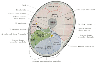
The sartorius muscle is the longest muscle in the human body. It is a long, thin, superficial muscle that runs down the length of the thigh in the anterior compartment.

Piriformis muscle

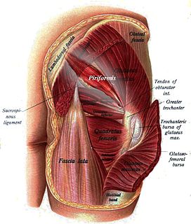
The piriformis is a muscle in the gluteal region of the lower limbs. It is one of the six muscles in the lateral rotator group.

Hip replacement surgery replacing hip joint with prosthetic implant

Hip replacement is a surgical procedure in which the hip joint is replaced by a prosthetic implant, that is, a hip prosthesis. Hip replacement surgery can be performed as a total replacement or a hemi (half) replacement. Such joint replacement orthopaedic surgery is generally conducted to relieve arthritis pain or in some hip fractures. A total hip replacement consists of replacing both the acetabulum and the femoral head while hemiarthroplasty generally only replaces the femoral head. Hip replacement is currently one of the most common orthopaedic operations, though patient satisfaction short- and long-term varies widely. Approximately 58% of total hip replacements are estimated to last 25 years. The average cost of a total hip replacement in 2012 was $40,364 in the United States, and about $7,700 to $12,000 in most European countries.

Slipped capital femoral epiphysis rare disease

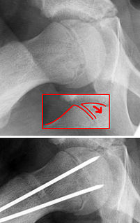
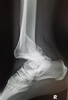
Slipped capital femoral epiphysis is a medical term referring to a fracture through the growth plate (physis), which results in slippage of the overlying end of the femur (metaphysis).

Iliopsoas

The iliopsoas refers to the joined psoas and the iliacus muscles. The two muscles are separate in the abdomen, but usually merge in the thigh. As such, they are usually given the common name iliopsoas. The iliopsoas muscle joins to the femur at the lesser trochanter, and acts as the strongest flexor of the hip.

Inferior gluteal nerve

The inferior gluteal nerve is the main motor neuron that innervates the gluteus maximus muscle. It is responsible for the movement of the gluteus maximus in activities requiring the hip to extend the thigh, such as climbing stairs. Injury to this nerve is rare but often occurs as a complication of posterior approach to the hip during hip replacement. When damaged, one would develop gluteus maximus lurch, which is a gait abnormality which causes the individual to 'lurch' backwards to compensate lack in hip extension.

The knee examination, in medicine and physiotherapy, is performed as part of a physical examination, or when a patient presents with knee pain or a history that suggests a pathology of the knee joint.

Iliofemoral ligament

The iliofemoral ligament is a ligament of the hip joint which extends from the ilium to the femur in front of the joint. It is also referred to as the Y-ligament or the ligament of Bigelow, and any combinations of these names.

In medicine, physiotherapy, chiropractic, and osteopathy the hip examination, or hip exam, is undertaken when a patient has a complaint of hip pain and/or signs and/or symptoms suggestive of hip joint pathology. It is a physical examination maneuver.

Acetabular labrum

The acetabular labrum is a ring of cartilage that surrounds the acetabulum of the hip. The anterior portion is most vulnerable when the labrum tears.

Femur neck

The femur neck is a flattened pyramidal process of bone, connecting the femoral head with the femoral shaft, and forming with the latter a wide angle opening medialward.

Ortolani test

The Ortolani test is part of the physical examination for developmental dysplasia of the hip, along with the Barlow maneuver. Specifically, the Ortolani test is positive when a posterior dislocation of the hip is reducible with this maneuver. This is part of the standard infant exam performed preferably in early infancy.

Femoral fracture

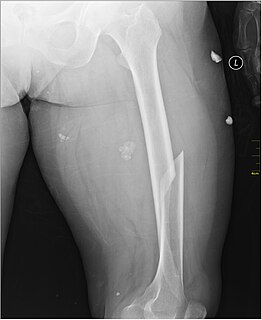
A femoral fracture is a bone fracture that involves the femur. They are typically sustained in high-impact trauma, such as car crashes, due to the large amount of force needed to break the bone. Fractures of the diaphysis, or middle of the femur, are managed differently from those at the head, neck, and trochanter.

Acetabular fracture

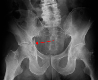
Fractures of the acetabulum occur when the head of the femur is driven into the pelvis. This injury is caused by a blow to either the side or front of the knee and often occurs as a dashboard injury accompanied by a fracture of the femur.

Pelvis lower part of the trunk of the human body between the abdomen and the thighs (sometimes also called pelvic region of the trunk

The pelvis is either the lower part of the trunk of the human body between the abdomen and the thighs or the skeleton embedded in it.

X-rays of hip dysplasia are one of the two main methods of medical imaging to diagnose hip dysplasia, the other one being medical ultrasonography.. Ultrasound imaging yields better results defining the anatomy until the cartilage is ossified. When the infant is around 3 months old a clear roentgenographic image can be achieved. Unfortunately the time the joint gives a good x-ray image is also the point at which nonsurgical treatment methods cease to give good results.

References

  1. 1 2 3 4 5 6 7 Essentials of musculoskeletal care. Sarwark, John F. Rosemont, Ill.: American Academy of Orthopaedic Surgeons. 2010. ISBN   9780892035793. OCLC   706805938.
  2. 1 2 3 4 1967-, Egol, Kenneth A., (2015). Handbook of fractures. Koval, Kenneth J., Zuckerman, Joseph D. (Joseph David), 1952-, Ovid Technologies, Inc. (5th ed.). Philadelphia: Wolters Kluwer Health. ISBN   9781451193626. OCLC   960851324.
  3. Jackson, Jonathan C.; Runge, Melissa M.; Nye, Nathaniel S. (2014-12-15). "Common Questions About Developmental Dysplasia of the Hip". American Family Physician. 90 (12). ISSN   0002-838X.
  4. 1 2 Thompson, Vernon P.; Epstein, Herman C. (July 1951). "TRAUMATIC DISLOCATION OF THE HIP: A Survey of Two Hundred and Four Cases Covering a Period of Twenty-one Years". JBJS. 33 (3): 746. doi:10.2106/00004623-195133030-00023. ISSN   0021-9355.
  5. 1 2 3 4 5 6 7 8 Skeletal trauma : basic science, management, and reconstruction. Browner, Bruce D.,, Jupiter, Jesse B.,, Krettek, Christian, 1953-, Anderson, Paul, 1952-, ClinicalKey. (Fifth ed.). Philadelphia, PA. ISBN   9781455776283. OCLC   898159499.
  6. 1 2 "Hip Dislocation-OrthoInfo - AAOS". orthoinfo.aaos.org. Retrieved 2017-10-01.
  7. Goddard, N. J. (August 2000). "Classification of traumatic hip dislocation". Clinical Orthopaedics and Related Research (377): 11–14. doi:10.1097/00003086-200008000-00004. ISSN   0009-921X. PMID   10943180.
  8. Hip Dislocation in Emergency Medicine at eMedicine
  9. Floyd, R.T. (2009). Manual of structural kinesiology. New York, NY: McGraw-Hill[ page needed ]
  10. 1 2 "Hip Dislocation-OrthoInfo - AAOS". Orthoinfo.aaos.org. 2014-06-01. Retrieved 2015-03-01.
  11. Stimson, Lewis Atterbury (1883). A treatise on fractures. The Library of Congress. Philadelphia, H.C. Lea's son & co.
  12. 1 2 3 4 5 6 7 8 9 10 11 12 13 Hip Dislocation Treatment & Management at eMedicine
  13. Pietro PERSIANI; Iakov MOLAYEM; Alessandro CALISTRI; Stefano ROSI; Marco BOVE; Ciro VILLANI (2008). "Hip subluxation and dislocation in cerebral palsy: Outcome of bone surgery in 21 hips" (PDF). Acta Orthop. Belg.
Classification
D
External resources