Hexokinase II, also known as Hexokinase B and HK2, is an enzyme which in humans is encoded by the HK2 gene on chromosome 2. [5] [6] Hexokinases phosphorylate glucose to produce glucose 6-phosphate, the first step in most glucose metabolism pathways. Hexokinase II is the predominant hexokinase form found in skeletal muscle. It localizes to the outer membrane of mitochondria. Expression of the HK2 gene is insulin-responsive, and studies in rat suggest that it is involved in the increased rate of glycolysis seen in rapidly growing cancer cells. [provided by RefSeq, Apr 2009] [6]
Hexokinase II is one of four homologous hexokinase isoforms in mammalian cells. [7] [8] [9] [10] [11]
The HK2 gene spans approximately 50 kb and consists of 18 exons. There is also an HK2 pseudogene integrated into a long interspersed nuclear repetitive DNA element located on the X chromosome. Though its DNA sequence is similar to the cDNA product of the actual HK2 mRNA transcript, it lacks an open reading frame for gene expression. [10]
This gene encodes a 100-kDa, 917-residue enzyme with highly similar N-terminal and C-terminal domains that each form half of the protein. [10] [12] This high similarity, along with the existence of a 50-kDa hexokinase (HK4), suggests that the 100-kDa hexokinases originated from a 50-kDa precursor via gene duplication and tandem ligation. [10] [11] Both N- and C-terminal domains possess catalytic ability and can be inhibited by glucose 6-phosphate, though the C-terminal domain demonstrates lower affinity for ATP and is only inhibited at higher concentrations of glucose 6-phosphate. [10] Despite there being two binding sites for glucose, it is proposed that glucose binding at one site induces a conformational change which prevents a second glucose from binding the other site. [13] Meanwhile, the first 12 amino acids of the highly hydrophobic N-terminal serve to bind the enzyme to the mitochondria, while the first 18 amino acids contribute to the enzyme’s stability. [9] [11]
As an isoform of hexokinase and a member of the sugar kinase family, hexokinase II catalyzes the rate-limiting and first obligatory step of glucose metabolism, which is the ATP-dependent phosphorylation of glucose to glucose 6-phosphate. [11] Physiological levels of glucose 6-phosphate can regulate this process by inhibiting hexokinase II as negative feedback, though inorganic phosphate (Pi) can relieve glucose 6-phosphate inhibition. [8] [10] [11] Pi can also directly regulate hexokinase II, and the double regulation may better suit its anabolic functions. [8] By phosphorylating glucose, hexokinase II effectively prevents glucose from leaving the cell and, thus, commits glucose to energy metabolism. [10] [12] Moreover, its localization and attachment to the OMM promotes the coupling of glycolysis to mitochondrial oxidative phosphorylation, which greatly enhances ATP production to meet the cell’s energy demands. [14] [15] Specifically, hexokinase II binds VDAC to trigger opening of the channel and release mitochondrial ATP to further fuel the glycolytic process. [8] [15]
Another critical function for OMM-bound hexokinase II is mediation of cell survival. [8] [9] Activation of Akt kinase maintains HK2-VDAC coupling, which subsequently prevents cytochrome c release and apoptosis, though the exact mechanism remains to be confirmed. [8] One model proposes that hexokinase II competes with the pro-apoptotic proteins BAX to bind VDAC, and in the absence of hexokinase II, BAX induces cytochrome c release. [8] [15] In fact, there is evidence that hexokinase II restricts BAX and BAK oligomerization and binding to the OMM. In a similar mechanism, the pro-apoptotic creatine kinase binds and opens VDAC in the absence of hexokinase II. [8] An alternative model proposes the opposite, that hexokinase II regulates binding of the anti-apoptotic protein Bcl-Xl to VDAC. [15]
In particular, hexokinase II is ubiquitously expressed in tissues, though it is majorly found in muscle and adipose tissue. [8] [10] [15] In cardiac and skeletal muscle, hexokinase II can be found bound to both the mitochondrial and sarcoplasmic membrane. [16] HK2 gene expression is regulated by a phosphatidylinositol 3-kinaselp70 S6 protein kinase-dependent pathway and can be induced by factors such as insulin, hypoxia, cold temperatures, and exercise. [10] [17] Its inducible expression indicates its adaptive role in metabolic responses to changes in the cellular environment. [17]
Hexokinase II is highly expressed in several cancers, including breast cancer and colon cancer. [9] [15] [18] Its role in coupling ATP from oxidative phosphorylation to the rate-limiting step of glycolysis may help drive the tumor cells’ growth. [15] Notably, inhibition of hexokinase II has demonstrably improved the effectiveness of anticancer drugs., [18] Thus, hexokinase II stands as a promising therapeutic target, though considering its ubiquitous expression and crucial role in energy metabolism, a reduction rather than complete inhibition of its activity should be pursued. [15] [18]
A study on non-insulin-dependent diabetes mellitus (NIDDM) revealed low basal glucose 6-phosphate levels in NIDDM patients that failed to increase with the addition of insulin. One possible cause is decreased phosphorylation of glucose due to a defect in hexokinase II, which was confirmed in further experiments. However, the study could not establish any links between NIDDM and mutations in the HK2 gene, indicating that the defect may lie in hexokinase II regulation. [10]
HK2 is known to interact with:
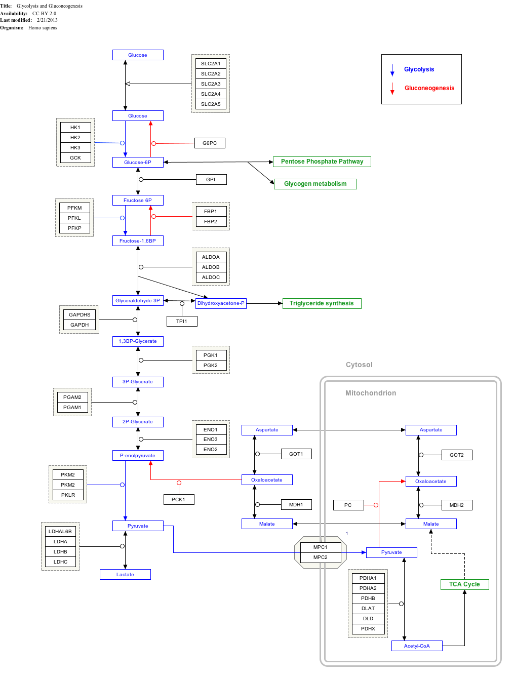
Click on genes, proteins and metabolites below to link to respective articles. [§ 1]
Glycolysis is the metabolic pathway that converts glucose into pyruvate and, in most organisms, occurs in the liquid part of cells. The free energy released in this process is used to form the high-energy molecules adenosine triphosphate (ATP) and reduced nicotinamide adenine dinucleotide (NADH). Glycolysis is a sequence of ten reactions catalyzed by enzymes.
In biochemistry, a kinase is an enzyme that catalyzes the transfer of phosphate groups from high-energy, phosphate-donating molecules to specific substrates. This process is known as phosphorylation, where the high-energy ATP molecule donates a phosphate group to the substrate molecule. As a result, kinase produces a phosphorylated substrate and ADP. Conversely, it is referred to as dephosphorylation when the phosphorylated substrate donates a phosphate group and ADP gains a phosphate group. These two processes, phosphorylation and dephosphorylation, occur four times during glycolysis.
A hexokinase is an enzyme that irreversibly phosphorylates hexoses, forming hexose phosphate. In most organisms, glucose is the most important substrate for hexokinases, and glucose-6-phosphate is the most important product. Hexokinase possesses the ability to transfer an inorganic phosphate group from ATP to a substrate.
Tumor hypoxia is the situation where tumor cells have been deprived of oxygen. As a tumor grows, it rapidly outgrows its blood supply, leaving portions of the tumor with regions where the oxygen concentration is significantly lower than in healthy tissues. Hypoxic microenvironments in solid tumors are a result of available oxygen being consumed within 70 to 150 μm of tumor vasculature by rapidly proliferating tumor cells thus limiting the amount of oxygen available to diffuse further into the tumor tissue. In order to support continuous growth and proliferation in challenging hypoxic environments, cancer cells are found to alter their metabolism. Furthermore, hypoxia is known to change cell behavior and is associated with extracellular matrix remodeling and increased migratory and metastatic behavior.
Pyruvate kinase is the enzyme involved in the last step of glycolysis. It catalyzes the transfer of a phosphate group from phosphoenolpyruvate (PEP) to adenosine diphosphate (ADP), yielding one molecule of pyruvate and one molecule of ATP. Pyruvate kinase was inappropriately named before it was recognized that it did not directly catalyze phosphorylation of pyruvate, which does not occur under physiological conditions. Pyruvate kinase is present in four distinct, tissue-specific isozymes in animals, each consisting of particular kinetic properties necessary to accommodate the variations in metabolic requirements of diverse tissues.
Glucokinase is an enzyme that facilitates phosphorylation of glucose to glucose-6-phosphate. Glucokinase occurs in cells in the liver and pancreas of humans and most other vertebrates. In each of these organs it plays an important role in the regulation of carbohydrate metabolism by acting as a glucose sensor, triggering shifts in metabolism or cell function in response to rising or falling levels of glucose, such as occur after a meal or when fasting. Mutations of the gene for this enzyme can cause unusual forms of diabetes or hypoglycemia.
In oncology, the Warburg effect is the observation that most cancer use aerobic glycolysis for energy generation rather than the mechanisms used by non-cancerous cells. This observation was first published by Otto Heinrich Warburg, who was awarded the 1931 Nobel Prize in Physiology for his "discovery of the nature and mode of action of the respiratory enzyme". The existence of the Warburg effect has fuelled popular misconceptions that cancer can be treated by dietary reductions in sugar and carbohydrate.
Phosphofructokinase-2 (6-phosphofructo-2-kinase, PFK-2) or fructose bisphosphatase-2 (FBPase-2), is an enzyme indirectly responsible for regulating the rates of glycolysis and gluconeogenesis in cells. It catalyzes formation and degradation of a significant allosteric regulator, fructose-2,6-bisphosphate (Fru-2,6-P2) from substrate fructose-6-phosphate. Fru-2,6-P2 contributes to the rate-determining step of glycolysis as it activates enzyme phosphofructokinase 1 in the glycolysis pathway, and inhibits fructose-1,6-bisphosphatase 1 in gluconeogenesis. Since Fru-2,6-P2 differentially regulates glycolysis and gluconeogenesis, it can act as a key signal to switch between the opposing pathways. Because PFK-2 produces Fru-2,6-P2 in response to hormonal signaling, metabolism can be more sensitively and efficiently controlled to align with the organism's glycolytic needs. This enzyme participates in fructose and mannose metabolism. The enzyme is important in the regulation of hepatic carbohydrate metabolism and is found in greatest quantities in the liver, kidney and heart. In mammals, several genes often encode different isoforms, each of which differs in its tissue distribution and enzymatic activity. The family described here bears a resemblance to the ATP-driven phospho-fructokinases; however, they share little sequence similarity, although a few residues seem key to their interaction with fructose 6-phosphate.
Glyceraldehyde 3-phosphate dehydrogenase is an enzyme of about 37kDa that catalyzes the sixth step of glycolysis and thus serves to break down glucose for energy and carbon molecules. In addition to this long established metabolic function, GAPDH has recently been implicated in several non-metabolic processes, including transcription activation, initiation of apoptosis, ER-to-Golgi vesicle shuttling, and fast axonal, or axoplasmic transport. In sperm, a testis-specific isoenzyme GAPDHS is expressed.
The study of the tumor metabolism, also known as tumor metabolome describes the different characteristic metabolic changes in tumor cells. The characteristic attributes of the tumor metabolome are high glycolytic enzyme activities, the expression of the pyruvate kinase isoenzyme type M2, increased channeling of glucose carbons into synthetic processes, such as nucleic acid, amino acid and phospholipid synthesis, a high rate of pyrimidine and purine de novo synthesis, a low ratio of Adenosine triphosphate and Guanosine triphosphate to Cytidine triphosphate and Uridine triphosphate, low Adenosine monophosphate levels, high glutaminolytic capacities, release of immunosuppressive substances and dependency on methionine.
Voltage-dependent anion channels, or mitochondrial porins, are a class of porin ion channel located on the outer mitochondrial membrane. There is debate as to whether or not this channel is expressed in the cell surface membrane.
Hexokinase I, also known as hexokinase A and HK1, is an enzyme that in humans is encoded by the HK1 gene on chromosome 10. Hexokinases phosphorylate glucose to produce glucose-6-phosphate (G6P), the first step in most glucose metabolism pathways. This gene encodes a ubiquitous form of hexokinase which localizes to the outer membrane of mitochondria. Mutations in this gene have been associated with hemolytic anemia due to hexokinase deficiency. Alternative splicing of this gene results in five transcript variants which encode different isoforms, some of which are tissue-specific. Each isoform has a distinct N-terminus; the remainder of the protein is identical among all the isoforms. A sixth transcript variant has been described, but due to the presence of several stop codons, it is not thought to encode a protein. [provided by RefSeq, Apr 2009]
Pyruvate dehydrogenase lipoamide kinase isozyme 4, mitochondrial (PDK4) is an enzyme that in humans is encoded by the PDK4 gene. It codes for an isozyme of pyruvate dehydrogenase kinase.
PFKFB3 is a gene that encodes the 6-phosphofructo-2-kinase/fructose-2,6-biphosphatase 3 enzyme in humans. It is one of 4 tissue-specific PFKFB isoenzymes identified currently (PFKFB1-4).
Aspartate aminotransferase, mitochondrial is an enzyme that in humans is encoded by the GOT2 gene. Glutamic-oxaloacetic transaminase is a pyridoxal phosphate-dependent enzyme which exists in cytoplasmic and inner-membrane mitochondrial forms, GOT1 and GOT2, respectively. GOT plays a role in amino acid metabolism and the urea and Kreb's cycle. Also, GOT2 is a major participant in the malate-aspartate shuttle, which is a passage from the cytosol to the mitochondria. The two enzymes are homodimeric and show close homology. GOT2 has been seen to have a role in cell proliferation, especially in terms of tumor growth.
Pyruvate kinase isozymes M1/M2 (PKM1/M2), also known as pyruvate kinase muscle isozyme (PKM), pyruvate kinase type K, cytosolic thyroid hormone-binding protein (CTHBP), thyroid hormone-binding protein 1 (THBP1), or opa-interacting protein 3 (OIP3), is an enzyme that in humans is encoded by the PKM2 gene.
Voltage-dependent anion-selective channel protein 2 is a protein that in humans is encoded by the VDAC2 gene on chromosome 10. This protein is a voltage-dependent anion channel and shares high structural homology with the other VDAC isoforms. VDACs are generally involved in the regulation of cell metabolism, mitochondrial apoptosis, and spermatogenesis. Additionally, VDAC2 participates in cardiac contractions and pulmonary circulation, which implicate it in cardiopulmonary diseases. VDAC2 also mediates immune response to infectious bursal disease (IBD).
Voltage-dependent anion-selective channel protein 3 (VDAC3) is a protein that in humans is encoded by the VDAC3 gene on chromosome 8. The protein encoded by this gene is a voltage-dependent anion channel and shares high structural homology with the other VDAC isoforms. Nonetheless, VDAC3 demonstrates limited pore-forming ability and, instead, interacts with other proteins to perform its biological functions, including sperm flagella assembly and centriole assembly. Mutations in VDAC3 have been linked to male infertility, as well as Parkinson's disease.
Hexokinase III, also known as hexokinase C, is an enzyme which in humans is encoded by the Hk3 gene on chromosome 5. Hexokinases phosphorylate glucose to produce glucose-6-phosphate, the first step in most glucose metabolism pathways. Similar to hexokinases I and II, this allosteric enzyme is inhibited by its product glucose-6-phosphate. [provided by RefSeq, Apr 2009]
The TP53-inducible glycolysis and apoptosis regulator (TIGAR) also known as fructose-2,6-bisphosphatase TIGAR is an enzyme that in humans is encoded by the C12orf5 gene.
This article incorporates text from the United States National Library of Medicine, which is in the public domain.