Lymphoma is a group of blood cancers that develop from lymphocytes. The name often refers to just the cancerous versions rather than all such tumors. Signs and symptoms may include enlarged lymph nodes, fever, drenching sweats, unintended weight loss, itching, and constantly feeling tired. The enlarged lymph nodes are usually painless. The sweats are most common at night.
Cancer is a group of diseases involving abnormal cell growth with the potential to invade or spread to other parts of the body. These contrast with benign tumors, which do not spread. Possible signs and symptoms include a lump, abnormal bleeding, prolonged cough, unexplained weight loss, and a change in bowel movements. While these symptoms may indicate cancer, they can also have other causes. Over 100 types of cancers affect humans.
White blood cells (WBCs), also called leukocytes or leucocytes, are the cells of the immune system that are involved in protecting the body against both infectious disease and foreign invaders. All white blood cells are produced and derived from multipotent cells in the bone marrow known as hematopoietic stem cells. Leukocytes are found throughout the body, including the blood and lymphatic system.
The Epstein–Barr virus, formally called Human gammaherpesvirus 4, is one of eight known human herpesvirus types in the herpes family, and is one of the most common viruses in humans.
Human immunodeficiency virus infection and acquired immune deficiency syndrome (HIV/AIDS) is a spectrum of conditions caused by infection with the human immunodeficiency virus (HIV). Following initial infection a person may not notice any symptoms, or may experience a brief period of influenza-like illness. Typically, this is followed by a prolonged period with no symptoms. As the infection progresses, it interferes more with the immune system, increasing the risk of developing common infections such as tuberculosis, as well as other opportunistic infections, and tumors that rarely affect people who have uncompromised immune systems. These late symptoms of infection are referred to as acquired immunodeficiency syndrome (AIDS). This stage is often also associated with unintended weight loss.
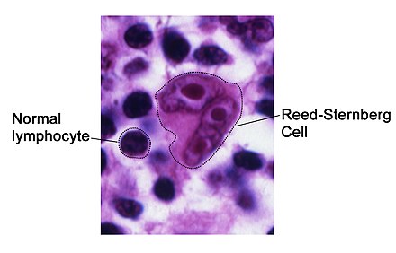
Reed–Sternberg cells are distinctive, giant cells found with light microscopy in biopsies from individuals with Hodgkin lymphoma. They are usually derived from B lymphocytes, classically considered crippled germinal center B cells, meaning they have not undergone hypermutation to express their antibody. Seen against a sea of B cells, they give the tissue a moth-eaten appearance.
Hodgkin lymphoma may be treated with chemotherapy, radiation therapy, and stem cell transplant.[4] The choice of treatment often depends on how advanced the cancer has become and whether or not it has favorable features.[4] In early disease, a cure is often possible.[10] The percentage of people who survive five years in the United States is 86%.[5] For those under the age of 20, rates of survival are 97%.[11] Radiation and some chemotherapy drugs, however, increase the risk of other cancers, heart disease, or lung disease over the subsequent decades.[10]
Chemotherapy is a type of cancer treatment that uses one or more anti-cancer drugs as part of a standardized chemotherapy regimen. Chemotherapy may be given with a curative intent, or it may aim to prolong life or to reduce symptoms. Chemotherapy is one of the major categories of the medical discipline specifically devoted to pharmacotherapy for cancer, which is called medical oncology.
Radiation therapy or radiotherapy, often abbreviated RT, RTx, or XRT, is therapy using ionizing radiation, generally as part of cancer treatment to control or kill malignant cells and normally delivered by a linear accelerator. Radiation therapy may be curative in a number of types of cancer if they are localized to one area of the body. It may also be used as part of adjuvant therapy, to prevent tumor recurrence after surgery to remove a primary malignant tumor. Radiation therapy is synergistic with chemotherapy, and has been used before, during, and after chemotherapy in susceptible cancers. The subspecialty of oncology concerned with radiotherapy is called radiation oncology.
Hematopoietic stem cell transplantation (HSCT) is the transplantation of multipotent hematopoietic stem cells, usually derived from bone marrow, peripheral blood, or umbilical cord blood. It may be autologous, allogeneic or syngeneic.
In 2015, about 574,000 people had Hodgkin's lymphoma, and 23,900 died.[6][7] In the United States, 0.2% of people are affected at some point in their life.[5] The most common age of diagnosis is between 20 and 40 years old.[5] It was named after the English physician Thomas Hodgkin, who first described the condition in 1832.[10][12]
Thomas Hodgkin was a British physician, considered one of the most prominent pathologists of his time and a pioneer in preventive medicine. He is now best known for the first account of Hodgkin's disease, a form of lymphoma and blood disease, in 1832. Hodgkin's work marked the beginning of times when a pathologist was actively involved in the clinical process. He was a contemporary of Thomas Addison and Richard Bright at Guy's Hospital.
Signs and symptoms
People with Hodgkin's lymphoma may present with the following symptoms:
Lymph nodes: the most common symptom of Hodgkin's is the painless enlargement of one or more lymph nodes, or lymphadenopathy. The nodes may also feel rubbery and swollen when examined. The nodes of the neck and shoulders (cervical and supraclavicular) are most frequently involved (80–90% of the time, on average). The lymph nodes of the chest are often affected, and these may be noticed on a chest radiograph.
Splenomegaly: enlargement of the spleen occurs in about 30% of people with Hodgkin's lymphoma. The enlargement, however, is seldom massive, and the size of the spleen may fluctuate during the course of treatment.
Hepatomegaly: enlargement of the liver, due to liver involvement, is present in about 5% of cases.
Hepatosplenomegaly: the enlargement of both the liver and spleen caused by the same disease.
Pain following alcohol consumption: classically, involved nodes are painful after alcohol consumption, though this phenomenon is very uncommon,[13] occurring in only two to three percent of people with Hodgkin's lymphoma,[14] thus having a low sensitivity. On the other hand, its positive predictive value is high enough for it to be regarded as a pathognomonic sign of Hodgkin’s lymphoma.[14] The pain typically has an onset within minutes after ingesting alcohol, and is usually felt as coming from the vicinity where there is an involved lymph node.[14] The pain has been described as either sharp and stabbing or dull and aching.[14]
Back pain: nonspecific back pain (pain that cannot be localised or its cause determined by examination or scanning techniques) has been reported in some cases of Hodgkin's lymphoma. The lower back is most often affected.[citation needed]
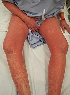
Red-coloured patches on the skin, easy bleeding and petechiae due to low platelet count (as a result of bone marrow infiltration, increased trapping in the spleen etc.—i.e. decreased production, increased removal)
Systemic symptoms: about one-third of people with Hodgkin's disease may also present with systemic symptoms, including low-grade fever; night sweats; unexplained weight loss of at least 10% of the person's total body mass in six months or less, itchy skin (pruritus) due to increased levels of eosinophils in the bloodstream; or fatigue (lassitude). Systemic symptoms such as fever, night sweats, and weight loss are known as B symptoms; thus, presence of fever, weight loss, and night sweats indicate that the person's stage is, for example, 2B instead of 2A.[15]
Cyclical fever: people may also present with a cyclical high-grade fever known as the Pel–Ebstein fever,[16] or more simply "P-E fever". However, there is debate as to whether the P-E fever truly exists.[17]
Lymphadenopathy or adenopathy is disease of the lymph nodes, in which they are abnormal in size or consistency. Lymphadenopathy of an inflammatory type is lymphadenitis, producing swollen or enlarged lymph nodes. In clinical practice, the distinction between lymphadenopathy and lymphadenitis is rarely made and the words are usually treated as synonymous. Inflammation of the lymphatic vessels is known as lymphangitis. Infectious lymphadenitis affecting lymph nodes in the neck is often called scrofula.
Cervical lymph nodes are lymph nodes found in the neck. Of the 800 lymph nodes in the human body, 300 are in the neck. Cervical lymph nodes are subject to a number of different pathological conditions including tumours, infection and inflammation.
Supraclavicular lymph nodes are lymph nodes found above to the clavicle, that can be felt in the supraclavicular fossa. The supraclavicular lymph nodes on the left side are called Virchow's nodes.
Diagnosis
Hodgkin's lymphoma must be distinguished from non-cancerous causes of lymph node swelling (such as various infections) and from other types of cancer. Definitive diagnosis is by lymph node biopsy (usually excisional biopsy with microscopic examination). Blood tests are also performed to assess function of major organs and to assess safety for chemotherapy. Positron emission tomography (PET) is used to detect small deposits that do not show on CT scanning. PET scans are also useful in functional imaging (by using a radiolabeled glucose to image tissues of high metabolism). In some cases a Gallium scan may be used instead of a PET scan.
A biopsy is a medical test commonly performed by a surgeon, interventional radiologist, or an interventional cardiologist involving extraction of sample cells or tissues for examination to determine the presence or extent of a disease. The tissue is generally examined under a microscope by a pathologist, and can also be analyzed chemically. When an entire lump or suspicious area is removed, the procedure is called an excisional biopsy. An incisional biopsy or core biopsy samples a portion of the abnormal tissue without attempting to remove the entire lesion or tumor. When a sample of tissue or fluid is removed with a needle in such a way that cells are removed without preserving the histological architecture of the tissue cells, the procedure is called a needle aspiration biopsy. Biopsies are most commonly performed for insight into possible cancerous and inflammatory conditions.
A blood test is a laboratory analysis performed on a blood sample that is usually extracted from a vein in the arm using a hypodermic needle, or via fingerprick. Multiple tests for specific blood components, such as a glucose test or a cholesterol test, are often grouped together into one test panel called a blood panel or blood work. Blood tests are often used in health care to determine physiological and biochemical states, such as disease, mineral content, pharmaceutical drug effectiveness, and organ function. Typical clinical blood panels include a basic metabolic panel or a complete blood count. Blood tests are also used in drug tests to detect drug abuse. In some of the United States, a blood test is required before marriage.
Positron-emission tomography (PET) is a nuclear medicine functional imaging technique that is used to observe metabolic processes in the body as an aid to the diagnosis of disease. The system detects pairs of gamma rays emitted indirectly by a positron-emitting radioligand, most commonly fluorine-18, which is introduced into the body on a biologically active molecule called a radioactive tracer. Different ligands are used for different imaging purposes, depending on what the radiologist/researcher wants to detect. Three-dimensional images of tracer concentration within the body are then constructed by computer analysis. In modern PET computed tomography scanners, three-dimensional imaging is often accomplished with the aid of a computed tomography X-ray scan performed on the patient during the same session, in the same machine.
Is the most common subtype and is composed of large tumornodules showing scattered lacunar classical RS cells set in a background of reactive lymphocytes, eosinophils and plasma cells with varying degrees of collagen fibrosis/sclerosis.
Is a common subtype and is composed of numerous classic RS cells admixed with numerous inflammatory cells including lymphocytes, histiocytes, eosinophils, and plasma cells without sclerosis. This type is most often associated with EBV infection and may be confused with the early, so-called 'cellular' phase of nodular sclerosing CHL.
Is a rare subtype, show many features which may cause diagnostic confusion with nodular lymphocyte predominant B-cell Non-Hodgkin's Lymphoma (B-NHL). This form also has the most favorable prognosis.
Is a rare subtype, composed of large numbers of often pleomorphic RS cells with only few reactive lymphocytes which may easily be confused with diffuse large cell lymphoma. Many cases previously classified within this category would now be reclassified under anaplastic large cell lymphoma.[19]
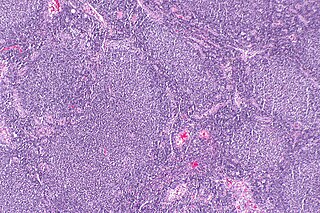
Lymph node biopsy showing Hodgkin's lymphoma, mixed-cellularity typeCT image of a 46-year-old person with Hodgkin's lymphoma, image at neck height. On the left side of the person's neck enlarged lymph nodes are visible (marked in red).
For the other forms, although the traditional B-cell markers (such as CD20) are not expressed on all cells,[19] Reed–Sternberg cells are usually of B cell origin.[20][21] Although Hodgkin's is now frequently grouped with other B-cell malignancies, some T-cell markers (such as CD2 and CD4) are occasionally expressed.[22] However, this may be an artifact of the ambiguity inherent in the diagnosis.
Hodgkin cells produce interleukin-21 (IL-21), which was once thought to be exclusive to T-cells. This feature may explain the behavior of classical Hodgkin's lymphoma, including clusters of other immune cells gathered around HL cells (infiltrate) in cultures.[23]
Staging
The staging is the same for both Hodgkin's and non-Hodgkin's lymphomas.
After Hodgkin lymphoma is diagnosed, a person will be staged: that is, they will undergo a series of tests and procedures that will determine what areas of the body are affected. These procedures may include documentation of their histology, a physical examination, blood tests, chest X-ray radiographs, computed tomography (CT)/Positron emission tomography (PET)/magnetic resonance imaging (MRI) scans of the chest, abdomen and pelvis, and usually a bone marrow biopsy. Positron emission tomography (PET) scan is now used instead of the gallium scan for staging. On the PET scan, sites involved with lymphoma light up very brightly enabling accurate and reproducible imaging.[24] In the past, a lymphangiogram or surgical laparotomy (which involves opening the abdominal cavity and visually inspecting for tumors) were performed. Lymphangiograms or laparotomies are very rarely performed, having been supplanted by improvements in imaging with the CT scan and PET scan.
On the basis of this staging, the person will be classified according to a staging classification (the Ann Arbor staging classification scheme is a common one):
Stage I is involvement of a single lymph node region (I) (mostly the cervical region) or single extralymphatic site (Ie);
Stage II is involvement of two or more lymph node regions on the same side of the diaphragm (II) or of one lymph node region and a contiguous extralymphatic site (IIe);
Stage III is involvement of lymph node regions on both sides of the diaphragm, which may include the spleen (IIIs) or limited contiguous extralymphatic organ or site (IIIe, IIIes);
Stage IV is disseminated involvement of one or more extralymphatic organs.
The absence of systemic symptoms is signified by adding "A" to the stage; the presence of systemic symptoms is signified by adding "B" to the stage. For localised extranodal extension from mass of nodes that does not advance the stage, subscript "E" is added. Splenic involvement is signified by adding "S" to the stage. The inclusion of "bulky disease" is signified by "X".
Stage 1 Hodgkin's lymphoma
Stage 2 Hodgkin's lymphoma
Stage 3 Hodgkin's lymphoma
Stage 4 Hodgkin's lymphoma
Pathology
Macroscopy
Affected lymph nodes (most often, laterocervical lymph nodes) are enlarged, but their shape is preserved because the capsule is not invaded. Usually, the cut surface is white-grey and uniform; in some histological subtypes (e.g. nodular sclerosis) a nodular aspect may appear.
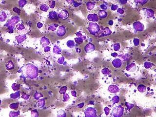
Microscopic examination of the lymph node biopsy reveals complete or partial effacement of the lymph node architecture by scattered large malignant cells known as Reed-Sternberg cells (RSC) (typical and variants) admixed within a reactive cell infiltrate composed of variable proportions of lymphocytes, histiocytes, eosinophils, and plasma cells. The Reed–Sternberg cells are identified as large often bi-nucleated cells with prominent nucleoli and an unusual CD45-, CD30+, CD15+/- immunophenotype. In approximately 50% of cases, the Reed–Sternberg cells are infected by the Epstein–Barr virus.[25]
Characteristics of classic Reed–Sternberg cells include large size (20–50 micrometres), abundant, amphophilic, finely granular/homogeneous cytoplasm; two mirror-image nuclei (owl eyes) each with an eosinophilic nucleolus and a thick nuclear membrane (chromatin is distributed close to the nuclear membrane).
Variants:
Hodgkin cell (atypical mononuclear RSC) is a variant of RS cell, which has the same characteristics, but is mononucleated.
Lacunar RSC is large, with a single hyperlobulated nucleus, multiple, small nucleoli and eosinophilic cytoplasm which is retracted around the nucleus, creating an empty space ("lacunae").
Pleomorphic RSC has multiple irregular nuclei.
"Popcorn" RSC (lympho-histiocytic variant) is a small cell, with a very lobulated nucleus, small nucleoli.
"Mummy" RSC has a compact nucleus with no nucleolus and basophilic cytoplasm.
Hodgkin's lymphoma can be sub-classified by histological type. The cell histology in Hodgkin's lymphoma is not as important as it is in non-Hodgkin's lymphoma: the treatment and prognosis in classic Hodgkin's lymphoma usually depends on the stage of disease rather than the histotype.
Management
People with early stage disease (IA or IIA) are effectively treated with radiation therapy or chemotherapy. The choice of treatment depends on the age, sex, bulk and the histological subtype of the disease.[citation needed] Adding localised radiation therapy after the chemotherapy regimen may provide a longer progression-free survival compared with chemotherapy treatment alone.[26] People with later disease (III, IVA, or IVB) are treated with combination chemotherapy alone. People of any stage with a large mass in the chest are usually treated with combined chemotherapy and radiation therapy.[citation needed]
The original treatment for Hodgkin's was MOPP. The abbreviation stands for the four drugs Mustargen (also known as chlormethine), Oncovin (also known as vincristine), Prednisone and Procarbazine (also known as Matulane). The treatment is usually administered in four week cycles, often for six cycles. MSD and VCR are administered intravenously, while procarbazine and prednisone are pills taken orally. MOPP was the first combination chemotherapy brought in that achieved a high success rate. It was developed at the National Cancer Institute in the 1960s by a team that included Vincent DeVita, Jr..
Although no longer the most effective combination, MOPP is still used after relapse or where the person has certain allergies or lung or heart problems which prevents the use of another regimen.
Currently, the ABVDchemotherapy regimen is the standard treatment of Hodgkin's disease in the US. The abbreviation stands for the four drugs Adriamycin, bleomycin, vinblastine, and dacarbazine. Developed in Italy in the 1970s, the ABVD treatment typically takes between six and eight months, although longer treatments may be required.
The newer Stanford V regimen is typically only half as long as the ABVD but involves a more intensive chemotherapy schedule and incorporates radiation therapy. In a randomised controlled study in Italy, Stanford V was inferior to ABVD;[27] however, this study has been heavily criticized due to its incorrect administration of radiotherapy, diverging from the original Stanford V protocol.[28]
BEACOPP is a form of treatment for stages > II mainly used in Europe. The cure rate with the BEACOPP esc. regimen is approximately 10–15% higher than with standard ABVD in advanced stages. This was shown in a paper in The New England Journal of Medicine (Diehl et al.), but US physicians still favor ABVD, maybe because some physicians think that BEACOPP induces more secondary leukemia. However, this seems negligible compared to the higher cure rates. BEACOPP is more expensive because of the requirement for concurrent treatment with GCSF to increase production of white blood cells. Currently, the German Hodgkin Study Group tests 8 cycles (8x) BEACOPP esc vs. 6x BEACOPP esc vs. 8x BEACOPP-14 baseline (HD15-trial).[29]
The common non-Hodgkin's treatment, rituximab (which is a monoclonal antibody against CD20) is not routinely used to treat Hodgkin's lymphoma due to the lack of CD20 surface antigens in most cases. The use of rituximab in Hodgkin's lymphoma, including the lymphocyte predominant subtype has been recently reviewed.[30]
Although increased age is an adverse risk factor for Hodgkin's lymphoma, in general elderly people without major comorbidities are sufficiently fit to tolerate standard therapy, and have a treatment outcome comparable to that of younger people. However, the disease is a different entity in older people and different considerations enter into treatment decisions.[31]
For Hodgkin's lymphomas, radiation oncologists typically use external beam radiation therapy (sometimes shortened to EBRT or XRT). Radiation oncologists deliver external beam radiation therapy to the lymphoma from a machine called a linear accelerator which produces high energy X-rays and electrons. People usually describe treatments as painless and similar to getting an X-ray. Treatments last less than 30 minutes each.
For lymphomas, there are a few different ways radiation oncologists target the cancer cells. Involved site radiation is when the radiation oncologists give radiation only to those parts of the person's body known to have the cancer.[32] Very often, this is combined with chemotherapy. Radiation therapy directed above the diaphragm to the neck, chest or underarms is called mantle field radiation. Radiation to below the diaphragm to the abdomen, spleen or pelvis is called inverted-Y field radiation. Total nodal irradiation is when the therapist gives radiation to all the lymph nodes in the body to destroy cells that may have spread.[33]
Adverse effects
The high cure rates and long survival of many people with Hodgkin's lymphoma has led to a high concern with late adverse effects of treatment, including cardiovascular disease and second malignancies such as acute leukemias, lymphomas, and solid tumors within the radiation therapy field. Most people with early-stage disease are now treated with abbreviated chemotherapy and involved site radiation therapy rather than with radiation therapy alone. Clinical research strategies are exploring reduction of the duration of chemotherapy and dose and volume of radiation therapy in an attempt to reduce late morbidity and mortality of treatment while maintaining high cure rates. Hospitals are also treating those who respond quickly to chemotherapy with no radiation.
In childhood cases of Hodgkin's lymphoma, long-term endocrine adverse effects are a major concern, mainly gonadal dysfunction and growth retardation. Gonadal dysfunction seems to be the most severe endocrine long-term effect, especially after treatment with alkylating agents or pelvic radiotherapy.[34]
Prognosis
Treatment of Hodgkin's disease has been improving over the past few decades. Recent trials that have made use of new types of chemotherapy have indicated higher survival rates than have previously been seen. In one recent European trial, the 5-year survival rate for those people with a favorable prognosis (FFP) was 98%, while that for people with worse outlooks was at least 85%.[35]
In 1998, an international effort[36] identified seven prognostic factors that accurately predict the success rate of conventional treatment in people with locally extensive or advanced stage Hodgkin's lymphoma. Freedom from progression (FFP) at 5 years was directly related to the number of factors present in a person. The 5-year FFP for people with zero factors is 84%. Each additional factor lowers the 5-year FFP rate by 7%, such that the 5-year FFP for a person with 5 or more factors is 42%.
The adverse prognostic factors identified in the international study are:
Other studies have reported the following to be the most important adverse prognostic factors: mixed-cellularity or lymphocyte-depleted histologies, male sex, large number of involved nodal sites, advanced stage, age of 40 years or more, the presence of B symptoms, high erythrocyte sedimentation rate, and bulky disease (widening of the mediastinum by more than one third, or the presence of a nodal mass measuring more than 10cm in any dimension.)
More recently, use of positron emission tomography (PET) early after commencing chemotherapy has demonstrated to have powerful prognostic ability.[37] This enables assessment of an individual's response to chemotherapy as the PET activity switches off rapidly in people who are responding. In this study,[37] after two cycles of ABVD chemotherapy, 83% of people were free of disease at 3 years if they had a negative PET versus only 28% in those with positive PET scans. This prognostic power exceeds conventional factors discussed above. Several trials are underway to see if PET-based risk adapted response can be used to improve a persons outcomes by changing chemotherapy early in people who are not responding.
Unlike some other lymphomas, whose number of new cases per year increases with age, Hodgkin's lymphoma has a bimodal curve for the number of cases; that is, it occurs most frequently in two separate age groups, the first being young adulthood (age 15–35) and the second being in those over 55 years old although these peaks may vary slightly with nationality.[39] Overall, it is more common in males, except for the nodular sclerosis variant, which is slightly more common in females. The annual number of cases of Hodgkin's lymphoma is 2.7 per 100,000 per persons per year, and the disease accounts for slightly less than 1% of all cancers worldwide.[40]
In 2010, globally it resulted in about 18,000 deaths down from 19,000 in 1990.[1]
The number of cases of Hodgkin's lymphoma is increased in people with HIV infection.[41] In contrast to many other lymphomas associated with HIV infection it occurs most commonly in people with higher CD4 T cell counts.
Canada
Hodgkin lymphoma accounts for 0.6% of all male cancer cases, and 0.4% of all female cancer cases in Canada. In 2017, approximately 990 Canadians will be diagnosed with Hodgkin lymphoma, and 140 will die of the disease.[42]
UK
Hodgkin lymphoma accounts for less than 1% of all cancer cases and deaths in the UK. Around 1,800 people were diagnosed with the disease in 2011, and around 330 people died in 2012.[43]
History
Photograph labeled of Hodgkin's disease, from a 1938 medical textbook
Hodgkin's lymphoma was first described in an 1832 report by Thomas Hodgkin, although Hodgkin noted that perhaps the earliest reference to the condition was provided by Marcello Malpighi in 1666.[44][12] While occupied as museum curator at Guy's Hospital, London, Hodgkin studied seven with painless lymph node enlargement. Of the seven cases, two were under the care of Richard Bright, one was of Thomas Addison, and one was of Robert Carswell.[44] Carswell's report of this seventh person was accompanied by numerous illustrations that aided early descriptions of the disease.[45]
Hodgkin's report on these seven people, entitled "On some morbid appearances of the absorbent glands and spleen", was presented to the Medical and Chirurgical Society of London in January 1832 and was subsequently published in the society's journal, Medical-Chirurgical Society Transactions.[44] Hodgkin's paper went largely unnoticed, however, even despite Bright highlighting it in an 1838 publication.[44] Indeed, Hodgkin himself did not view his contribution as particularly significant.[46]
In 1856, Samuel Wilks independently reported on a series of people with the same disease that Hodgkin had previously described.[46] Wilks, a successor to Hodgkin at Guy's Hospital, was unaware of Hodgkin's prior work on the subject. Bright made Wilks aware of Hodgkin's contribution and in 1865, Wilks published a second paper, entitled "Cases of enlargement of the lymphatic glands and spleen", in which he called the disease "Hodgkin's disease" in honor of his predecessor.[46]
Theodor Langhans and WS Greenfield first described the microscopic characteristics of Hodgkin's lymphoma in 1872 and 1878, respectively.[44] In 1898 and 1902, respectively, Carl Sternberg and Dorothy Reed independently described the cytogenetic features of the malignant cells of Hodgkin's lymphoma, now called Reed–Sternberg cells.[44]
Tissue specimens from Hodgkin's seven people remained at Guy's Hospital for a number of years. Nearly 100 years after Hodgkin's initial publication, histopathologic reexamination confirmed Hodgkin's lymphoma in only three of seven of these people.[46] The remaining cases included non-Hodgkin lymphoma, tuberculosis, and syphilis.[46]
Hodgkin's lymphoma was one of the first cancers which could be treated using radiation therapy and, later, it was one of the first to be treated by combination chemotherapy.
Notable cases
Paul Allen, co-founder of Microsoft was diagnosed with Hodgkin's lymphoma in 1982. He later died from non-Hodgkin lymphoma on October 15, 2018.[47]
Starchild Abraham Cherrix, a teenager whose refusal to undergo further conventional treatment after relapsing in 2006 resulted in a court battle and a change to Virginia laws about medical neglect.[51]
Michael C. Hall (born February 1, 1971), American actor, best known for his lead role as Dexter Morgan, in Showtime's crime series Dexter. In 2010, aged 38, Hall announced he was undergoing treatment for Hodgkin's Lymphoma; within two years, the disease was in full remission.[58]
Dinu Lipatti (1917–1950), Romanian classical pianist and composer. Diagnosed in 1947, received cortisone treatment in 1949; died from a burst abscess on his one lung.[63]
Brandon Tartikoff, American television executive, diagnosed around 1974, died in 1997.[68]
Ethan Zohn, American professional soccer player and a winner of the Survivor reality television series. Zohn was diagnosed twice (in 2009 and 2011).[69]
Related Research Articles
Non-Hodgkin lymphoma (NHL) is a group of blood cancers that includes all types of lymphoma except Hodgkin's lymphomas. Symptoms include enlarged lymph nodes, fever, night sweats, weight loss and tiredness. Other symptoms may include bone pain, chest pain or itchiness. Some forms are slow-growing, while others are fast-growing.
The lymphatic system is part of the vascular system and an important part of the immune system, composed of a large network of lymphatic vessels that carry a clear fluid called lymph directionally towards the heart. The lymphatic system was first described in the seventeenth century independently by Olaus Rudbeck and Thomas Bartholin. Unlike the circulatory system, the lymphatic system is not a closed system. The human circulatory system processes an average of 20 litres of blood per day through capillary filtration, which removes plasma while leaving the blood cells. Roughly 17 litres of the filtered plasma is reabsorbed directly into the blood vessels, while the remaining three litres remain in the interstitial fluid. One of the main functions of the lymph system is to provide an accessory return route to the blood for the surplus three litres.
Testicular cancer is cancer that develops in the testicles, a part of the male reproductive system. Symptoms may include a lump in the testicle, or swelling or pain in the scrotum. Treatment may result in infertility.
Chronic lymphocytic leukemia (CLL) is a type of cancer in which the bone marrow makes too many lymphocytes. Early on there is typically no symptoms. Later non-painful lymph node swelling, feeling tired, fever, night sweats, or weight loss for no clear reason may occur. Enlargement of the spleen and low red blood cells (anemia) may also occur. It typically worsens gradually over years.
This is a list of terms related to oncology. The original source for this list was the US National Cancer Institute's public domain Dictionary of Cancer Terms.
Ann Arbor staging is the staging system for lymphomas, both in Hodgkin's lymphoma and non-Hodgkin lymphoma. It was initially developed for Hodgkin's, but has some use in NHL. It has roughly the same function as TNM staging in solid tumors.
Follicular lymphoma (FL) is a cancer that involves certain types of white blood cells known as lymphocytes. The cancer originates from the uncontrolled division of specific types of B-cells known as centrocytes and centroblasts. These cells normally occupy the follicles in the germinal centers of lymphoid tissues such as lymph nodes. The cancerous cells in FL typically form follicular or follicle-like structures in the tissues they invade. These structures are usually the dominant histological feature of this cancer.
Sézary disease is a type of cutaneous lymphoma that was first described by Albert Sézary. The affected cells are T-cells that have pathological quantities of mucopolysaccharides. Sézary disease is sometimes considered a late stage of mycosis fungoides with lymphadenopathy.
Lymphoma (lymposarcoma) in animals is a type of cancer defined by a proliferation of malignant lymphocytes within solid organs such as the lymph nodes, bone marrow, liver and spleen. The disease also may occur in the eye, skin, and gastrointestinal tract.
The B-cell lymphomas are types of lymphoma affecting B cells. Lymphomas are "blood cancers" in the lymph nodes. They develop more frequently in older adults and in immunocompromised individuals.
Angioimmunoblastic T-cell lymphoma is a mature T-cell lymphoma of blood or lymph vessel immunoblasts characterized by a polymorphous lymph node infiltrate showing a marked increase in follicular dendritic cells (FDCs) and high endothelial venules (HEVs) and systemic involvement.
A micrometastasis is a small collection of cancer cells that has been shed from the original tumor and spread to another part of the body through the lymphovascular system. Micrometastases are too few, in size and quantity, to be picked up in a screening or diagnostic test, and therefore cannot be seen with imaging tests such as a mammogram, MRI, ultrasound, PET, or CT scans. These migrant cancer cells may group together to form a second tumor, which is so small that it can only be seen under a microscope. Approximately ninety percent of people who die from cancer die from metastatic disease, since these cells are so challenging to detect. It is important for these cancer cells to be treated immediately after discovery, in order to prevent the relapse and the likely death of the patient.
Mantle cell lymphoma (MCL) is a type of non-Hodgkin's lymphoma (NHL), comprising about 6% of NHL cases. There are only about 15,000 patients presently in the U.S with mantle cell lymphoma.
Marginal zone B-cell lymphomas, now commonly termed marginal zone lymphomas, are a heterogeneous group of lymphomas that derive from the malignant transformation of marginal zone B-cells. Marginal zone B cells are innate lymphoid cells that normally function by rapidly mounting IgM antibody immune responses to antigens such as those presented by infectious agents and damaged tissues. They are lymphocytes of the B-cell line that originate and mature in secondary lypmphoid follicles and then move to the marginal zones of mucosa-associated lymphoid tissue, the spleen, or lymph nodes. Mucosa-associated lymphoid tissue is a diffuse system of small concentrations of lymphoid tissue found in various submucosal membrane sites of the body such as the gastrointestinal tract, mouth, nasal cavity, pharynx, thyroid gland, breast, lung, salivary glands, eye, skin and the human spleen.
Nodular lymphocyte predominant Hodgkin lymphoma (NLPHL) is an indolent CD20(+) form of lymphoma.
Lutzner cells were discovered by Marvin A. Lutzner, Lucien-Marie Pautrier, and Albert Sézary. These cells are also referred to as Pautrier’s abscess, Sézary’s cell, or Sézary-Lutzner cells. They are a form of T-lymphocytes that has been mutated This atypical form of T-lymphocytes contains T-cell receptors on the surface and is found in both the dermis and epidermis layers of the skin. Since Lutzner cells are a mutated form of T-lymphocytes, they develop in bone marrow and are transported to the thymus is order to mature. The production and maturation stages occur before the cell has developed a mutation. Lutzner cells can form cutaneous T-cell lymphoma, which is a form of skin cancer.
Thyroid lymphoma is a rare cancer constituting 1% to 2% of all thyroid cancers and less than 2% of lymphomas. Thyroid lymphomas are classified as non–Hodgkin's B-cell lymphomas in a majority of cases, although Hodgkin's lymphoma of the thyroid has also been identified.
Carcinoma of the tonsil is a type of squamous cell carcinoma. The tonsil is the most common site of squamous cell carcinoma in the oropharynx. It comprises 23.1% of all malignancies of the oropharynx. The tumors frequently present at advanced stages, and around 70% of patients present with metastasis to the cervical lymph nodes. . The most reported complaints include sore throat, otalgia or dysphagia. Some patients may complain of feeling the presence of a lump in the throat. Approximately 20% patients present with a node in the neck as the only symptom.
Epstein–Barr virus-associated lymphoproliferative diseases are a group of disorders in which one or more types of lymphoid cells, i.e. B cells, T cells, NK cells, and histiocytic-dendritic cells, are infected with the Epstein–Barr virus (EBV). This causes the infected cells to divide excessively, and is associated with the development of various non-cancerous, pre-cancerous, and cancerous lymphoproliferative disorders (LPDs). These LPDs include the well-known disorder occurring during the initial infection with the EBV, infectious mononucleosis, and the large number of subsequent disorders that may occur thereafter. The virus is usually involved in the development and/or progression of these LPDs although in some cases it may be an "innocent" bystander, i.e. present in, but not contributing to, the disease.
References
1 2 Lozano R, Naghavi M, Foreman K, Lim S, Shibuya K, Aboyans V, et al. (Dec 15, 2012). "Global and regional mortality from 235 causes of death for 20 age groups in 1990 and 2010: a systematic analysis for the Global Burden of Disease Study 2010". Lancet. 380 (9859): 2095–128. doi:10.1016/S0140-6736(12)61728-0. OCLC23245604. PMID23245604.
↑ Ward, E; DeSantis, C; Robbins, A; Kohler, B; Jemal, A (2014). "Childhood and adolescent cancer statistics, 2014". CA: A Cancer Journal for Clinicians. 64 (2): 83–103. doi:10.3322/caac.21219. PMID24488779.
1 2 Hodgkin T (1832). "On some morbid experiences of the absorbent glands and spleen". Med Chir Trans. 17: 69–97.
↑ Bräuninger A, Schmitz R, Bechtel D, Renné C, Hansmann ML, Küppers R (April 2006). "Molecular biology of Hodgkin's and Reed–Sternberg cells in Hodgkin's lymphoma". Int. J. Cancer. 118 (8): 1853–61. doi:10.1002/ijc.21716. PMID16385563.
↑ Tzankov A, Bourgau C, Kaiser A, Zimpfer A, Maurer R, Pileri SA, Went P, Dirnhofer S (December 2005). "Rare expression of T-cell markers in classical Hodgkin's lymphoma". Mod. Pathol. 18 (12): 1542–9. doi:10.1038/modpathol.3800473. PMID16056244.
↑ Lamprecht B, Kreher S, Anagnostopoulos I, Jöhrens K, Monteleone G, Jundt F, Stein H, Janz M, Dörken B, Mathas S (2008). "Aberrant expression of the Th2 cytokine IL-21 in Hodgkin lymphoma cells regulates STAT3 signaling and attracts Treg cells via regulation of MIP-3a". Blood. 112 (Oct 2008): 3339–3347. doi:10.1182/blood-2008-01-134783. PMID18684866.
↑ Gobbi PG, Levis A, Chisesi T, Broglia C, Vitolo U, Stelitano C, Pavone V, Cavanna L, Santini G, Merli F, Liberati M, Baldini L, Deliliers GL, Angelucci E, Bordonaro R, Federico M (2005). "ABVD versus modified stanford V versus MOPPEBVCAD with optional and limited radiotherapy in intermediate- and advanced-stage Hodgkin's lymphoma: final results of a multicenter randomised trial by the Intergruppo Italiano Linfomi". J. Clin. Oncol. 23 (36): 9198–207. doi:10.1200/JCO.2005.02.907. PMID16172458.
↑ Edwards-Bennett SM, Jacks LM, Moskowitz CH, Wu EJ, Zhang Z, Noy A, Portlock CS, Straus DJ, Zelenetz AD, Yahalom J (March 2010). "Stanford V program for locally extensive and advanced Hodgkin lymphoma: the Memorial Sloan-Kettering Cancer Center experience". Annals of Oncology. 21 (3): 574–81. doi:10.1093/annonc/mdp337. PMID19759185.
↑ Saini KS, Azim HA, Cocorocchio E, Vanazzi A, Saini ML, Raviele PR, Pruneri G, Peccatori FA (2011). "Rituximab in Hodgkin lymphoma: Is the target always a hit?". Cancer Treat Rev. 37 (5): 385–90. doi:10.1016/j.ctrv.2010.11.005. PMID21183282.
↑ Specht, Lena; Yahalom, Joachim; Illidge, Tim; Berthelsen, Anne Kiil; Constine, Louis S.; Eich, Hans Theodor; Girinsky, Theodore; Hoppe, Richard T.; Mauch, Peter (2013-06-25). "Modern Radiation Therapy for Hodgkin Lymphoma: Field and Dose Guidelines From the International Lymphoma Radiation Oncology Group (ILROG)". International Journal of Radiation Oncology*Biology*Physics. 89 (4): 854–862. doi:10.1016/j.ijrobp.2013.05.005. ISSN0360-3016. PMID23790512.
↑ van Dorp W, van Beek RD, Laven JS, Pieters R, de Muinck Keizer-Schrama SM, van den Heuvel-Eibrink MM (2011). "Long-term endocrine side effects of childhood Hodgkin's lymphoma treatment: A review". Human Reproduction Update. 18 (1): 12–28. doi:10.1093/humupd/dmr038. PMID21896559.
↑ Fermé C, Eghbali H, Meerwaldt JH, Rieux C, Bosq J, Berger F, Girinsky T, Brice P, van't Veer MB, Walewski JA, Lederlin P, Tirelli U, Carde P, Van den Neste E, Gyan E, Monconduit M, Diviné M, Raemaekers JM, Salles G, Noordijk EM, Creemers GJ, Gabarre J, Hagenbeek A, Reman O, Blanc M, Thomas J, Vié B, Kluin-Nelemans JC, Viseu F, Baars JW, Poortmans P, Lugtenburg PJ, Carrie C, Jaubert J, Henry-Amar M (November 2007). "Chemotherapy plus involved-field radiation in early-stage Hodgkin's disease". The New England Journal of Medicine. 357 (19): 1916–27. doi:10.1056/NEJMoa064601. PMID17989384.
↑ Hasenclever D, Diehl V (November 19, 1998). "A Prognostic Score for Advanced Hodgkin's Disease". New England Journal of Medicine. 339 (21): 1506–14. doi:10.1056/NEJM199811193392104. PMID9819449.
1 2 Biggi A, Gallamini A, Chauvie S, Hutchings M, Kostakoglu L, Gregianin M, Meignan M, Malkowski B, Hofman MS, Barrington SF (2013). "International Validation Study for Interim PET in ABVD-Treated, Advanced-Stage Hodgkin Lymphoma: Interpretation Criteria and Concordance Rate Among Reviewers". Journal of Nuclear Medicine. 54 (5): 683–690. doi:10.2967/jnumed.112.110890. PMID23516309.
Charlotte DeCroes Jacobs. Henry Kaplan and the Story of Hodgkin's Disease (Stanford University Press; 2010) 456 pages; combines a biography of the American radiation oncologist (1918–84) with a history of the lymphatic cancer whose treatment he helped to transform.
This page is based on this Wikipedia article Text is available under the CC BY-SA 4.0 license; additional terms may apply. Images, videos and audio are available under their respective licenses.