Lassa mammarenavirus

Last updated
Lassa mammarenavirus
Lassa virus.JPG
TEM micrograph of Lassa mammarenavirus virions
Virus classification OOjs UI icon edit-ltr.svg
(unranked): Virus
Realm: Riboviria
Kingdom: Orthornavirae
Phylum: Negarnaviricota
Class: Ellioviricetes
Order: Bunyavirales
Family: Arenaviridae
Genus: Mammarenavirus
Species:
Lassa mammarenavirus
Synonyms
  • Lassa virus

Lassa mammarenavirus (LASV) is an arenavirus that causes Lassa hemorrhagic fever, [1] a type of viral hemorrhagic fever (VHF), in humans and other primates. Lassa mammarenavirus is an emerging virus and a select agent, requiring Biosafety Level 4-equivalent containment. It is endemic in West African countries, especially Sierra Leone, the Republic of Guinea, Nigeria, and Liberia, where the annual incidence of infection is between 300,000 and 500,000 cases, resulting in 5,000 deaths per year. [2]

Contents

As of 2012 discoveries within the Mano River region of west Africa have expanded the endemic zone between the two known Lassa endemic regions, indicating that LASV is more widely distributed throughout the tropical wooded savannah ecozone in west Africa. [3] There are no approved vaccines against Lassa fever for use in humans. [4]

Discovery

In 1969, missionary nurse Laura Wine fell ill with a mysterious disease she contracted from an obstetrical patient in Lassa, a village in Borno State, Nigeria. [5] [6] [1] She was then transported to Jos, where she died. Subsequently, two others became infected, one of whom was fifty-two-year-old nurse Lily Pinneo who had cared for Laura Wine. [7] Samples from Pinneo were sent to Yale University in New Haven where a new virus, that would later be known as Lassa mammarenavirus, was isolated for the first time by Jordi Casals-Ariet, Sonja Buckley, and others. [8] [9] [10] Casals contracted the fever, and nearly lost his life; one technician died from it. [8] By 1972, the multimammate rat, Mastomys natalensis, was found to be the main reservoir of the virus in West Africa, able to shed virus in its urine and feces without exhibiting visible symptoms. [11] [12]

Virology

Structure and genome

Lassa virus structure and genome.Figure by Fehling et al., 2012 Viruses-04-02973-g002.webp
Lassa virus structure and genome.Figure by Fehling et al., 2012

Lassa viruses [14] [15] are enveloped, single-stranded, bisegmented, ambisense RNA viruses. Their genome [16] is contained in two RNA segments that code for two proteins each, one in each sense, for a total of four viral proteins. [17] The large segment encodes a small zinc finger protein (Z) that regulates transcription and replication, [18] [19] and the RNA polymerase (L). The small segment encodes the nucleoprotein (NP) and the surface glycoprotein precursor (GP, also known as the viral spike), which is proteolytically cleaved into the envelope glycoproteins GP1 and GP2 that bind to the alpha-dystroglycan receptor and mediate host cell entry. [20]

Lassa fever causes hemorrhagic fever frequently shown by immunosuppression. Lassa mammarenavirus replicates very rapidly, and demonstrates temporal control in replication. [21] The first replication step is transcription of mRNA copies of the negative- or minus-sense genome. This ensures an adequate supply of viral proteins for subsequent steps of replication, as the NP and L proteins are translated from the mRNA. The positive- or plus-sense genome, then makes viral complementary RNA (vcRNA) copies of itself. The RNA copies are a template for producing negative-sense progeny, but mRNA is also synthesized from it. The mRNA synthesized from vcRNA are translated to make the GP and Z proteins. This temporal control allows the spike proteins to be produced last, and therefore, delay recognition by the host immune system.[ citation needed ]

Nucleotide studies of the genome have shown that Lassa has four lineages: three found in Nigeria and the fourth in Guinea, Liberia, and Sierra Leone. The Nigerian strains seem likely to have been ancestral to the others but additional work is required to confirm this. [22]

Receptors

Entry mechanisms of Old World and New World arenaviruses. Viruses-12-00784-g002.png
Entry mechanisms of Old World and New World arenaviruses.

Lassa mammarenavirus gains entry into the host cell by means of the cell-surface receptor the alpha-dystroglycan (alpha-DG), [20] a versatile receptor for proteins of the extracellular matrix. It shares this receptor with the prototypic Old World arenavirus lymphocytic choriomeningitis virus. Receptor recognition depends on a specific sugar modification of alpha-dystroglycan by a group of glycosyltransferases known as the LARGE proteins. Specific variants of the genes encoding these proteins appear to be under positive selection in West Africa where Lassa is endemic. [23] Alpha-dystroglycan is also used as a receptor by viruses of the New World clade C arenaviruses (Oliveros and Latino viruses). In contrast, the New World arenaviruses of clades A and B, which include the important viruses Machupo, Guanarito, Junin, and Sabia in addition to the non pathogenic Amapari virus, use the transferrin receptor 1. A small aliphatic amino acid at the GP1 glycoprotein amino acid position 260 is required for high-affinity binding to alpha-DG. In addition, GP1 amino acid position 259 also appears to be important, since all arenaviruses showing high-affinity alpha-DG binding possess a bulky aromatic amino acid (tyrosine or phenylalanine) at this position.[ citation needed ]

Unlike most enveloped viruses which use clathrin coated pits for cellular entry and bind to their receptors in a pH dependent fashion, Lassa and lymphocytic choriomeningitis virus instead use an endocytotic pathway independent of clathrin, caveolin, dynamin and actin. Once within the cell the viruses are rapidly delivered to endosomes via vesicular trafficking albeit one that is largely independent of the small GTPases Rab5 and Rab7. On contact with the endosome pH-dependent membrane fusion occurs mediated by the envelope glycoprotein, which at the lower pH of the endosome binds the lysosome protein LAMP1 which results in membrane fusion and escape from the endosome.[ citation needed ]

Life cycle

Lassa virus life cycle. Figure by Fehling et al., 2012 Viruses-04-02973-g003.webp
Lassa virus life cycle. Figure by Fehling et al., 2012

The life cycle of Lassa mammarenavirus is similar to the Old World arenaviruses. Lassa mammarenavirus enters the cell by the receptor-mediated endocytosis. Which endocytotic pathway is used is not known yet, but at least the cellular entry is sensitive to cholesterol depletion. It was reported that virus internalization is limited upon cholesterol depletion. The receptor used for cell entry is alpha-dystroglycan, a highly conserved and ubiquitously expressed cell surface receptor for extracellular matrix proteins. Dystroglycan, which is later cleaved into alpha-dystroglycan and beta-dystroglycan is originally expressed in most cells to mature tissues, and it provides molecular link between the ECM and the actin-based cytoskeleton. [24] After the virus enters the cell by alpha-dystroglycan mediated endocytosis, the low-pH environment triggers pH-dependent membrane fusion and releases RNP (viral ribonucleoprotein) complex into the cytoplasm. Viral RNA is unpacked, and replication and transcription initiate in the cytoplasm. [24] As replication starts, both S and L RNA genomes synthesize the antigenomic S and L RNAs, and from the antigenomic RNAs, genomic S and L RNA are synthesized. Both genomic and antigenomic RNAs are needed for transcription and translation. The S RNA encodes GP and NP (viral nucleocapsid protein) proteins, while L RNA encodes Z and L proteins. The L protein most likely represents the viral RNA-dependent RNA polymerase. [25] When the cell is infected by the virus, L polymerase is associated with the viral RNP and initiates the transcription of the genomic RNA. The 5’ and 3’ terminal 19 nt viral promoter regions of both RNA segments are necessary for recognition and binding of the viral polymerase. The primary transcription first transcribes mRNAs from the genomic S and L RNAs, which code NP and L proteins, respectively. Transcription terminates at the stem-loop (SL) structure within the intergenomic region. Arenaviruses use a cap snatching strategy to gain the cap structures from the cellular mRNAs, and it is mediated by the endonuclease activity of the L polymerase and the cap binding activity of NP. Antigenomic RNA transcribes viral genes GPC and Z, encoded in genomic orientation, from S and L segments respectively. The antigenomic RNA also serves as the template for the replication. [4] After translation of GPC, it is posttranslationally modified in the endoplasmic reticulum. GPC is cleaved into GP1 and GP2 at the later stage of the secretory pathway. It has been reported that the cellular protease SKI-1/S1P is responsible for this cleavage. The cleaved glycoproteins are incorporated into the virion envelope when the virus buds and release from the cell membrane. [25]

Pathogenesis

False color Scanning electron micrograph of Lassa virus (in orange) budding off an infected cell Lassa Virus (51719108882).jpg
False color Scanning electron micrograph of Lassa virus (in orange) budding off an infected cell

Lassa fever is caused by the Lassa mammarenavirus. The symptoms include flu-like illness characterized by fever, general weakness, cough, sore throat, headache, and gastrointestinal manifestations. Hemorrhagic manifestations include vascular permeability. [4]

Upon entry, the Lassa mammarenavirus infects almost every tissue in the human body. It starts with the mucosa, intestine, lungs and urinary system, and then progresses to the vascular system. [5]

The main targets of the virus are antigen-presenting cells, mainly dendritic cells and endothelial cells. [26] [27] [28] In 2012 it was reported how Lassa mammarenavirus nucleoprotein (NP) sabotages the host's innate immune system response. Generally, when a pathogen enters into a host, innate defense system recognizes the pathogen-associated molecular patterns (PAMP) and activates an immune response. One of the mechanisms detects double stranded RNA (dsRNA), which is only synthesized by negative-sense viruses. In the cytoplasm, dsRNA receptors, such as RIG-I (retinoic acid-inducible gene I) and MDA-5 (melanoma differentiation associated gene 5), detect dsRNAs and initiate signaling pathways that translocate IRF-3 (interferon regulatory factor 3) and other transcription factors to the nucleus. Translocated transcription factors activate expression of interferons 𝛂 and 𝛃, and these initiate adaptive immunity. NP encoded in Lassa mammarenavirus is essential in viral replication and transcription, but it also suppresses host innate IFN response by inhibiting translocation of IRF-3. NP of Lassa mammarenavirus is reported to have an exonuclease activity to only dsRNAs. [29] the NP dsRNA exonuclease activity counteracts IFN responses by digesting the PAMPs thus allowing the virus to evade host immune responses. [30]

See also

Related Research Articles

<i>Flaviviridae</i> Family of viruses

Flaviviridae is a family of enveloped positive-strand RNA viruses which mainly infect mammals and birds. They are primarily spread through arthropod vectors. The family gets its name from the yellow fever virus; flavus is Latin for "yellow", and yellow fever in turn was named because of its propensity to cause jaundice in victims. There are 89 species in the family divided among four genera. Diseases associated with the group include: hepatitis (hepaciviruses), hemorrhagic syndromes, fatal mucosal disease (pestiviruses), hemorrhagic fever, encephalitis, and the birth defect microcephaly (flaviviruses).

<i>Rhabdoviridae</i> Family of viruses in the order Mononegavirales

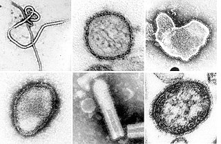
Rhabdoviridae is a family of negative-strand RNA viruses in the order Mononegavirales. Vertebrates, invertebrates, plants, fungi and protozoans serve as natural hosts. Diseases associated with member viruses include rabies encephalitis caused by the rabies virus, and flu-like symptoms in humans caused by vesiculoviruses. The name is derived from Ancient Greek rhabdos, meaning rod, referring to the shape of the viral particles. The family has 40 genera, most assigned to three subfamilies.

<span class="mw-page-title-main">Rabies virus</span> Species of virus

Rabies virus, scientific name Rabies lyssavirus, is a neurotropic virus that causes rabies in animals, including humans. Rabies transmission can occur through the saliva of animals and less commonly through contact with human saliva. Rabies lyssavirus, like many rhabdoviruses, has an extremely wide host range. In the wild it has been found infecting many mammalian species, while in the laboratory it has been found that birds can be infected, as well as cell cultures from mammals, birds, reptiles and insects. Rabies is reported in more than 150 countries and on all continents except Antarctica. The main burden of disease is reported in Asia and Africa, but some cases have been reported also in Europe in the past 10 years, especially in returning travellers.

<i>Bunyavirales</i> Order of RNA viruses

Bunyavirales is an order of segmented negative-strand RNA viruses with mainly tripartite genomes. Member viruses infect arthropods, plants, protozoans, and vertebrates. It is the only order in the class Ellioviricetes. The name Bunyavirales derives from Bunyamwera, where the original type species Bunyamwera orthobunyavirus was first discovered. Ellioviricetes is named in honor of late virologist Richard M. Elliott for his early work on bunyaviruses.

<span class="mw-page-title-main">Viral protein</span>

The term viral protein refers to both the products of the genome of a virus and any host proteins incorporated into the viral particle. Viral proteins are grouped according to their functions, and groups of viral proteins include structural proteins, nonstructural proteins, regulatory proteins, and accessory proteins. Viruses are non-living and do not have the means to reproduce on their own, instead depending on their host cell's machinery to do this. Thus, viruses do not code for most of the proteins required for their replication and the translation of their mRNA into viral proteins, but use proteins encoded by the host cell for this purpose.

Pseudoviridae is a family of viruses, which includes three genera.

<span class="mw-page-title-main">Arenavirus</span> Family of RNA viruses

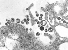
An arenavirus is a bi- or trisegmented ambisense RNA virus that is a member of the family Arenaviridae. These viruses infect rodents and occasionally humans. A class of novel, highly divergent arenaviruses, properly known as reptarenaviruses, have also been discovered which infect snakes to produce inclusion body disease. At least eight arenaviruses are known to cause human disease. The diseases derived from arenaviruses range in severity. Aseptic meningitis, a severe human disease that causes inflammation covering the brain and spinal cord, can arise from the lymphocytic choriomeningitis virus. Hemorrhagic fever syndromes, including Lassa fever, are derived from infections such as Guanarito virus, Junin virus, Lassa virus, Lujo virus, Machupo virus, Sabia virus, or Whitewater Arroyo virus. Because of the epidemiological association with rodents, some arenaviruses and bunyaviruses are designated as roboviruses.

Lymphocytic choriomeningitis (LCM) is a rodent-borne viral infectious disease that presents as aseptic meningitis, encephalitis or meningoencephalitis. Its causative agent is lymphocytic choriomeningitis mammarenavirus (LCMV), a member of the family Arenaviridae. The name was coined by Charles Armstrong in 1934.

Venezuelan hemorrhagic fever (VHF) is a zoonotic human illness first identified in 1989. The disease is most prevalent in several rural areas of central Venezuela and is caused by Guanarito mammarenavirus (GTOV) which belongs to the Arenaviridae family. The short-tailed cane mouse is the main host for GTOV which is spread mostly by inhalation of aerosolized droplets of saliva, respiratory secretions, urine, or blood from infected rodents. Person-to-person spread is possible, but uncommon.

The genome and proteins of HIV (human immunodeficiency virus) have been the subject of extensive research since the discovery of the virus in 1983. "In the search for the causative agent, it was initially believed that the virus was a form of the Human T-cell leukemia virus (HTLV), which was known at the time to affect the human immune system and cause certain leukemias. However, researchers at the Pasteur Institute in Paris isolated a previously unknown and genetically distinct retrovirus in patients with AIDS which was later named HIV." Each virion comprises a viral envelope and associated matrix enclosing a capsid, which itself encloses two copies of the single-stranded RNA genome and several enzymes. The discovery of the virus itself occurred two years following the report of the first major cases of AIDS-associated illnesses.

<i>Argentinian mammarenavirus</i> Species of virus

Argentinian mammarenavirus, better known as the Junin virus or Junín virus (JUNV), is an arenavirus in the Mammarenavirus genus that causes Argentine hemorrhagic fever (AHF). The virus took its original name from the city of Junín, around which the first cases of infection were reported, in 1958.

<i>Orthobunyavirus</i> Genus of viruses

Orthobunyavirus is a genus of the Peribunyaviridae family in the order Bunyavirales. There are currently ~170 viruses recognised in this genus. These have been assembled into 103 species and 20 serogroups.

Plectrovirus is a genus of viruses, in the family Plectroviridae. Bacteria in the phylum Mycoplasmatota serve as natural hosts, making these viruses bacteriophages. Acholeplasma virus L51 is the only species in the genus.

<i>Zaire ebolavirus</i> Species of virus affecting humans and animals

Zaire ebolavirus, more commonly known as Ebola virus, is one of six known species within the genus Ebolavirus. Four of the six known ebolaviruses, including EBOV, cause a severe and often fatal hemorrhagic fever in humans and other mammals, known as Ebola virus disease (EVD). Ebola virus has caused the majority of human deaths from EVD, and was the cause of the 2013–2016 epidemic in western Africa, which resulted in at least 28,646 suspected cases and 11,323 confirmed deaths.

Batai orthobunyavirus (BATV) is a RNA virus belonging to order Bunyavirales, genus Orthobunyavirus.

Aquaparamyxovirus is a genus of viruses in the family Paramyxoviridae, order Mononegavirales. The genus includes two species. Fish serve as the natural hosts for AsaPV, in which the virus may cause proliferative gill inflammation.

<span class="mw-page-title-main">Negative-strand RNA virus</span> Phylum of viruses

Negative-strand RNA viruses are a group of related viruses that have negative-sense, single-stranded genomes made of ribonucleic acid (RNA). They have genomes that act as complementary strands from which messenger RNA (mRNA) is synthesized by the viral enzyme RNA-dependent RNA polymerase (RdRp). During replication of the viral genome, RdRp synthesizes a positive-sense antigenome that it uses as a template to create genomic negative-sense RNA. Negative-strand RNA viruses also share a number of other characteristics: most contain a viral envelope that surrounds the capsid, which encases the viral genome, −ssRNA virus genomes are usually linear, and it is common for their genome to be segmented.

Bat mumps orthorubulavirus, formerly Bat mumps rubulavirus (BMV), is a member of genus Orthorubulavirus, family Paramyxoviridae, and order Mononegavirales. Paramyxoviridae viruses were first isolated from bats using heminested PCR with degenerate primers. This process was then followed by Sanger sequencing. A specific location of this virus is not known because it was isolated from bats worldwide. Although multiple paramyxoviridae viruses have been isolated worldwide, BMV specifically has not been isolated thus far. However, BMV was detected in African fruit bats, but no infectious form has been isolated to date. It is known that BMV is transmitted through saliva in the respiratory system of bats. While the virus was considered its own species for a few years, phylogenetic analysis has since shown that it is a member of Mumps orthorubulavirus.

<i>Orthornavirae</i> Kingdom of viruses

Orthornavirae is a kingdom of viruses that have genomes made of ribonucleic acid (RNA), including genes which encode an RNA-dependent RNA polymerase (RdRp). The RdRp is used to transcribe the viral RNA genome into messenger RNA (mRNA) and to replicate the genome. Viruses in this kingdom share a number of characteristics which promote rapid evolution, including high rates of genetic mutation, recombination, and reassortment.

Rio Negro virus is an alphavirus that was first isolated in Argentina in 1980. The virus was first called Ag80-663 but was renamed to Rio Negro virus in 2005. It is a former member of the Venezuelan equine encephalitis complex (VEEC), which are a group of alphaviruses in the Americas that have the potential to emerge and cause disease. Río Negro virus was recently reclassified as a distinct species. Closely related viruses include Mucambo virus and Everglades virus.

References

  1. 1 2 Frame JD, Baldwin JM, Gocke DJ, Troup JM (July 1970). "Lassa fever, a new virus disease of man from West Africa. I. Clinical description and pathological findings". The American Journal of Tropical Medicine and Hygiene. 19 (4): 670–6. doi:10.4269/ajtmh.1970.19.670. PMID   4246571.
  2. "Lassa fever". www.who.int.
  3. Sogoba N, Feldmann H, Safronetz D (September 2012). "Lassa fever in West Africa: evidence for an expanded region of endemicity". Zoonoses and Public Health. 59 Suppl 2 (59): 43–7. doi: 10.1111/j.1863-2378.2012.01469.x . PMID   22958249. S2CID   27063493.
  4. 1 2 3 Yun NE, Walker DH (October 2012). "Pathogenesis of Lassa fever". Viruses. 4 (10): 2031–48. doi: 10.3390/v4102031 . PMC   3497040 . PMID   23202452.
  5. 1 2 Donaldson RI (2009). The Lassa Ward:One Man's Fight Against One of the World's Deadliest Diseases. St. Martin's Press. ISBN   978-0-312-37700-7.
  6. "Lassa Fever | CDC". www.cdc.gov. Retrieved 2016-09-23.
  7. Frame JD (May 1992). "The story of Lassa fever. Part I: Discovering the disease". New York State Journal of Medicine. 92 (5): 199–202. PMID   1614671.
  8. 1 2 Prono L (9 January 2008). Zhang Y (ed.). Encyclopedia of Global Health. Vol. 1. SAGE. p. 354. ISBN   978-1-4129-4186-0. OCLC   775277696.
  9. Buckley SM, Casals J, Downs WG (July 1970). "Isolation and antigenic characterization of Lassa virus". Nature. 227 (5254): 174. Bibcode:1970Natur.227..174B. doi: 10.1038/227174a0 . PMID   5428406. S2CID   4211129.
  10. Oransky I (2005-03-12). "Sonja Buckley". Lancet. 365 (9463): 932. doi: 10.1016/S0140-6736(05)71068-0 . PMID   15786567. S2CID   5188080.
  11. Fraser DW, Campbell CC, Monath TP, Goff PA, Gregg MB (November 1974). "Lassa fever in the Eastern Province of Sierra Leone, 1970-1972. I. Epidemiologic studies". The American Journal of Tropical Medicine and Hygiene. 23 (6): 1131–9. doi:10.4269/ajtmh.1974.23.1131. PMID   4429182.
  12. Monath TP, Maher M, Casals J, Kissling RE, Cacciapuoti A (November 1974). "Lassa fever in the Eastern Province of Sierra Leone, 1970-1972. II. Clinical observations and virological studies on selected hospital cases". The American Journal of Tropical Medicine and Hygiene. 23 (6): 1140–9. doi:10.4269/ajtmh.1974.23.1140. PMID   4429183.
  13. 1 2 Fehling SK, Lennartz F, Strecker T (November 2012). "Multifunctional nature of the arenavirus RING finger protein Z". Viruses. 4 (11): 2973–3011. doi: 10.3390/v4112973 . PMC   3509680 . PMID   23202512.
  14. Jamie Dyal and Ben Fohner Lassa virus Stanford University Humans and Viruses Class of 2005, n.d. accessed 9 May 2018
  15. Lashley, Felissa R., and Jerry D. Durham. Emerging Infectious Diseases: Trends and Issues. New York: Springer Pub., 2002. Print.
  16. Ridley, Matt. Genome: The Autobiography of a Species in 23 Chapters. New York: HarperCollins, 1999. Print.
  17. "Lassa virus RefSeq Genome".
  18. Cornu TI, de la Torre JC (October 2001). "RING finger Z protein of lymphocytic choriomeningitis virus (LCMV) inhibits transcription and RNA replication of an LCMV S-segment minigenome". Journal of Virology. 75 (19): 9415–26. doi:10.1128/JVI.75.19.9415-9426.2001. PMC   114509 . PMID   11533204.
  19. Djavani M, Lukashevich IS, Sanchez A, Nichol ST, Salvato MS (September 1997). "Completion of the Lassa fever virus sequence and identification of a RING finger open reading frame at the L RNA 5' End". Virology. 235 (2): 414–8. doi: 10.1006/viro.1997.8722 . PMID   9281522.
  20. 1 2 Cao W, Henry MD, Borrow P, Yamada H, Elder JH, Ravkov EV, et al. (December 1998). "Identification of alpha-dystroglycan as a receptor for lymphocytic choriomeningitis virus and Lassa fever virus". Science. 282 (5396): 2079–81. Bibcode:1998Sci...282.2079C. doi:10.1126/science.282.5396.2079. PMID   9851928.
  21. Lashley F (2002). Emerging Infectious Diseases Trends and Issues. Springer Publishing Company.
  22. Bowen MD, Rollin PE, Ksiazek TG, Hustad HL, Bausch DG, Demby AH, et al. (August 2000). "Genetic diversity among Lassa virus strains". Journal of Virology. 74 (15): 6992–7004. doi:10.1128/JVI.74.15.6992-7004.2000. PMC   112216 . PMID   10888638.
  23. "Endemic: MedlinePlus Medical Encyclopedia".
  24. 1 2 Rojek JM, Kunz S (April 2008). "Cell entry by human pathogenic arenaviruses". Cellular Microbiology. 10 (4): 828–35. doi: 10.1111/j.1462-5822.2007.01113.x . PMID   18182084.
  25. 1 2 Drosten C, Kümmerer BM, Schmitz H, Günther S (January 2003). "Molecular diagnostics of viral hemorrhagic fevers". Antiviral Research. 57 (1–2): 61–87. doi:10.1016/s0166-3542(02)00201-2. PMID   12615304.
  26. Levene MI, Gibson NA, Fenton AC, Papathoma E, Barnett D (July 1990). "The use of a calcium-channel blocker, nicardipine, for severely asphyxiated newborn infants". Developmental Medicine and Child Neurology. 32 (7): 567–74. doi:10.1111/j.1469-8749.1990.tb08540.x. PMID   2391009. S2CID   38807245.
  27. Mahanty S, Hutchinson K, Agarwal S, McRae M, Rollin PE, Pulendran B (March 2003). "Cutting edge: impairment of dendritic cells and adaptive immunity by Ebola and Lassa viruses". Journal of Immunology. 170 (6): 2797–801. doi: 10.4049/jimmunol.170.6.2797 . PMID   12626527.
  28. Baize S, Kaplon J, Faure C, Pannetier D, Georges-Courbot MC, Deubel V (March 2004). "Lassa virus infection of human dendritic cells and macrophages is productive but fails to activate cells". Journal of Immunology. 172 (5): 2861–9. doi: 10.4049/jimmunol.172.5.2861 . PMID   14978087.
  29. Hastie KM, King LB, Zandonatti MA, Saphire EO (Aug 2012). "Structural basis for the dsRNA specificity of the Lassa virus NP exonuclease". PLOS ONE. 7 (8): e44211. Bibcode:2012PLoSO...744211H. doi: 10.1371/journal.pone.0044211 . PMC   3429428 . PMID   22937163.
  30. Hastie KM, Bale S, Kimberlin CR, Saphire EO (April 2012). "Hiding the evidence: two strategies for innate immune evasion by hemorrhagic fever viruses". Current Opinion in Virology. 2 (2): 151–6. doi:10.1016/j.coviro.2012.01.003. PMC   3758253 . PMID   22482712.