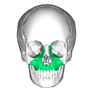
In vertebrates, the maxilla is the upper fixed bone of the jaw formed from the fusion of two maxillary bones. In humans, the upper jaw includes the hard palate in the front of the mouth. The two maxillary bones are fused at the intermaxillary suture, forming the anterior nasal spine. This is similar to the mandible, which is also a fusion of two mandibular bones at the mandibular symphysis. The mandible is the movable part of the jaw.

The cavernous sinus within the human head is one of the dural venous sinuses creating a cavity called the lateral sellar compartment bordered by the temporal bone of the skull and the sphenoid bone, lateral to the sella turcica.

The superior ophthalmic vein is a vein of the orbit that drains venous blood from structures of the upper orbit. It is formed by the union of the angular vein, and supraorbital vein. It passes backwards within the orbit alongside the ophthalmic artery, then exits the orbit through the superior orbital fissure to drain into the cavernous sinus.
The inferior ophthalmic vein is a vein of the orbit that - together with the superior ophthalmic vein - represents the principal drainage system of the orbit. It begins from a venous network in the front of the orbit, then passes backwards through the lower orbit. It drains several structures of the orbit. It may end by splitting into two branches, one draining into the pterygoid venous plexus and the other ultimately into the cavernous sinus.

The supraorbital vein is a vein of the forehead. It communicates with the frontal branch of the superficial temporal vein. It passes through the supraorbital notch, and merges with the angular vein to form the superior ophthalmic vein. The supraorbital vein helps to drain blood from the forehead, eyebrow, and upper eyelid.
The superficial temporal vein is a vein of the side of the head which collects venous blood from the region of the temple. It arises from an anastomosing venous plexus on the side and vertex of the head. The superficial temporal vein terminates within the substance of the parotid gland by uniting with the maxillary vein to form the retromandibular vein.
The angular vein is a vein of the face. It is the upper part of the facial vein, above its junction with the superior labial vein. It is formed by the junction of the supratrochlear vein and supraorbital vein, and joins with the superior labial vein. It drains the medial canthus, and parts of the nose and the upper lip. It can be a route of spread of infection from the danger triangle of the face to the cavernous sinus.
The retromandibular vein is a major vein of the face. It is formed within the parotid gland by the confluence of the maxillary vein, and superficial temporal vein. It descends in the gland and splits into two branches upon emerging from the gland. Its anterior branch then joins the (anterior) facial vein forming the common facial vein, while its posterior branch joins the posterior auricular vein forming the external jugular vein.

The lacrimal artery is an artery of the orbit. It is a branch of the ophthalmic artery. It accompanies the lacrimal nerve along the upper border of the lateral rectus muscle, travelling forward to reach the lacrimal gland. It supplies the lacrimal gland, two rectus muscles of the eye, the eyelids, and the conjunctiva.

The posterior superior alveolar artery is a branch of the maxillary artery. It is one of two or three superior alveolar arteries. It provides arterial suply to the molar and premolar teeth, maxillary sinus and adjacent bone, and the gingiva.

The infratemporal fossa is an irregularly shaped cavity that is a part of the skull. It is situated below and medial to the zygomatic arch. It is not fully enclosed by bone in all directions. It contains superficial muscles, including the lower part of the temporalis muscle, the lateral pterygoid muscle, and the medial pterygoid muscle. It also contains important blood vessels such as the middle meningeal artery, the pterygoid plexus, and the retromandibular vein, and nerves such as the mandibular nerve (CN V3) and its branches.

The lacrimal sac or lachrymal sac is the upper dilated end of the nasolacrimal duct, and is lodged in a deep groove formed by the lacrimal bone and frontal process of the maxilla. It connects the lacrimal canaliculi, which drain tears from the eye's surface, and the nasolacrimal duct, which conveys this fluid into the nasal cavity. Lacrimal sac occlusion leads to dacryocystitis.

The infraorbital nerve is a branch of the maxillary nerve. It arises in the pterygopalatine fossa. It passes through the inferior orbital fissure to enter the orbit. It travels through the orbit, then enters and traverses the infraorbital canal, exiting the canal at the infraorbital foramen to reach the face. It provides sensory innervation to the skin and mucous membranes around the middle of the face.

The angular artery is an artery of the face. It is the terminal part of the facial artery. It ascends to the medial angle of the eye's orbit. It is accompanied by the angular vein. It ends by anastomosing with the dorsal nasal branch of the ophthalmic artery. It supplies the lacrimal sac, the orbicularis oculi muscle, and the outer side of the nose.
The supraorbital artery is a branch of the ophthalmic artery. It passes anteriorly within the orbit to exit the orbit through the supraorbital foramen or notch alongside the supraorbital nerve, splitting into two terminal branches which go on to form anastomoses with arteries of the head.

The long posterior ciliary arteries are arteries of the orbit. There are long posterior ciliary arteries two on each side of the body. They are branches of the ophthalmic artery. They pass forward within the eye to reach the ciliary body where they ramify and anastomose with the anterior ciliary arteries, thus forming the major arterial circle of the iris.The long posterior ciliary arteries contribute arterial supply to the choroid, ciliary body, and iris.
The short posterior ciliary arteries are a number of branches of the ophthalmic artery. They pass forward with the optic nerve to reach the eyeball, piercing the sclera around the entry of the optic nerve into the eyeball.
The anterior ciliary arteries are seven arteries in each eye-socket that arise from muscular branches of the ophthalmic artery and supply the conjunctiva, sclera, rectus muscles, and the ciliary body. The arteries end by anastomosing with branches of the long posterior ciliary arteries to form the circulus arteriosus major.

The vorticose veins, referred to clinically as the vortex veins, are veins that drain the choroid of the eye. There are usually 4-5 vorticose veins in each eye, with at least one vorticose vein per each quadrant of the eye. Vorticose veins drain into the superior ophthalmic vein, and inferior ophthalmic vein.
The central retinal vein is a vein that drains the retina of the eye. It travels backwards through the centre of the optic nerve accompanied by the central retinal artery before exiting the optic nerve together with the central retinal artery to drain into either the superior ophthalmic vein or the cavernous sinus.