Ovine rinderpest

Last updated
Morbillivirus caprinae
Virus classification OOjs UI icon edit-ltr.svg
(unranked): Virus
Realm: Riboviria
Kingdom: Orthornavirae
Phylum: Negarnaviricota
Class: Monjiviricetes
Order: Mononegavirales
Family: Paramyxoviridae
Genus: Morbillivirus
Species:
Morbillivirus caprinae
Synonyms [1]
  • Peste-des-petits-ruminants virus
  • Pest des petits ruminants virus

Ovine rinderpest, also commonly known as peste des petits ruminants (PPR), is a contagious disease primarily affecting goats and sheep; however, camels and wild small ruminants can also be affected. [2] PPR is currently present in North, Central, West and East Africa, the Middle East, and South Asia. [3] It is caused by Morbillivirus caprinae in the genus Morbillivirus, and is closely related to, among others, Morbillivirus pecoris, Morbillivirus hominis (Measles virus), and Morbillivirus canis (also known as canine distemper virus). The disease is highly contagious, and can have an 80–100% mortality rate in acute cases in an epizootic setting. The virus does not infect humans.

Contents

The disease was first described in 1942 in Côte d'Ivoire, and has since been detected in more than 70 countries in the world. [4]

In 2017, the disease was reported to be affecting saiga antelope in Mongolia, causing near-catastrophic herd depletion for the endangered species. [5]

In 2018, it was stated that the disease was reported to be in Bulgaria close to the border with Turkey. [6] In a flock of 540 sheep and goats, two animals tested positive and one died, with disease confirmed by only one laboratory without any further tests. [7] Nevertheless, over 4000 sheep and goats were killed. [8]

Synonyms

PPR is also known as goat plague, kata, syndrome of stomatitis-pneumoenteritis, and ovine rinderpest. [9]

Official agencies such as the FAO and OIE use the French name "peste des petits ruminants" with several spelling variants.

Signs and symptoms

Symptoms are similar to those of rinderpest in cattle and involves oral necrosis, mucopurulent nasal and ocular discharges, cough, pneumonia, and diarrhea, [10] though they vary according to the previous immune status of the sheep, the geographic location, the time of year, or if the infection is new or chronic. They also vary according to the breed of sheep. However, fever in addition to either diarrhea or signs of oral discomfort is sufficient to suspect the diagnosis. [10] Incubation period is 3–5 days. [11]

Hyperacute cases

Hyperacute cases are found dead without previous symptoms. They die with a serous, foamy, or haemorrhagic discharge coming out of the nose.

Acute cases at onset

In acute cases, animals are recumbent, sometimes in self-auscultation position. Body temperature is high (40.5 to 41 °C) in the beginning of the onset in acute cases. The most typical signs are seen in the digestive tract. When entering an affected flock, one sees many animals with hind limbs stained by sticky faeces. Some sheep have an arched back and show pain when defecating. Tenesmus may be noticed when taking rectal temperature. Fluid faeces are olive green to brown. Examination of the mouth shows ulceration of the buccal mucosae, especially on the inner face of the lips, and neighboring gum. There can be periodontitis or serous nasal exudate and conjunctivitis.

Evolution of acute cases

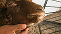
Nasal discharge becomes mucopurulent and may obstruct the nose. A dry, fitful coughing develops. Death occurs from 5 to 10 days after the onset of the fever. Some animals may recover, but a dry, stertorous coughing often persists for some days. [12] Besides coughing, there is intensive labial dermatitis with scab formation, resembling orf. [13] Miscarriages may occur. [14]

Cause

Peste des petits ruminants is caused by a Morbillivirus Morbillivirus caprinae – which is related to but distinct from Rinderpest virus. Four genotypes (lineages) of the virus are described. [10] Their classification is based on the nucleoprotein (N) or previously the fusion (F) protein gene. Lineages I and II are found mainly in West Africa. Lineage III is generally found in East Africa. Lineage IV was long known as the Asian lineage, but has now spread to the African continent and become the most prevalent lineage of all. [15]

Epidemiology

Origin and spread

This virus appears to have evolved at the start of the 20th century in Nigeria. [16] The extant genotypes subsequently appeared in West Africa (lineages I and II), East Africa and Arabia (lineage III), and Pakistan–India (lineage IV). [10]

The first description of the disease was published in 1942 and relates to an outbreak in Côte d’Ivoire, West Africa, in goats and sheep in 1940. [17] [10] It spread to East Africa and Arabia at the beginning of the 1980s and to Pakistan and India in the early 1990s (Calcutta goat markets) finally reaching Tibet in 2007. [10] The first description of this virus in India was in 1987.

The outbreak in Burkina Faso in 1999 was caused by the lineage I group. Genotype III has caused outbreaks in Ethiopia (1996) and also in Arabia, southern India, and Tamil Nadu (1992). This lineage was found in Yemen in 2001. Genotype IV has been isolated in Kuwait in 1999.

Geographical repartition

As of 2017, the disease is present in West Africa, part of Central Africa (Gabon, Central African Republic), East Africa (north of the Equator), the Middle East and the Indian subcontinent including Nepal and Myanmar. The disease is endemic in the Indian subcontinent and is a major threat to fast-growing goat husbandry in India, causing an annual loss of around 1800 million Indian rupees.

In North Africa, only Egypt was once hit, but since summer 2008, Morocco is suffering a generalized outbreak with 133 known cases in 129 provinces, mostly affecting sheep. [18] The outbreak has precipitated the vaccination of a large number of the 17 million sheep and five million goats in the country. [19]

Dissemination

The disease is transmitted by infected aerosols in situation of close contact of animals. The long-distance spread is by sick animals. [10] As the virus soon becomes inactive outside the body, indirect contamination is generally limited.

In an affected flock, even in pest-free regions, the disease does not progress very rapidly, in spite of the close contact between animals. New clinical cases may be observed daily for a 1-month period. [20]

Post mortem lesions

The lesions are situated in the digestive tract. Quick post mortem examination will lead to the discovery of many haemorrhagic patches on the serous membranes, and intense pneumonia. A risk exists that it may conclude with enzootic pneumonia, inability to open the mouth, and problems with the oesophagus and different parts of the intestine.

Erosions and inflammation are widespread on buccal mucosa. The same lesions are also present in pharynx, oesophagus, and on mucus-producing epithelia of the gut, from abomasum to rectum. Zebra-striped lesions on coecum and colon are said to be typical in some cases. Rarely, also petechiae are on the rumen mucosa. [21]

Microscopic lesions

Microscopic study of both natural and experimental cases revealed congestion and edema of the lungs, while in other cases it revealed a network of fibrin infiltrated with neutrophils, the formation of syncytia and giant cells, and the presence of a pink-colored bacterial colony.[ citation needed ]  There was infiltration of neutrophils and mononuclear cells within the alveoli, bronchioles, alveolar wall, and interstitium of the lung.[ citation needed ]  Microscopic analysis also revealed that there was interstitial pneumonia followed by sloughing of the bronchial epithelium in the lungs and the prominence of type II pneumocytes.[ citation needed ] Numerous inflammatory cells were found in the submucosa and lamina propria of intestinal samples, and vacuolar degeneration and syncytial cells, which are microscopic features, were observed in hepatocytes of liver samples from goats. Periportal multifocal lymphocytic infiltrations were common in the liver parenchyma.[ citation needed ]

Leptomeningitis and nonsuppurative encephalitis were visible in brain sections under a microscope, and these conditions were characterized by vascular congestion, hemorrhages in the parenchyma, perivascular cuffing with mild to moderate mononuclear cells (mostly lymphocytes and few macrophages), focal to diffuse microgliosis, neuronal degeneration, satellitosis, and neuronophagia. [22]

In summary Epithelial cells, alveolar macrophages, lungs, and hepatocytes all showed histopathological alterations, primarily infiltrations of inflammatory cells, syncytia, and presence of intranuclear and/or intracytoplasmic eosinophillic inclusions. [23]

Diagnosis

History and clinical signs enable a presumptive diagnosis to be made in endemic regions. The virus can be detected in acute cases from various swabs and blood samples, using PCR and ELISA. Antibodies can also be detected by ELISA. [14]

Treatment and control

Antibiotics such as chloramphenicol, penicillin, and streptomycin can be used and supportive treatment may be helpful. [14] Additionally, a vaccine has been developed that may decrease death in the flock. [14] Movement restrictions and slaughter of affected flocks may be required in an attempt to eradicate the disease. [14] A global eradication programme has been developed by the Food and Agriculture Organization of the United Nations and the World Organisation for Animal Health. [24] More information can be found on FAO's website on the implementation of this global plan. [25] It is considered feasible to eradicate ovine rinderpest in 15 years, starting in the year 2016. [10]

Related Research Articles

<span class="mw-page-title-main">Bluetongue disease</span> Viral disease in animals

Bluetongue disease is a noncontagious, insect-borne, viral disease of ruminants, mainly sheep and less frequently cattle, yaks, goats, buffalo, deer, dromedaries, and antelope. It is caused by Bluetongue virus (BTV). The virus is transmitted by the midges Culicoides imicola, Culicoides variipennis, and other culicoids.

<i>Morbillivirus</i> Genus of viruses

Morbillivirus is a genus of viruses in the order Mononegavirales, in the family Paramyxoviridae. Humans, dogs, cats, cattle, seals, and cetaceans serve as natural hosts. This genus includes six species, with a seventh species being extinct. Diseases in humans associated with viruses classified in this genus include measles; in animals, they include acute febrile respiratory tract infection and Canine distemper. In 2013, a wave of increased death among the Common bottlenose dolphin population was attributed to morbillivirus.

<span class="mw-page-title-main">Rinderpest</span> Eradicated morbillivirus disease

Rinderpest was an infectious viral disease of cattle, domestic buffalo, and many other species of even-toed ungulates, including gaurs, buffaloes, large antelope, deer, giraffes, wildebeests, and warthogs. The disease was characterized by fever, oral erosions, diarrhea, lymphoid necrosis, and high mortality. Death rates during outbreaks were usually extremely high, approaching 100% in immunologically naïve populations. Rinderpest was mainly transmitted by direct contact and by drinking contaminated water, although it could also be transmitted by air.

Walter Plowright CMG FRS FRCVS was an English veterinary scientist who devoted his career to the eradication of the cattle plague rinderpest. Plowright received the 1999 World Food Prize for his development of tissue culture rinderpest vaccine (TCRV), the key element in the quest to eliminate rinderpest. Rinderpest became the first animal disease to be eliminated worldwide.

<span class="mw-page-title-main">Ovine pulmonary adenocarcinoma</span>

Ovine pulmonary adenocarcinoma (OPA), also known as ovine pulmonary adenomatosis, or jaagsiekte, is a chronic and contagious disease of the lungs of sheep and goats. OPA is caused by a retrovirus called jaagsiekte sheep retrovirus (JSRV).

PPR may refer to:

<i>Jaagsiekte sheep retrovirus</i> Species of virus

Jaagsiekte sheep retrovirus (JSRV) is a betaretrovirus which is the causative agent of a contagious lung cancer in sheep, called ovine pulmonary adenocarcinoma.

<i>African swine fever virus</i> Species of virus

African swine fever virus (ASFV) is a large, double-stranded DNA virus in the Asfarviridae family. It is the causative agent of African swine fever (ASF). The virus causes a hemorrhagic fever with high mortality rates in domestic pigs; some isolates can cause death of animals as quickly as a week after infection. It persistently infects its natural hosts, warthogs, bushpigs, and soft ticks of the genus Ornithodoros, which likely act as a vector, with no disease signs. It does not cause disease in humans. ASFV is endemic to sub-Saharan Africa and exists in the wild through a cycle of infection between ticks and wild pigs, bushpigs, and warthogs. The disease was first described after European settlers brought pigs into areas endemic with ASFV, and as such, is an example of an emerging infectious disease.

<span class="mw-page-title-main">Bovine malignant catarrhal fever</span> Species of virus

Bovine malignant catarrhal fever (BMCF) is a fatal lymphoproliferative disease caused by a group of ruminant gamma herpes viruses including Alcelaphine gammaherpesvirus 1 (AlHV-1) and Ovine gammaherpesvirus 2 (OvHV-2) These viruses cause unapparent infection in their reservoir hosts, but are usually fatal in cattle and other ungulates such as deer, antelope, and buffalo. In Southern Africa the disease is known as snotsiekte, from the Afrikaans.

Visna-maedi virus from the genus Lentivirus and subfamily Orthoretrovirinae, is a retrovirus that causes encephalitis and chronic pneumonitis in sheep. It is known as visna when found in the brain, and maedi when infecting the lungs. Lifelong, persistent infections in sheep occur in the lungs, lymph nodes, spleen, joints, central nervous system, and mammary glands; The condition is sometimes known as ovine progressive pneumonia (OPP), particularly in the United States, or Montana sheep disease. White blood cells of the monocyte/macrophage lineage are the main target of the virus.

<span class="mw-page-title-main">Eradication of infectious diseases</span> Elimination of a disease from all hosts

The eradication of infectious diseases is the reduction of the prevalence of an infectious disease in the global host population to zero.

<span class="mw-page-title-main">Sheep</span> Domesticated ruminant bred for meat, wool, and milk

Sheep or domestic sheep are a domesticated, ruminant mammal typically kept as livestock. Although the term sheep can apply to other species in the genus Ovis, in everyday usage it almost always refers to domesticated sheep. Like all ruminants, sheep are members of the order Artiodactyla, the even-toed ungulates. Numbering a little over one billion, domestic sheep are also the most numerous species of sheep. An adult female is referred to as a ewe, an intact male as a ram, occasionally a tup, a castrated male as a wether, and a young sheep as a lamb.

<span class="mw-page-title-main">Caseous lymphadenitis</span> Medical condition

Caseous lymphadenitis (CLA) is an infectious disease caused by the bacterium Corynebacterium pseudotuberculosis, that affects the lymphatic system, resulting in abscesses in the lymph nodes and internal organs. It is found mostly in goats and sheep and at the moment it has no cure.

<span class="mw-page-title-main">Social history of viruses</span> Influence of viruses and viral infections on human history

The social history of viruses describes the influence of viruses and viral infections on human history. Epidemics caused by viruses began when human behaviour changed during the Neolithic period, around 12,000 years ago, when humans developed more densely populated agricultural communities. This allowed viruses to spread rapidly and subsequently to become endemic. Viruses of plants and livestock also increased, and as humans became dependent on agriculture and farming, diseases such as potyviruses of potatoes and rinderpest of cattle had devastating consequences.

Cache Valley orthobunyavirus (CVV) is a member of the order Bunyavirales, genus Orthobunyavirus, and serogroup Bunyamwera, which was first isolated in 1956 from Culiseta inornata mosquitos collected in Utah's Cache Valley. CVV is an enveloped arbovirus, nominally 80–120 nm in diameter, whose genome is composed of three single-stranded, negative-sense RNA segments. The large segment of related bunyaviruses is approximately 6800 bases in length and encodes a probable viral polymerase. The middle CVV segment has a 4463-nucleotide sequence and the smallest segment encodes for the nucleocapsid, and a second non-structural protein. CVV has been known to cause outbreaks of spontaneous abortion and congenital malformations in ruminants such as sheep and cattle. CVV rarely infects humans, but when they are infected it has caused encephalitis and multiorgan failure.

Feline morbillivirus comes from the genus Morbillivirus, specifically influencing wild and domestic cats. The first report of a Feline morbillivirus outbreak occurred in Hong Kong in 2012. Approximately 10% of stray cats in Hong Kong and mainland China were reported to possess the virus at the time with additional infections found in Japan as well. 40% of cats tested in Japan were Fmo-PV positive and exhibited early symptoms of kidney failure. While the first cases of Feline morbillivirus were found in China, Hong Kong and Japan, the virus can also be found in Italy, Germany, and the United States. Feline morbillivirus exhibits a substantial amount of genetic diversity, yet cases in Japan and Hong Kong proved to have identical nucleotide sequences. It is also hypothesized that the morbillivirus has high adaptability due to its presence in multiple species. It is often found in dogs, cats, cattle, whales, dolphins, porpoises, and even humans. It likely originated from an ancestral version and underwent viral evolution to adapt to transmission in different species. Other common morbilliviruses include measles, rinderpest virus, canine distemper virus and peste des petits ruminants virus.

A Foreign animal disease (FAD) is an animal disease or pest, whether terrestrial or aquatic, not known to exist in the United States or its territories. When these diseases can significantly affect human health or animal production and when there is significant economic cost for disease control and eradication efforts, they are considered a threat to the United States. Another term gaining preference to be used is Transboundary Animal Disease (TAD), which is defined as those epidemic diseases which are highly contagious or transmissible and have the potential for very rapid spread, irrespective of national borders, causing serious socio-economic and possibly public health consequences. An Emerging Animal Disease "may be defined as any terrestrial animal, aquatic animal, or zoonotic disease not yet known or characterized, or any known or characterized terrestrial animal or aquatic animal disease in the United States or its territories that changes or mutates in pathogenicity, communicability, or zoonotic potential to become a threat to terrestrial animals, aquatic animals, or humans."

Gopal Dhinakar Raj is an Indian veterinary scientist, an academic and the project director of the Translational Research Platform for Veterinary Biologicals, a partnership program between the Department of Biotechnology and the Tamil Nadu Veterinary and Animal Sciences University. Known for the development of diagnostic test kits for animal and poultry diseases such as Leptospirosis and Egg drop syndrome, Raj is a member of the DBT Task Force on Animal Biotechnology. The Department of Biotechnology of the Government of India awarded him the National Bioscience Award for Career Development, one of the highest Indian science awards, for his contributions to biosciences in 2007.

The National Veterinary Research Institute (NVRI) is a research institute in Nigeria that was established in 1924 and has the mandate to conduct research into how to ably identify, treat and control animal diseases as well as the development of vaccines for such and training and the provision of support services to livestock and poultry farmers. The institute is under the supervision of Federal Ministry of Agriculture and Rural Development. The chief executive is Maryam Muhammad, a veterinary doctor with research interests in the molecular epidemiology of Salmonella in poultry, public health and environment and development.

References

  1. "ICTV Taxonomy history: Small ruminant morbillivirus". International Committee on Taxonomy of Viruses (ICTV). Retrieved 15 January 2019.
  2. Munir, M. (2014-10-01). "Role of Wild Small Ruminants in the Epidemiology of Peste Des Petits Ruminants". Transboundary and Emerging Diseases. 61 (5): 411–424. doi: 10.1111/tbed.12052 . ISSN   1865-1682. PMID   23305511.
  3. Banyard, Ashley C.; Parida, Satya; Batten, Carrie; Oura, Chris; Kwiatek, Olivier; Libeau, Genevieve (2010). "Global distribution of peste des petits ruminants virus and prospects for improved diagnosis and control". Journal of General Virology. 91 (12): 2885–2897. doi: 10.1099/vir.0.025841-0 . PMID   20844089.
  4. "Peste des Petits Ruminants". Food and Agriculture Organization of The United Nations .
  5. "PESTE DES PETITS RUMINANTS – MONGOLIA (03): (HOVD) SAIGA ANTELOPE". ProMED-mail. 9 March 2017. Retrieved 9 March 2017.
  6. "Bulgaria reports another case of ovine rinderpest". Reuters. 19 July 2018. Retrieved 2 August 2018.
  7. Prevalence of Antimicrobial Resistance in Ruminants in Bulgaria: A Pilot Study
  8. Protests amid outrage over killing of sheep and goats over rinderpest outbreak in Bulgaria
  9. Parida, S.; Muniraju, M.; Mahapatra, M.; Muthuchelvan, D.; Buczkowski, H.; Banyard, A.C. (2015). "Peste des petits ruminants". Veterinary Microbiology. 181 (1–2): 90–106. doi:10.1016/j.vetmic.2015.08.009. PMC   4655833 . PMID   26443889.
  10. 1 2 3 4 5 6 7 8 Taylor, William (2015). "The global eradication of peste des petits ruminants (PPR) within 15 years—is this a pipe dream?". Tropical Animal Health and Production. 48 (3): 559–667. doi:10.1007/s11250-016-0993-x. PMID   26851956. S2CID   255107410.
  11. "Rinderpest | animal disease". Encyclopedia Britannica. Retrieved 2019-12-07.
  12. J. Berrada, Observations des premiers cas confirmés de peste des petits ruminants au Maroc, oral presentation, El Jadida, 31-07-2008.
  13. Handbook of Animal Diseases in the Tropics, op cit.
  14. 1 2 3 4 5 Peste des Petits Ruminants Archived 2013-02-22 at archive.today reviewed and published by WikiVet, accessed 10 October 2011.
  15. Kwiatek, Olivier; Ali, Yahia Hassan; Saeed, Intisar Kamil; Khalafalla, Abdelmelik Ibrahim; Mohamed, Osama Ishag; Abu Obeida, Ali; Abdelrahman, Magdi Badawi; Osman, Halima Mohamed; Taha, Khalid Mohamed (2011). "Asian Lineage of Peste des Petits Ruminants Virus, Africa". Emerging Infectious Diseases. 17 (7): 1223–1231. doi:10.3201/eid1707.101216. ISSN   1080-6040. PMC   3381390 . PMID   21762576.
  16. Muniraju, M; Munir, M; Parthiban, AR; Banyard, AC; Bao, J; Wang, Z; Ayebazibwe, C; Ayelet, G; El Harrak, M; Mahapatra, M; Libeau, G; Batten, C; Parida, S (2014). "Molecular evolution of peste des petits ruminants virus". Emerging Infectious Diseases. 20 (12): 2023–2033. doi:10.3201/eid2012.140684. PMC   4257836 . PMID   25418782.
  17. Dhar, Pronab; Sreenivasa, B.P; Barrett, Thomas; Corteyn, Mandy; Singh, R.P; Bandyopadhyay, S.K (2002). "Recent epidemiology of peste des petits ruminants virus (PPRV)". Veterinary Microbiology. 88 (2): 153–159. doi:10.1016/s0378-1135(02)00102-5. PMID   12135634.
  18. "Outbreak of 'peste des petits ruminants' in Morocco". FAO Newsroom. FAO. September 9, 2008. Retrieved 2008-09-10.
  19. "Morocco to vaccinate all livestock after virus outbreak". AFP . September 9, 2008. Archived from the original on May 20, 2011. Retrieved 2008-09-10.
  20. L. Mahin (2008) Observations sur un foyer de Peste des petits ruminants, unpublished data.
  21. Tligui, Observations nécropsiques sur les premiers cas confirmés de peste des petits ruminants au Maroc, oral presentation, El Jadida, 31-07-2008.
  22. Seki, Fumio; Takeda, Makoto (2022). "Novel and classical morbilliviruses: Current knowledge of three divergent morbillivirus groups". Microbiology and Immunology. 66 (12): 552–563. doi:10.1111/1348-0421.13030. PMID   36151905. S2CID   252497033. This review cites this research. Sahoo, Monalisa; M, Dinesh; Thakor, Jigarji Chaturji; Baloni, Suraj; Saxena, Sonal; Shrivastava, Sameer; Dhama, Kuldeep; Singh, Karampal; Singh, Rajendra (2020-03-01). "Neuropathology mediated through caspase dependent extrinsic pathway in goat kids naturally infected with PPRV". Microbial Pathogenesis . 140: 103949. doi:10.1016/j.micpath.2019.103949. ISSN   0882-4010. PMID   31875517. S2CID   209482267.
  23. Maes, Piet; et al. (2018). "Taxonomy of the family Arenaviridae and the order Bunyavirales: Update 2018". Archives of Virology. 163 (8): 2295–2310. doi: 10.1007/s00705-018-3843-5 . hdl: 2263/64947 . PMID   29680923. S2CID   254054051. This review cites this research. Blasdell, Kim R.; Duong, Veasna; Eloit, Marc; Chretien, Fabrice; Ly, Sowath; Hul, Vibol; Deubel, Vincent; Morand, Serge; Buchy, Philippe (2016). "Evidence of human infection by a new mammarenavirus endemic to Southeastern Asia". eLife. 5. doi: 10.7554/elife.13135 . PMC   4900801 . PMID   27278118.
  24. FAO and WHO. 2016. Peste des petits ruminants GLOBAL ERADICATION PROGRAM. Rome.
  25. FAO. Peste des Petits Ruminants. http://www.fao.org/ppr/en/ accessed Nov. 2016