Preaortic lymph nodes

Last updated
Preaortic lymph node
Illu lymph chain07.jpg
Left lumbar lymph nodes (paraaortic lymph nodes)
  1. Lateral aortic
  2. Preaortic
  3. Postaortic
  4. Intermediate lumbar
Right lumbar lymph nodes (paracaval lymph nodes)
  1. Lateral caval
  2. Precaval
  3. Postcaval
Gray611.png
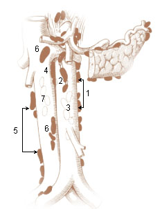
The parietal lymph glands of the pelvis.
Details
System Lymphatic system
Source inferior mesenteric lymph nodes
Drains toprimarily intestinal trunk
Identifiers
Latin nodi lymphoidei praeaortici
Anatomical terminology

The preaortic lymph nodes lie in front of the aorta, and may be divided into celiac lymph nodes, superior mesenteric lymph nodes, and inferior mesenteric lymph nodes groups, arranged around the origins of the corresponding arteries.

Contents

The celiac lymph nodes are grouped into three sets: the gastric, hepatic and splenic lymph nodes. These groups also form their own subgroups.

The superior mesenteric lymph nodes are grouped into three sets: the mesenteric, ileocolic and mesocolic lymph nodes.

The inferior mesenteric lymph nodes have a subgroup of pararectal lymph nodes.

The preaortic lymph nodes receive a few vessels from the lateral aortic lymph nodes, but their principal afferents are derived from the organs supplied by the three arteries with which they are associated–the celiac, superior and inferior mesenteric arteries.

Some of their efferents pass to the retroaortic lymph nodes, but the majority unite to form the intestinal lymph trunk, which enters the cisterna chyli.

Additional images

Related Research Articles

<span class="mw-page-title-main">Duodenum</span> First section of the small intestine

The duodenum is the first section of the small intestine in most higher vertebrates, including mammals, reptiles, and birds. In mammals it may be the principal site for iron absorption. The duodenum precedes the jejunum and ileum and is the shortest part of the small intestine.

<span class="mw-page-title-main">Thoracic duct</span> Lymphatic vessel

In human anatomy, the thoracic duct is the larger of the two lymph ducts of the lymphatic system. The thoracic duct usually begins from the upper aspect of the cisterna chyli, passing out of the abdomen through the aortic hiatus into first the posterior mediastinum and then the superior mediastinum, extending as high up as the root of the neck before descending to drain into the systemic (blood) circulation at the venous angle.

<span class="mw-page-title-main">Abdominal aorta</span> Largest artery in the abdomen

In human anatomy, the abdominal aorta is the largest artery in the abdominal cavity. As part of the aorta, it is a direct continuation of the descending aorta.

<span class="mw-page-title-main">Periaortic lymph nodes</span> Human lymph nodes

The periaortic lymph nodes are a group of lymph nodes that lie in front of the lumbar vertebrae near the aorta. These lymph nodes receive drainage from the gastrointestinal tract and the abdominal organs.

<span class="mw-page-title-main">Celiac artery</span> First major branch of the abdominal aorta

The celiacartery, also known as the celiac trunk or truncus coeliacus, is the first major branch of the abdominal aorta. It is about 1.25 cm in length. Branching from the aorta at thoracic vertebra 12 (T12) in humans, it is one of three anterior/ midline branches of the abdominal aorta.

<span class="mw-page-title-main">Superior mesenteric artery</span> Artery which supplies blood to the intestines and pancreas

In human anatomy, the superior mesenteric artery (SMA) is an artery which arises from the anterior surface of the abdominal aorta, just inferior to the origin of the celiac trunk, and supplies blood to the intestine from the lower part of the duodenum through two-thirds of the transverse colon, as well as the pancreas.

<span class="mw-page-title-main">Inferior mesenteric artery</span>

In human anatomy, the inferior mesenteric artery (IMA) is the third main branch of the abdominal aorta and arises at the level of L3, supplying the large intestine from the distal transverse colon to the upper part of the anal canal. The regions supplied by the IMA are the descending colon, the sigmoid colon, and part of the rectum.

<span class="mw-page-title-main">Nerve plexus</span> Network of nerve fibres

A nerve plexus is a plexus of intersecting nerves. A nerve plexus is composed of afferent and efferent fibers that arise from the merging of the anterior rami of spinal nerves and blood vessels. There are five spinal nerve plexuses, except in the thoracic region, as well as other forms of autonomic plexuses, many of which are a part of the enteric nervous system. The nerves that arise from the plexuses have both sensory and motor functions. These functions include muscle contraction, the maintenance of body coordination and control, and the reaction to sensations such as heat, cold, pain, and pressure. There are several plexuses in the body, including:

<span class="mw-page-title-main">Middle suprarenal arteries</span> Arteries of the abdomen

The middle suprarenal artery is a paired artery in the abdomen. It is a branch of the aorta. It supplies the adrenal gland.

<span class="mw-page-title-main">Intestinal arteries</span>

The intestinal arteries arise from the convex side of the superior mesenteric artery. They are usually from twelve to fifteen in number, and are distributed to the jejunum and ileum.

<span class="mw-page-title-main">Celiac ganglia</span> Two large masses of nerve tissue in the upper abdomen

The celiac ganglia or coeliac ganglia are two large irregularly shaped masses of nerve tissue in the upper abdomen. Part of the sympathetic subdivision of the autonomic nervous system (ANS), the two celiac ganglia are the largest ganglia in the ANS, and they innervate most of the digestive tract.

<span class="mw-page-title-main">Tracheobronchial lymph nodes</span>

The tracheobronchial lymph nodes are lymph nodes that are located around the division of trachea and main bronchi.

<span class="mw-page-title-main">Inferior mesenteric lymph nodes</span>

The inferior mesenteric lymph nodes consist of:

<span class="mw-page-title-main">Outline of human anatomy</span> Overview of and topical guide to human anatomy

The following outline is provided as an overview of and topical guide to human anatomy:

<span class="mw-page-title-main">Superior mesenteric lymph nodes</span>

The superior mesenteric lymph nodes may be divided into three principal groups:

<span class="mw-page-title-main">Celiac lymph nodes</span>

The celiac lymph nodes are associated with the branches of the celiac artery. Other lymph nodes in the abdomen are associated with the superior and inferior mesenteric arteries. The celiac lymph nodes are grouped into three sets: the gastric, hepatic and splenic lymph nodes. They receive lymph from the stomach, duodenum, pancreas, spleen, liver, and gall bladder.

<span class="mw-page-title-main">Hepatic lymph nodes</span> Lymph nodes on the common hepatic artery

The hepatic lymph nodes consist of the following groups:

<span class="mw-page-title-main">Gastric lymph nodes</span>

The gastric lymph nodes are lymph nodes which drain the stomach and consist of two sets, superior and inferior:

The superior mesenteric vessels are composed of the superior mesenteric artery and the superior mesenteric vein.

References

PD-icon.svgThis article incorporates text in the public domain from page 705 of the 20th edition of Gray's Anatomy (1918)