
The pancreas is an organ of the digestive system and endocrine system of vertebrates. In humans, it is located in the abdomen behind the stomach and functions as a gland. The pancreas is a mixed or heterocrine gland, i.e., it has both an endocrine and a digestive exocrine function. 99% of the pancreas is exocrine and 1% is endocrine. As an endocrine gland, it functions mostly to regulate blood sugar levels, secreting the hormones insulin, glucagon, somatostatin and pancreatic polypeptide. As a part of the digestive system, it functions as an exocrine gland secreting pancreatic juice into the duodenum through the pancreatic duct. This juice contains bicarbonate, which neutralizes acid entering the duodenum from the stomach; and digestive enzymes, which break down carbohydrates, proteins and fats in food entering the duodenum from the stomach.

The left and right brachiocephalic veins are major veins in the upper chest, formed by the union of the ipsilateral internal jugular vein and subclavian vein behind the sternoclavicular joint. The left brachiocephalic vein is more than twice the length of the right brachiocephalic vein.

Great vessels are the large vessels that bring blood to and from the heart. These are:
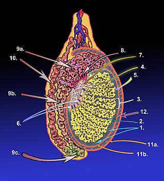
The tunica albuginea is a dense, blue-white layer of fibrous tissue surrounding the testis. It is the middle of three envelopes forming the capsule of the testis; it is deep to the visceral layer of tunica vaginalis, and superficial to the tunica vasculosa testis.

The sigmoid arteries are 2–5 branches of the inferior mesenteric artery that are distributed to the distal descending colon and the sigmoid colon.

The right colic artery is an artery of the abdomen, a branch of the superior mesenteric artery supplying the ascending colon. It divides into two terminal branches - an ascending branch and a descending branch - which form anastomoses with the middle colic artery, and ileocolic artery (respectively).

The middle colic artery is an artery of the abdomen; a branch of the superior mesenteric artery distributed to parts of the ascending and transverse colon. It usually divides into two terminal branches - a left one and a right one - which go on to form anastomoses with the left colic artery, and right colic artery (respectively), thus participating in the formation of the marginal artery of the colon.

The infratrochlear nerve is a branch of the nasociliary nerve (itself a branch of the ophthalmic nerve (CN V1)) in the orbit. It exits the orbit inferior to the trochlea of superior oblique. It provides sensory innervation to structures of the orbit and skin of adjacent structures.

The retropharyngeal space is a potential space and deep compartment of the head and neck situated posterior to the pharynx. The RPS is bounded anteriorly by the buccopharyngeal fascia, posteriorly by the alar fascia, and laterally by the carotid sheath. It extends between the base of the skull superiorly, and the mediastinum inferiorly. It contains the retropharyngeal lymph nodes. Its function is to facilitate movements in the superoinferior axis of the larynx, pharynx, and esophagus in relation to the cervical spine.

The femoral canal is the medial compartment of the three compartments of the femoral sheath. It is conical in shape. The femoral canal contains lymphatic vessels, and adipose and loose connective tissue, as well as - sometimes - a deep inguinal lymph node. The function of the femoral canal is to accommodate the distension of the femoral vein when venous return from the leg is increased or temporarily restricted.
The femoral sheath is a funnel-shaped downward extension of abdominal fascia within which the femoral artery and femoral vein pass between the abdomen and the thigh. The femoral sheath is subdivided by two vertical partitions to form three compartments ; the medial compartment is known as the femoral canal and contains lymphatic vessels and a lymph node, whereas the intermediate canal and the lateral canal accommodate the femoral vein and the femoral artery (respectively). Some neurovascular structures perforate the femoral sheath. Topographically, the femoral sheath is contained within the femoral triangle.

The dorsal nasal artery is an artery of the face. It is one of the two terminal branches of the ophthalmic artery. It contributes arterial supply to the lacrimal sac, and outer surface of the nose.

The supratrochlear artery is one of the terminal branches of the ophthalmic artery. It arises within the orbit. It exits the orbit alongside the supratrochlear nerve. It contributes arterial supply to the skin, muscles and pericranium of the forehead.

The crista terminalis is a vertical ridge on the posterolateral inner surface of the adult right atrium extending between the superior vena cava, and the inferior vena cava. The crista terminalis denotes where the junction of the embryologic sinus venosus and the right atrium occurred during embryonic development. It forms a boundary between the rough trabecular portion and the smooth, sinus venosus-derived portion of the internal surface of the right atrium. The sinoatrial node is located within the crista terminalis.

The dorsal pancreatic artery is a branch of the splenic artery. It anastomoses with the superior pancreaticoduodenal artery and continues as the inferior pancreatic artery on its lower border.

The inferior mesenteric lymph nodes consist of:

The deep cervical lymph nodes are a group of cervical lymph nodes in the neck that form a chain along the internal jugular vein within the carotid sheath.

The inferior deep cervical lymph nodes are one of the two groups of the deep cervical lymph nodes.
The superior deep cervical lymph nodes are the deep cervical lymph nodes that are situated adjacent to the superior portion of the internal jugular vein. They drain either to the inferior deep cervical lymph nodes or into the jugular trunk.
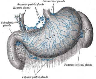
The celiac lymph nodes are associated with the branches of the celiac artery. Other lymph nodes in the abdomen are associated with the superior and inferior mesenteric arteries. The celiac lymph nodes are grouped into three sets: the gastric, hepatic and splenic lymph nodes. They receive lymph from the stomach, duodenum, pancreas, spleen, liver, and gall bladder.


