Delta-aminolevulinic acid dehydratase

Last updated
ALAD
Protein ALAD PDB 1e51.png ALADwithZinc 1ylv ALADwithLead 1qnv.png
Available structures
PDB Ortholog search: PDBe RCSB
Identifiers
Aliases ALAD , ALADH, PBGS, aminolevulinate dehydratase, ALA dehydratase
External IDs OMIM: 125270 MGI: 96853 HomoloGene: 16 GeneCards: ALAD
Orthologs
SpeciesHumanMouse
Entrez
Ensembl
UniProt
RefSeq (mRNA)

NM_000031
NM_001003945
NM_001317745

NM_001276446
NM_008525

RefSeq (protein)

NP_000022
NP_001003945
NP_001304674

NP_001263375
NP_032551

Location (UCSC) Chr 9: 113.39 – 113.4 Mb Chr 4: 62.43 – 62.44 Mb
PubMed search [3] [4]
Wikidata
View/Edit Human View/Edit Mouse
porphobilinogen synthase
1e51.jpg
DALA dehydratase
Identifiers
EC no. 4.2.1.24
CAS no. 9036-37-7
Databases
IntEnz IntEnz view
BRENDA BRENDA entry
ExPASy NiceZyme view
KEGG KEGG entry
MetaCyc metabolic pathway
PRIAM profile
PDB structures RCSB PDB PDBe PDBsum
Gene Ontology AmiGO / QuickGO
Search
PMC articles
PubMed articles
NCBI proteins
Delta-aminolevulinic acid dehydratase
Identifiers
Symbol ALAD
NCBI gene 210
HGNC 395
OMIM 125270
RefSeq NM_001003945
UniProt P13716
Other data
EC number 4.2.1.24
Locus Chr. 9 q32
Search for
Structures Swiss-model
Domains InterPro
ALAD
PDB 1b4k EBI.jpg
high resolution crystal structure of a mg2-dependent 5-aminolevulinic acid dehydratase
Identifiers
SymbolALAD
Pfam PF00490
Pfam clan CL0036
InterPro IPR001731
PROSITE PDOC00153
SCOP2 1aw5 / SCOPe / SUPFAM
Available protein structures:
Pfam   structures / ECOD  
PDB RCSB PDB; PDBe; PDBj
PDBsum structure summary

Aminolevulinic acid dehydratase (porphobilinogen synthase, or ALA dehydratase, or aminolevulinate dehydratase) is an enzyme (EC 4.2.1.24) that in humans is encoded by the ALAD gene. [5] [6] Porphobilinogen synthase (or ALA dehydratase, or aminolevulinate dehydratase) synthesizes porphobilinogen through the asymmetric condensation of two molecules of aminolevulinic acid. All natural tetrapyrroles, including hemes, chlorophylls and vitamin B12, share porphobilinogen as a common precursor. Porphobilinogen synthase is the prototype morpheein. [7]

Contents

Function

It catalyzes the following reaction, the second step of the biosynthesis of porphyrin:

2 5-Aminolevulinic acid porphobilinogen + 2 H2O

It therefore catalyzes the condensation of 2 molecules of 5-aminolevulinate to form porphobilinogen (a precursor of heme, cytochromes and other hemoproteins). This reaction is the first common step in the biosynthesis of all biological tetrapyrroles. Zinc is essential for enzymatic activity.

Structure

The structural basis for allosteric regulation of Porphobilinogen synthase (PBGS) is modulation of a quaternary structure equilibrium between octamer and hexamer (via dimers), which is represented schematically as 6mer* ↔ 2mer* ↔ 2mer ↔ 8mer. The * represents a reorientation between two domains of each subunit that occurs in the dissociated state because it is sterically forbidden in the larger multimers. [7]

The PBGS quaternary structure equilibrium includes an inactive hexamer (PDB id 1PV8) that does not have subunit interactions necessary for an ordered active site lid. Dissociation to the pro-hexamer dimer can be followed by a conformational change that reorients the two ab-barrel domains to form the pro-octamer dimer. Association of pro-octamer dimer to octamer (PDB id 1E51) includes formation of subunit interfaces that support order in the active site lid. PBGS Quaternary Structure Equlibrium.jpg
The PBGS quaternary structure equilibrium includes an inactive hexamer (PDB id 1PV8) that does not have subunit interactions necessary for an ordered active site lid. Dissociation to the pro-hexamer dimer can be followed by a conformational change that reorients the two αβ-barrel domains to form the pro-octamer dimer. Association of pro-octamer dimer to octamer (PDB id 1E51) includes formation of subunit interfaces that support order in the active site lid.

PBGS is encoded by a single gene and each PBGS multimer is composed of multiple copies of the same protein. Each PBGS subunit consists of a ~300 residue αβ-barrel domain, which houses the enzyme's active site in its center, and a >25 residue N-terminal arm domain. Allosteric regulation of PBGS can be described in terms of the orientation of the αβ-barrel domain with respect to the N-terminal arm domain.

Each N-terminal arm has up to two interactions with other subunits in a PBGS multimer. One of these interactions helps to stabilize a "closed" conformation of the active site lid. The other interaction restricts solvent access from the other end of the αβ-barrel.

In the inactive multimeric state, the N-terminal arm domain is not involved in the lid-stabilizing interaction, and in the crystal structure of the inactive assembly, the active site lid is disordered.

Allosteric regulators

As a nearly universal enzyme with a highly conserved active site, PBGS would not be a prime target for the development of antimicrobials and/or herbicides. To the contrary, allosteric sites can be much more phylogenetically variable than active sites, thus presenting more drug development opportunities. [7]

Phylogenetic variation in PBGS allostery leads to the framing of discussion of PBGS allosteric regulation in terms of intrinsic and extrinsic factors.

Intrinsic allosteric regulators

Magnesium

The allosteric magnesium ion lies at the highly hydrated interface of two pro-octamer dimers. It appears to be easily dissociable, and it has been shown that hexamers accumulate when magnesium is removed in vitro . [8]

pH

Though it is not common to consider hydronium ions as allosteric regulators, in the case of PBGS, side chain protonation at locations other than the active site has been shown to affect the quaternary structure equilibrium, and thus to affect the rate of its catalyzed reaction as well.

Extrinsic allosteric regulators

Small molecule hexamer stabilization

Inspection of the PBGS 6mer* reveals a surface cavity that is not present in the 8mer. Small molecule binding to this phylogenetically variable cavity has been proposed to stabilize 6mer* of the targeted PBGS and consequently inhibit activity.

Such allosteric regulators are known as morphlocks because they lock PBGS in a specific morpheein form (6mer*). [9]

Lead poisoning

ALAD enzymatic activity is inhibited by lead, beginning at blood lead levels that were once considered to be safe (<10 μg/dL) and continuing to correlate negatively across the range from 5 to 95 μg/dL. [10] Inhibition of ALAD by lead leads to anemia primarily because it both inhibits heme synthesis and shortens the lifespan of circulating red blood cells, but also by stimulating the excessive production of the hormone erythropoietin, leading to inadequate maturation of red cells from their progenitors. A defect in the ALAD structural gene can cause increased sensitivity to lead poisoning and acute hepatic porphyria. Alternatively spliced transcript variants encoding different isoforms have been identified. [11]

Deficiency

A deficiency of porphobilinogen synthase is usually acquired (rather than hereditary) and can be caused by heavy metal poisoning, especially lead poisoning, as the enzyme is very susceptible to inhibition by heavy metals. [12]

Hereditary insufficiency of porphobilinogen synthase is called porphobilinogen synthase (or ALA dehydratase) deficiency poprhyria. It is an extremely rare cause of porphyria, [13] with less than 10 cases ever reported. [14] All disease associated protein variants favor hexamer formation relative to the wild type human enzyme. [13]

Heme synthesis--note that some reactions occur in the cytoplasm and some in the mitochondrion (yellow) Heme synthesis.png
Heme synthesisnote that some reactions occur in the cytoplasm and some in the mitochondrion (yellow)

PBGS as the prototype morpheein

The morpheein model of allostery exemplified by PBGS adds an additional layer of understanding to potential mechanisms for regulation of protein function and complements the increased focus that the protein science community is placing on protein structure dynamics. [7]

This model illustrates how the dynamics of phenomena such as alternate protein conformations, alternate oligomeric states, and transient protein-protein interactions can be harnessed for allosteric regulation of catalytic activity.

Related Research Articles

<span class="mw-page-title-main">Heme</span> Chemical coordination complex of an iron ion chelated to a porphyrin

Heme, or haem, is a precursor to hemoglobin, which is necessary to bind oxygen in the bloodstream. Heme is biosynthesized in both the bone marrow and the liver.

<span class="mw-page-title-main">Allosteric regulation</span> Regulation of enzyme activity

In biochemistry, allosteric regulation is the regulation of an enzyme by binding an effector molecule at a site other than the enzyme's active site.

<span class="mw-page-title-main">Phenylalanine hydroxylase</span> Mammalian protein found in Homo sapiens

Phenylalanine hydroxylase. (PAH) (EC 1.14.16.1) is an enzyme that catalyzes the hydroxylation of the aromatic side-chain of phenylalanine to generate tyrosine. PAH is one of three members of the biopterin-dependent aromatic amino acid hydroxylases, a class of monooxygenase that uses tetrahydrobiopterin (BH4, a pteridine cofactor) and a non-heme iron for catalysis. During the reaction, molecular oxygen is heterolytically cleaved with sequential incorporation of one oxygen atom into BH4 and phenylalanine substrate. In humans, mutations in its encoding gene, PAH, can lead to the metabolic disorder phenylketonuria.

<span class="mw-page-title-main">Aminolevulinic acid synthase</span> Class of enzymes

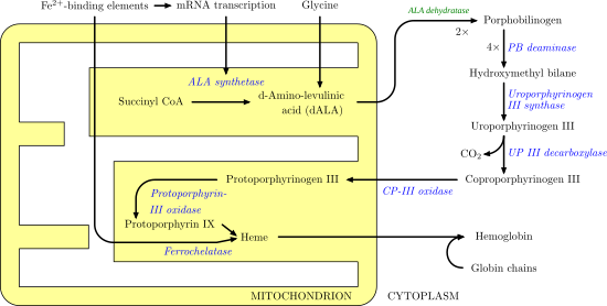
Aminolevulinic acid synthase (ALA synthase, ALAS, or delta-aminolevulinic acid synthase) is an enzyme (EC 2.3.1.37) that catalyzes the synthesis of δ-aminolevulinic acid (ALA) the first common precursor in the biosynthesis of all tetrapyrroles such as hemes, cobalamins and chlorophylls. The reaction is as follows:

Dehydratases are a group of lyase enzymes that form double and triple bonds in a substrate through the removal of water. They can be found in many places including the mitochondria, peroxisome and cytosol. There are more than 150 different dehydratase enzymes that are classified into four groups. Dehydratases can act on hydroxyacyl-CoA with or without cofactors, and some have a metal and non-metal cluster act as their active site.

<span class="mw-page-title-main">Acute intermittent porphyria</span> Medical condition

Acute intermittent porphyria (AIP) is a rare metabolic disorder affecting the production of heme resulting from a deficiency of the enzyme porphobilinogen deaminase. It is the most common of the acute porphyrias.

<span class="mw-page-title-main">Chromosome 9</span> Human chromosome

fChromosome 9 is one of the 23 pairs of chromosomes in humans. Humans normally have two copies of this chromosome, as they normally do with all chromosomes. Chromosome 9 spans about 150 million base pairs of nucleic acids and represents between 4.0 and 4.5% of the total DNA in cells.

<span class="mw-page-title-main">Protoporphyrinogen oxidase</span>

Protoporphyrinogen oxidase or protox is an enzyme that in humans is encoded by the PPOX gene.

<span class="mw-page-title-main">Purine nucleoside phosphorylase</span> Enzyme

Purine nucleoside phosphorylase, PNP, PNPase or inosine phosphorylase is an enzyme that in humans is encoded by the NP gene. It catalyzes the chemical reaction

<span class="mw-page-title-main">Porphobilinogen deaminase</span>

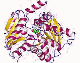
Porphobilinogen deaminase (hydroxymethylbilane synthase, or uroporphyrinogen I synthase) is an enzyme (EC 2.5.1.61) that in humans is encoded by the HMBS gene. Porphobilinogen deaminase is involved in the third step of the heme biosynthetic pathway. It catalyzes the head to tail condensation of four porphobilinogen molecules into the linear hydroxymethylbilane while releasing four ammonia molecules:

<span class="mw-page-title-main">Porphobilinogen</span> Chemical compound

Porphobilinogen (PBG) is an organic compound that occurs in living organisms as an intermediate in the biosynthesis of porphyrins, which include critical substances like hemoglobin and chlorophyll.

Erythropoietic porphyria is a type of porphyria associated with erythropoietic cells. In erythropoietic porphyrias, the enzyme deficiency occurs in the red blood cells.

<span class="mw-page-title-main">Aspartate kinase</span> Class of enzymes

Aspartate kinase or aspartokinase (AK) is an enzyme that catalyzes the phosphorylation of the amino acid aspartate. This reaction is the first step in the biosynthesis of three other amino acids: methionine, lysine, and threonine, known as the "aspartate family". Aspartokinases are present only in microorganisms and plants, but not in animals, which must obtain aspartate-family amino acids from their diet. Consequently, methionine, lysine and threonine are essential amino acids in animals.

<span class="mw-page-title-main">Cystathionine beta synthase</span> Mammalian protein found in humans

Cystathionine-β-synthase, also known as CBS, is an enzyme (EC 4.2.1.22) that in humans is encoded by the CBS gene. It catalyzes the first step of the transsulfuration pathway, from homocysteine to cystathionine:

<span class="mw-page-title-main">PCBD1</span> Protein-coding gene in the species Homo sapiens

Pterin-4-alpha-carbinolamine dehydratase is an enzyme that in humans is encoded by the PCBD1 gene.

<span class="mw-page-title-main">ALAS2</span> Protein-coding gene in humans

Delta-aminolevulinate synthase 2 also known as ALAS2 is a protein that in humans is encoded by the ALAS2 gene. ALAS2 is an aminolevulinic acid synthase.

<span class="mw-page-title-main">ALAS1</span> Protein-coding gene in the species Homo sapiens

Delta-aminolevulinate synthase 1 also known as ALAS1 is a protein that in humans is encoded by the ALAS1 gene. ALAS1 is an aminolevulinic acid synthase.

<span class="mw-page-title-main">Morpheein</span> Model of protein allosteric regulation

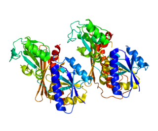
Morpheeins are proteins that can form two or more different homo-oligomers, but must come apart and change shape to convert between forms. The alternate shape may reassemble to a different oligomer. The shape of the subunit dictates which oligomer is formed. Each oligomer has a finite number of subunits (stoichiometry). Morpheeins can interconvert between forms under physiological conditions and can exist as an equilibrium of different oligomers. These oligomers are physiologically relevant and are not misfolded protein; this distinguishes morpheeins from prions and amyloid. The different oligomers have distinct functionality. Interconversion of morpheein forms can be a structural basis for allosteric regulation, an idea noted many years ago, and later revived. A mutation that shifts the normal equilibrium of morpheein forms can serve as the basis for a conformational disease. Features of morpheeins can be exploited for drug discovery. The dice image represents a morpheein equilibrium containing two different monomeric shapes that dictate assembly to a tetramer or a pentamer. The one protein that is established to function as a morpheein is porphobilinogen synthase, though there are suggestions throughout the literature that other proteins may function as morpheeins.

<span class="mw-page-title-main">Robert J. Desnick</span> American geneticist

Robert J. Desnick is an American human geneticist whose basic and translational research accomplishments include significant discoveries in genomics, pharmacogenetics, gene therapy, personalized medicine, and the treatment of genetic diseases. His translational research has led to the development the enzyme replacement therapy (ERT) and the chaperone therapy for Fabry disease, ERT for Niemann–Pick disease type B, and the RNA Interference Therapy for the Acute Hepatic Porphyrias.

<span class="mw-page-title-main">Aminolevulinic acid dehydratase deficiency porphyria</span> Medical condition

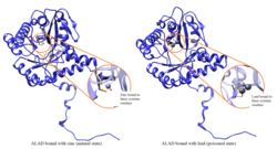
Aminolevulinic acid dehydratase deficiency porphyria is an extremely rare autosomal recessive metabolic disorder that results from inappropriately low levels of the enzyme delta-aminolevulinic acid dehydratase (ALAD), which is required for normal heme synthesis. This deficiency results in the accumulation of a toxic metabolic precursor in the heme synthesis pathway called aminolevulinic acid (ALA). Lead poisoning can also disrupt ALAD and result in elevated ALA causing the same symptoms. Heme is a component of hemoglobin which carries oxygen in red blood cells.

References

  1. 1 2 3 GRCh38: Ensembl release 89: ENSG00000148218 - Ensembl, May 2017
  2. 1 2 3 GRCm38: Ensembl release 89: ENSMUSG00000028393 - Ensembl, May 2017
  3. "Human PubMed Reference:". National Center for Biotechnology Information, U.S. National Library of Medicine.
  4. "Mouse PubMed Reference:". National Center for Biotechnology Information, U.S. National Library of Medicine.
  5. Eiberg H, Mohr J, Nielsen LS (February 1983). "delta-Aminolevulinatedehydrase: synteny with ABO-AK1-ORM (and assignment to chromosome 9)". Clinical Genetics. 23 (2): 150–4. doi:10.1111/j.1399-0004.1983.tb01864.x. PMID   6839527. S2CID   27267679.
  6. Beaumont C, Foubert C, Grandchamp B, Weil D, Gross MS, Nordmann Y (May 1984). "Assignment of the human gene for delta aminolevulinate dehydrase to chromosome 9 by somatic cell hybridization and specific enzyme immunoassay". Annals of Human Genetics. 48 (2): 153–9. doi:10.1111/j.1469-1809.1984.tb01010.x. PMID   6378062. S2CID   24098976.
  7. 1 2 3 4 Jaffe EK, Lawrence SH (March 2012). "Allostery and the dynamic oligomerization of porphobilinogen synthase". Archives of Biochemistry and Biophysics. 519 (2): 144–53. doi:10.1016/j.abb.2011.10.010. PMC   3291741 . PMID   22037356.
  8. Breinig S, Kervinen J, Stith L, Wasson AS, Fairman R, Wlodawer A, et al. (September 2003). "Control of tetrapyrrole biosynthesis by alternate quaternary forms of porphobilinogen synthase". Nature Structural Biology. 10 (9): 757–63. doi:10.1038/nsb963. PMID   12897770. S2CID   24188785.
  9. Lawrence SH, Jaffe EK (2008). "Expanding the Concepts in Protein Structure-Function Relationships and Enzyme Kinetics: Teaching using Morpheeins". Biochemistry and Molecular Biology Education. 36 (4): 274–283. doi:10.1002/bmb.20211. PMC   2575429 . PMID   19578473.
  10. Abadin H, Ashizawa A, Stevens YW, Llados F, Diamond G, Sage G, Citra M, Quinones A, Bosch SJ, Swarts SG (August 2007). Toxicological Profile for Lead (PDF). Atlanta, GA: Agency for Toxic Substances and Disease Registry (US). pp. 22, 30. PMID   24049859 . Retrieved 22 November 2015.
  11. "Entrez Gene: ALAD aminolevulinate, delta-, dehydratase".
  12. ALA dehydratase reaction, from NetBiochem at the University of Utah. Last modified 1/5/95
  13. 1 2 Jaffe EK, Stith L (February 2007). "ALAD porphyria is a conformational disease". American Journal of Human Genetics. 80 (2): 329–37. doi:10.1086/511444. PMC   1785348 . PMID   17236137.
  14. Overview of the Porphyrias Archived 2011-07-22 at the Wayback Machine at The Porphyrias Consortium (a part of NIH Rare Diseases Clinical Research Network (RDCRN)) Retrieved June 2011

Further reading