Foraminifera test

Last updated

Foraminiferal tests are the tests (or shells) of Foraminifera.

Contents

Foraminiferan tests (ventral view) Benthic foraminifera.jpg
Foraminiferan tests (ventral view)

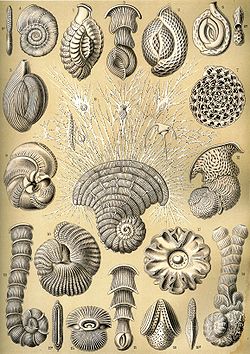
Foraminifera (forams for short) are single-celled predatory protists, mostly marine, and usually protected with shells. These shells, often called tests, can be single-chambered or have multiple interconnected chambers; the cellular machinery is contained within the shell. So important is the test to the biology of foraminifera that it provides the scientific name of the group—foraminifera, Latin for "hole bearers", referring to the pores connecting chambers of the shell in the multi-chambered species.

Foraminiferal tests are usually made of calcite, a form of calcium carbonate (CaCO
3
), but are sometimes made of aragonite, agglutinated sediment particles, chitin, or (rarely) of silica. [1] Other foraminifera lack tests altogether. [2]

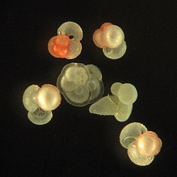
Over 50,000 species are recognized, both living (6,700 - 10,000) [3] [4] and fossil (40,000). [5] [6] They are usually less than 1 mm in size, but some are much larger, the largest species reaching up to 20 cm. [7] Most forams are benthic, but about 40 extant species are planktic. [8] The hard nature of most foraminiferal tests leads to an excellent fossil record, and they are widely researched to infer information about past climate and environments. [9]

External video
Nuvola apps kaboodle.svg foraminiferans
Nuvola apps kaboodle.svg Foraminiferal networks and growth

Background

Foraminiferal tests serve to protect the organism within. Owing to their generally hard and durable construction (compared to other protists), the tests of foraminifera are a major source of scientific knowledge about the group.

Openings in the test that allow the cytoplasm to extend outside are called apertures. [10] The primary aperture, leading to the exterior, take many different shapes in different species, including but not limited to rounded, crescent-shaped, slit-shaped, hooded, radiate (star-shaped), dendritic (branching). Some foraminifera have "toothed", flanged, or lipped primary apertures. There may be only one primary aperture or multiple; when multiple are present, they may be clustered or equatorial. In addition to the primary aperture, many foraminifera have supplemental apertures. These may form as relict apertures (past primary apertures from an earlier growth stage) or as unique structures.

Test shape is highly variable among different foraminifera; they may be single-chambered (unilocular) or multi-chambered (multilocular). In multilocular forms, new chambers are added as the organism grows. A wide variety of test morphologies is found in both unilocular and multilocular forms, including spiraled, serial, and milioline, among others. [11]

Many multi-chambered foraminifera follow a life cycle alternating between microspheric and megalospheric forms (see Foraminifera SS Reproduction). The name of each morph refers to the size of the initial chamber, rather than to the size of the entire organism. Megalosphere and Microsphere.png
Many multi-chambered foraminifera follow a life cycle alternating between microspheric and megalospheric forms (see Foraminifera § Reproduction). The name of each morph refers to the size of the initial chamber, rather than to the size of the entire organism.

Many foraminifera exhibit dimorphism in their tests, with microspheric and megalospheric individuals. These names should not be taken as referring to the size of the full organism; rather, they refer to the size of the first chamber, or proloculus. Tests as fossils are known from as far back as the Ediacaran period, [12] and many marine sediments are composed primarily of them. For instance, the limestone that makes up the pyramids of Egyp t is composed almost entirely of nummulitic benthic foraminifera. [13] It is estimated that reef foraminifera generate about 43 million tons of calcium carbonate per year. [14]

Genetic studies have identified the naked amoeba Reticulomyxa and the peculiar xenophyophores as foraminiferans without tests. A few other amoeboids produce reticulose pseudopodia, and were formerly classified with the forams as the Granuloreticulosa, but this is no longer considered a natural group, and most are now placed among the Cercozoa. [15]

Composition

The form and composition of their tests are the primary means by which forams are identified and classified. Most secrete calcareous tests, composed of calcium carbonate. [16] Calcareous tests may be composed of either aragonite or calcite depending on species; among those with calcite tests, the test may contain either a high or low fraction of magnesium substitution. [17] The test contains an organic matrix, which can sometimes be recovered from fossil samples. [17]

Some studies suggest a high amount of homoplasy in foraminifera, and that neither agglutinated nor calcareous foraminifera form monophyletic groupings. [18]

Soft

In some forams, the tests may be composed of organic material, typically the protein tectin. Tectin walls may have sediment particles loosely adhered onto the surface. [11] The foram Reticulomyxa entirely lacks a test, having only a membranous cell wall. [2] Organic-walled forams have traditionally been grouped as the "allogromiids"; however, genetic studies have found that this does not make up a natural group. [18]

Agglutinated

Other forams have tests made from small pieces of sediment cemented together (agglutinated) by either proteins (possibly collagen-related), calcium carbonate, or Iron (III) oxide. [11] [19] In the past these forms were grouped together as the single-chambered "astrorhizids" and the multi-chambered textulariids. However, recent genetic studies suggest that "astrorhizids" do not make up a natural grouping, instead forming a broad base of the foram tree. [18]

Textulariid foraminifera, unlike other living members of the globothalamea, have agglutinated tests; however, grains in these tests are cemented with a calcite cement. This calcite cement is made up of small (<100 nm) globular nanograins, similar to in other globothalameans. These tests may also have many pores, another feature uniting them with the globothalamea. [20]

Agglutinating foraminifera may be selective regarding what particles they incorporate into their shells. Some species prefer certain sizes and types of rock particles; other species are preferential towards certain biological materials. Certain species of foraminifera are known to have preferentially agglutinated coccoliths to form their tests; others preferentially utilise echinoderm plates, diatoms, or even other foraminiferans' tests. [21]

The foraminifera Spiculosiphon preferentially agglutinates silica sponge spicules using an organic cement; it shows strong selectivity also towards shape, utilising elongated spicules on its "stalk" and shortened ones on its "bulb". It is thought to use the spicules as both a means of elevating itself off the seabed as well as to lengthen the reach of its pseudopodia to capture prey. [19]

Xenophyophores create the largest agglutinated tests of any foraminifera XenophyophoreNOAA.jpg
Xenophyophores create the largest agglutinated tests of any foraminifera

The agglutinated tests of xenophyophores are the largest of any foraminifera, reaching up to 20 cm in diameter. The name "xenophyophore", meaning "bearer of foreign bodies", refers to this agglutinating habit. Xenophyophores selectively uptake sediment grains between 63 and 500 µm, avoiding larger pebbles and finer silts; type of sediment seems to be a strong factor in which particles are agglutinated, as particle type preferentially includes sulfides, oxides, volcanic glass, and especially tests of smaller foraminifera. Xenophyophores 1.5 cm in diameter have been recorded completely naked, with no test whatsoever. [22]

Calcareous

Of those foraminifera with calcareous tests, several different structures of calcite crystals are found.

Porcelaneous

SEM photomicrograph of a miliolid test wall, showing the nanogranular extrados (e) and needlelike porcelain (p) layers. Miliolid wall SEM.png
SEM photomicrograph of a miliolid test wall, showing the nanogranular extrados (e) and needlelike porcelain (p) layers.
The porcelanous test of the miliolid foraminiferan Quinqueloculina from the North Sea Quinqueloculina seminula.jpg
The porcelanous test of the miliolid foraminiferan Quinqueloculina from the North Sea

Porcelaneous walls are found in the Miliolida. These consist of high-magnesium calcite organized with an ordered outer and inner calcite lining (the "extrados" and "intrados", respectively) and randomly oriented needle-shaped calcite crystals forming a thick center layer (the "porcelain"). An organic inner lining is also present. The external surface may have a pitted structure, but it is not perforated by holes. "Cornuspirid" miliolids apparently lack any extrados. [23] [24] [20]

Monocrystalline

SEM photomicrograph of Patellina sp., showing cleavage of monocrystalline test Patellina cleavage.png
SEM photomicrograph of Patellina sp., showing cleavage of monocrystalline test

A "monocrystalline" test structure has traditionally been described for the Spirillinida. However, these tests remain poorly understood and poorly described. Some supposed "monocrystalline" spirillinids have been found to actually have tests consisting of a mosaic of very small crystals when observed with scanning electron microscope. SEM observation of Patellina sp. suggests that a truly monocrystalline test may indeed be present, with apparent cleavage faces. [20]

Fibre bundles

SEM photomicrograph of a lagenid test wall, showing fibre bundle structures. Internal organic layer can be seen to the far right of the image. Lagenid microstructure.png
SEM photomicrograph of a lagenid test wall, showing fibre bundle structures. Internal organic layer can be seen to the far right of the image.

Lagenid tests consist of "fibre bundles" that can reach tens of micrometres long; each "bundle" is formed from a single calcite crystal, is triangular in cross-section, and has a pore in the centre (thought to be an artefact of test deposition). There is also an internal organic layer, attached to the "cone" structure of the fibre bundles. As the crystalline structure varies significantly from that of other calcareous foraminifera, it is thought to represent a separate evolution of the calcareous test. The exact mineralisation process of lagenids remains unclear. [24]

Hyaline

Rotaliid tests are described as "hyaline". They are formed from low-to-high-magnesium calcite "nanograins" positioned with their C-axes perpendicular to the external surface of the test. Further, these nanograins can have higher-level structure, such as rows, columns, or bundles. [20] The test wall is characteristically bilamellar (two-layered) and perforated throughout with small pores. The outer calcite layer of the test wall is referred to as the "outer lamina" while the inner calcite layer is referred to as the "inner lining"; this should not be confused with the organic inner lining beneath the test. Sandwiched between the outer lamina and the inner lining is the "median layer", a protein layer that separates the two. The median layer is quite variable; depending on the species it may be well-defined while in others it is not sharply delineated. Some genera may contain sediment particles within the median layer. [11] [25] [24]

The now-extinct Fusulinids have traditionally been considered unique in having tests of homogenous microgranular crystals with no preferred orientation and almost no cement. However, a 2017 study found that the supposed microgranular structure was actually the result of diagenetic alteration of the fossils, and that unaltered fusulinid tests instead had a hyaline structure. This suggests that the group is affiliated with the Globothalamea. [26]

SEM photomicrograph showing cross section of a rotaliid wall. Note "globular nanograins" and two-layered test wall. Arrows point to pores. Rotaliid test wall SEM.png
SEM photomicrograph showing cross section of a rotaliid wall. Note "globular nanograins" and two-layered test wall. Arrows point to pores.

Robertinids have aragonitic tests with perforations; these are similar to the tests of rotaliids in that they are formed from nanograins, however, they differ in composition and in having well-organised columnar domains. As the earliest planktonic forams had aragonitic tests, it has been suggested that this may represent a separate evolution of a planktonic lifestyle within the Robertinida, rather than being close relatives of Globigerinans. [20]

Hyaline aragonitic tests are also present in the Involutinida. [24]

Spicules

SEM photomicrograph of a carterinid test wall, showing secreted calcite spicules in organic matrix Carterina spiculotesta wall.png
SEM photomicrograph of a carterinid test wall, showing secreted calcite spicules in organic matrix

The Carterinids, including the genera Carterina and Zaninettia, have a unique crystalline structure of the test which long complicated their classification. The test in this genus consists of spicules of low-magnesium calcite, bound together with an organic matrix and containing "blebs" of organic matter; this led some researchers to conclude that the test must be agglutinated. However, life studies have failed to find agglutination, and in fact the genus has been discovered on artificial substrate where sediment particles do not accumulate. [27] A 2014 genetic study found carterinids to be an independent lineage within the Globothalamea, and supported the idea of the spicules being secreted as spicule shape differed consistently between specimens of Carterina and Zaninettia collected from the same locality (ovoid in Carterina, rounded-rectangular in Zaninettia). [28]

Silicate

One genus, Miliamellus , has a non-perforated test made of opaline silica. [29] It is similar in shape and structure to the porcelanous tests of typical miliolids; the test consists of an internal and external organic layer, as well as a middle silica layer made of elongate rods. This silica layer is further divided into outer, middle, and inner subunits; the outer and inner subunits each are approximately 0.2μm thick and consist of subparallel sheets of silica rods with their long axes parallel to the test surface. The middle subunit is approximately 18μm in thickness and consists of a three-dimensional lattice of silica rods with no organic component in the open space. The ultrastructure differs from that of miliolids in that the rods are over twice as long and twice as thick on average, in that the rods of Miliamellus are hollow rather than solid, and of course in having a silica test rather than calcite. [30]

Test wall construction

Anatomy and types of secreted foraminiferal walls Foraminifera wall types.png
Anatomy and types of secreted foraminiferal walls

When a secreted test is present, walls of foraminiferal tests may be either nonlamellar or lamellar.

Nonlamellar walls are found in some foraminifera, such as Carterinida , Spirillinida , and Miliolida. In these forms, the secretion of a new chamber is not associated with any further deposition over previous chambers. As such there is no associated layering of calcite layers on the test. [25]

In foraminifera with lamellar walls, the deposition of a new chamber is accompanied by the deposition of a layer over previously-formed chambers. This layer may cover all previous chambers, or it may cover only some of them. These layers are known as secondary lamellae.

Foraminifera with lamellar walls can be further broken down into those with monolamellar walls and those with bilamellar walls. Monolamellar foraminifera secrete test walls which consist of a single layer, while those of bilamellar foraminifera are double-layered with an organic "median layer", sometimes containing sediment particles. In the case of bilamellar foraminifera, the outer layer is referred to as the "outer lamella" whilst the inner layer is referred to as the "inner lining". Monolamellar forams include the Lagenida, while bilamellar forms include the Rotaliida (including the major planktonic subgroup, the Globigerinina). [25]

Bilamellar test walls can be further divided into those with septal flaps (a layer of test wall covering the previously-secreted septum) and those lacking septal flaps. Septal flaps are not known to be present in any foraminifera other than those with bilamellar walls.

The presence of a septal flap is often, though not always, associated with the presence of an interlocular space. As the name suggests, this is a small space located between chambers; it may be open and form part of the outer surface of the test, or it may be enclosed to form a void. The layer enclosing the void is formed from different parts of the lamellae in different genera, suggesting an independent evolution of enclosed interlocular spaces in order to strengthen the test. [25]

Foraminiferans
Foram-globigerina hg.jpg
Empty globigerinid test, showing multiple test chambers with pores
G bulloides Brady 1884.jpg
...and pseudopodia emanating from pores in life
Benthic foraminifera Favulina hexagona, together with nanofossils enclosed inside the shell hexagons Favulina hexagona.png
Benthic foraminifera Favulina hexagona, together with nanofossils enclosed inside the shell hexagons
Foraminiferan shapes
Haeckel Thalamphora.jpg
Haeckel Thalamophora 12.jpg
      Drawings by Haeckel 1904 (click for details)

Related Research Articles

<span class="mw-page-title-main">Xenophyophorea</span> Clade of single-celled organisms

Xenophyophorea is a clade of foraminiferans. Members of this class are multinucleate unicellular organisms found on the ocean floor throughout the world's oceans, at depths of 500 to 10,600 metres. They are a kind of foraminiferan that extract minerals from their surroundings and use them to form an exoskeleton known as a test.

<span class="mw-page-title-main">Foraminifera</span> Phylum of amoeboid protists

Foraminifera are single-celled organisms, members of a phylum or class of amoeboid protists characterized by streaming granular ectoplasm for catching food and other uses; and commonly an external shell of diverse forms and materials. Tests of chitin are believed to be the most primitive type. Most foraminifera are marine, the majority of which live on or within the seafloor sediment, while a smaller number float in the water column at various depths, which belong to the suborder Globigerinina. Fewer are known from freshwater or brackish conditions, and some very few (nonaquatic) soil species have been identified through molecular analysis of small subunit ribosomal DNA.

<span class="mw-page-title-main">Microfossil</span> Fossil that requires the use of a microscope to see it

A microfossil is a fossil that is generally between 0.001 mm and 1 mm in size, the visual study of which requires the use of light or electron microscopy. A fossil which can be studied with the naked eye or low-powered magnification, such as a hand lens, is referred to as a macrofossil.

<span class="mw-page-title-main">Calcareous</span> Adjective meaning mostly or partly composed of calcium carbonate

Calcareous is an adjective meaning "mostly or partly composed of calcium carbonate", in other words, containing lime or being chalky. The term is used in a wide variety of scientific disciplines.

<span class="mw-page-title-main">Fusulinida</span> Extinct order of single-celled organisms

The Fusulinida is an extinct order within the Foraminifera in which the tests are traditionally considered to have been composed of microgranular calcite. Like all forams, they were single-celled organisms. In advanced forms the test wall was differentiated into two or more layers. Loeblich and Tappan, 1988, gives a range from the Lower Silurian to the Upper Permian, with the fusulinid foraminifera going extinct with the Permian–Triassic extinction event. While the latter is true, a more supported projected timespan is from the Mid-Carboniferous period.

<span class="mw-page-title-main">Textulariida</span> Order of single-celled organisms

The Textulariida are an order of foraminifera that produce agglutinated shells or tests. An agglutinated test is one made of foreign particles glued together with an organic or calcareous cement to form an external shell on the outside of the organism. Commonly, the order had been made up of all species of Foraminifera with these types of shells, but genetic studies indicate these organisms do not form an evolutionary group, and several superfamilies in the order have been moved to the order Allogromiida. The remaining forms are sometimes divided into three orders: the Trochamminida and Lituolida, which have organic cement, and the Textulariida sensu stricto, which use a calcareous cement. All three orders or superfamilies are known as fossils from the Cambrian onwards.

<span class="mw-page-title-main">Test (biology)</span> Hard shell of some spherical marine animals

In biology, a test is the hard shell of some spherical marine animals and protists, notably sea urchins and microorganisms such as testate foraminiferans, radiolarians, and testate amoebae. The term is also applied to the covering of scale insects. The related Latin term testa is used for the hard seed coat of plant seeds.

<span class="mw-page-title-main">Marine sediment</span>

Marine sediment, or ocean sediment, or seafloor sediment, are deposits of insoluble particles that have accumulated on the seafloor. These particles have their origins in soil and rocks and have been transported from the land to the sea, mainly by rivers but also by dust carried by wind and by the flow of glaciers into the sea. Additional deposits come from marine organisms and chemical precipitation in seawater, as well as from underwater volcanoes and meteorite debris.

Carterina is a genus in the family Trochamminidae, composing its own subfamily Carterininae. The genus is described from specimens gathered during the Challenger expedition's circumnavigation of the Earth from 1872-1876.

<span class="mw-page-title-main">Carterinida</span> Order of single-celled organisms

Carterinida is an order of multi-chambered foraminifera within the Globothalamea. Members of this order form hard tests out of thin calcite rods known as spicules, which are held together by a proteinaceous matrix. As of August 2023, the order contains a single family, Carterinidae.

Miliamellus is a genus of Cenozoic benthic foraminifera with tests made of imperforate opaline silica. It is the only genus in the order Silicoloculinida and the family Silicoloculinidae. It is sometimes referred to by the junior synonym Silicoloculina.

Clavulina is a genus of aggulinated benthic foraminiferans with an elongate test. The early stage is triserial and triangular in section, the later stage uniserial and rectilinear, with angular to rounded section. In some species agglutinated walls have considerable calcareous cement. Septa are secondarily doubled as a result of imperforate floors, which are added as new chambers are formed. Walls contain fine bifurcating canaliculi within, openings of which are sealed internally by an inner organic lining, and externally by the imperforate surface layer of the wall. The aperture is interiomarginal in the early triserial stage, terminal and rounded in the adult.

Clavulinopsis is a genus of foraminifera from the Upper Cretaceous of the United States, included in the Textulariida. The type species is Clavulinopsis hofkeri Banner and Desai, 1985.

<i>Amphistegina</i> Genus of single-celled organisms

Amphistegina is a genus of foraminiferal protists included in the Rotaliida with a stratigraphic range extending from the Eocene to recent and a cosmopolitan distribution. The test is an asymmetrically biconvex trochospiral that may be bi-involute or partially evolute on the spiral side. Chambers are numerous, broad. and low, strongly curved back at the periphery to form chamber prolongations. The umbilical side is stellate, like that of Asterigerina, and has a distinct umbilical plug. The wall is calcareous, optically radial; the surface finely perforate and smooth overall. The periphery angular to carinate (keeled); the aperture an interiomarginal slit on the umbilical side, bordered by a lip.

<span class="mw-page-title-main">Rotaliata</span> Class of single-celled organisms

Rotaliata is a class of Foraminifera characterized by tests that are exclusively multichambered, mostly planospiral or trochspiral, or derived from either. The aperture is commonly at the base of the apertural face, at least in early stages, but may be terminal, and single or complex. Test interior may be complex with secondary chambers and interconnecting canal system.

<span class="mw-page-title-main">Globothalamea</span> Class of single-celled organisms

Globothalamea comprises a class of multichambered foraminifera based in part on SSU rDNA evidence; the other is Tubothalamea.

Stercomata are extracellular pellets of waste material produced by some groups of foraminiferans, including xenophyophoreans and komokiaceans, Gromia, and testate amoebae. The pellets are ovoid (egg-shaped), brownish in color, and on average measure from 10-20 µm in length. Stercomata are composed of small mineral grains and undigested waste products held together by strands of glycosaminoglycans.

<span class="mw-page-title-main">Monothalamea</span> Taxonomic group of foraminifera

"Monothalamea" is a grouping of foraminiferans, traditionally consisting of all foraminifera with single-chambered tests. Recent work has shown that the grouping is paraphyletic, and as such does not constitute a natural group; nonetheless, the name "monothalamea" continues to be used by foraminifera workers out of convenience.

<i>Occultammina</i> Genus of single-celled organisms

Occultammina is a genus of xenophyophorean foraminifera known from the Atlantic and Pacific oceans. It is notable for being the first known infaunal xenophyophore as well as for being a possible identity for the enigmatic trace fossil Paleodictyon.

Global paleoclimate indicators are the proxies sensitive to global paleoclimatic environment changes. They are mostly derived from marine sediments. Paleoclimate indicators derived from terrestrial sediments, on the other hand, are commonly influenced by local tectonic movements and paleogeographic variations. Factors governing the earth climate system include plate tectonics, which controls the configuration of continents, the interplay between the atmosphere and the ocean, and the earth's orbital characteristics. Global paleoclimate indicators are established based on the information extracted from the analyses of geologic materials, including biological, geochemical and mineralogical data preserved in marine sediments. Indicators are generally grouped into three categories; paleontological, geochemical and lithological.

References

  1. Kennett, J.P.; Srinivasan, M.S. (1983). Neogene planktonic foraminifera: a phylogenetic atlas. Hutchinson Ross. ISBN   978-0-87933-070-5.
  2. 1 2 Pawlowski, Jan; Bolivar, Ignacio; Fahrni, Jose F.; Vargas, Colomban De; Bowser, Samuel S. (1999). "Molecular Evidence That Reticulomyxa Filosa Is A Freshwater Naked Foraminifer". Journal of Eukaryotic Microbiology. 46 (6): 612–617. doi:10.1111/j.1550-7408.1999.tb05137.x. ISSN   1550-7408. PMID   10568034. S2CID   36497475.
  3. Pawlowski, J.; Lejzerowicz, F.; Esling, P. (1 October 2014). "Next-Generation Environmental Diversity Surveys of Foraminifera: Preparing the Future". The Biological Bulletin. 227 (2): 93–106. doi:10.1086/BBLv227n2p93. ISSN   0006-3185. PMID   25411369. S2CID   24388876.
  4. Ald, S.M. et al. (2007) Diversity, Nomenclature, and Taxonomy of Protists, Syst. Biol. 56(4), 684–689, DOI: 10.1080/10635150701494127.
  5. Pawlowski, J., Lejzerowicz, F., & Esling, P. (2014). Next-generation environmental diversity surveys of foraminifera: preparing the future. The Biological Bulletin, 227(2), 93–106.
  6. "World Foraminifera Database".
  7. Marshall M (3 February 2010). "Zoologger: 'Living beach ball' is giant single cell". New Scientist .
  8. Hemleben, C.; Anderson, O.R.; Spindler, M. (1989). Modern Planktonic Foraminifera. Springer-Verlag. ISBN   978-3-540-96815-3.
  9. Wassilieff, Maggy (2006) "Plankton – Animal plankton", Te Ara – the Encyclopedia of New Zealand. Accessed: 2 November 2019.
  10. Lana, C (2001). "Cretaceous Carterina (Foraminifera)". Marine Micropaleontology. 41 (1–2): 97–102. Bibcode:2001MarMP..41...97L. doi:10.1016/S0377-8398(00)00050-5.
  11. 1 2 3 4 Saraswati, Pratul Kumar; Srinivasan, M. S. (2016), Saraswati, Pratul Kumar; Srinivasan, M.S. (eds.), "Calcareous-Walled Microfossils", Micropaleontology: Principles and Applications, Springer International Publishing, pp. 81–119, doi:10.1007/978-3-319-14574-7_6, ISBN   978-3-319-14574-7
  12. Kontorovich, A. E.; Varlamov, A. I.; Grazhdankin, D. V.; Karlova, G. A.; Klets, A. G.; Kontorovich, V. A.; Saraev, S. V.; Terleev, A. A.; Belyaev, S. Yu.; Varaksina, I. V.; Efimov, A. S. (1 December 2008). "A section of Vendian in the east of West Siberian Plate (based on data from the Borehole Vostok 3)". Russian Geology and Geophysics. 49 (12): 932–939. Bibcode:2008RuGG...49..932K. doi:10.1016/j.rgg.2008.06.012. ISSN   1068-7971.
  13. Foraminifera: History of Study, University College London, retrieved 20 September 2007
  14. Langer, M. R.; Silk, M. T. B.; Lipps, J. H. (1997). "Global ocean carbonate and carbon dioxide production: The role of reef Foraminifera". Journal of Foraminiferal Research. 27 (4): 271–277. doi:10.2113/gsjfr.27.4.271.
  15. Adl, S. M.; Simpson, A. G. B.; Farmer, M. A.; Anderson; et al. (2005). "The new higher level classification of Eukaryotes with emphasis on the taxonomy of Protists". Journal of Eukaryotic Microbiology. 52 (5): 399–451. doi: 10.1111/j.1550-7408.2005.00053.x . PMID   16248873. S2CID   8060916.
  16. Sen Gupta, Barun K. (1982). "Ecology of benthic Foraminifera". In Broadhead, T.W. (ed.). Foraminifera: notes for a short course organized by M.A. Buzas and B.K. Sen Gupta. Studies in Geology. Vol. 6. University of Tennessee, Dept. of Geological Sciences. pp. 37–50. ISBN   978-0910249058. OCLC   9276403.
  17. 1 2 Sen Gupta, Barun K. (2003), "Systematics of Modern Foraminifera", in Sen Gupta, Barun K. (ed.), Modern Foraminifera, Springer Netherlands, pp. 7–36, doi:10.1007/0-306-48104-9_2, ISBN   978-0-306-48104-8
  18. 1 2 3 Pawlowski, Jan; Holzmann, Maria; Tyszka, Jarosław (1 April 2013). "New supraordinal classification of Foraminifera: Molecules meet morphology". Marine Micropaleontology. 100: 1–10. Bibcode:2013MarMP.100....1P. doi:10.1016/j.marmicro.2013.04.002. ISSN   0377-8398.
  19. 1 2 Maldonado, Manuel; López-Acosta, María; Sitjà, Cèlia; Aguilar, Ricardo; García, Silvia; Vacelet, Jean (10 June 2013). "A giant foraminifer that converges to the feeding strategy of carnivorous sponges: Spiculosiphon oceana sp. nov. (Foraminifera, Astrorhizida)". Zootaxa. 3669 (4): 571–584. doi:10.11646/zootaxa.3669.4.9. hdl: 10261/92975 . ISSN   1175-5334. PMID   26312358.
  20. 1 2 3 4 5 Dubicka, Zofia (2019). "Chamber arrangement versus wall structure in the high-rank phylogenetic classification of Foraminifera". Acta Palaeontologica Polonica. 64. doi: 10.4202/app.00564.2018 . ISSN   0567-7920.
  21. Thomsen, Erik; Rasmussen, Tine L. (1 July 2008). "Coccolith-Agglutinating Foraminifera from the Early Cretaceous and How They Constructed Their Tests". Journal of Foraminiferal Research. 38 (3): 193–214. doi:10.2113/gsjfr.38.3.193. ISSN   0096-1191.
  22. Levin, Lisa A.; Thomas, Cynthia L. (1 December 1988). "The ecology of xenophyophores (Protista) on eastern Pacific seamounts". Deep Sea Research Part A. Oceanographic Research Papers. 35 (12): 2003–2027. Bibcode:1988DSRA...35.2003L. doi:10.1016/0198-0149(88)90122-7. ISSN   0198-0149.
  23. Jain, Sreepat (2020), Jain, Sreepat (ed.), "Benthic Foraminifera", Fundamentals of Invertebrate Palaeontology: Microfossils, Springer Geology, New Delhi: Springer India, pp. 171–192, doi:10.1007/978-81-322-3962-8_9, ISBN   978-81-322-3962-8, S2CID   242629553
  24. 1 2 3 4 Dubicka, Zofia; Owocki, Krzysztof; Gloc, Michał (1 April 2018). "Micro- and Nanostructures of Calcareous Foraminiferal Tests: Insight from Representatives of Miliolida, Rotaliida and Lagenida". Journal of Foraminiferal Research. 48 (2): 142–155. doi:10.2113/gsjfr.48.2.142. ISSN   0096-1191.
  25. 1 2 3 4 Hansen, Hans Jørgen (2003), Sen Gupta, Barun K. (ed.), "Shell construction in modern calcareous Foraminifera", Modern Foraminifera, Dordrecht: Springer Netherlands, pp. 57–70, doi:10.1007/0-306-48104-9_4, ISBN   978-0-306-48104-8
  26. Dubicka, Zofia; Gorzelak, Przemysław (9 November 2017). "Unlocking the biomineralization style and affinity of Paleozoic fusulinid foraminifera". Scientific Reports. 7 (1): 15218. Bibcode:2017NatSR...715218D. doi:10.1038/s41598-017-15666-1. ISSN   2045-2322. PMC   5680253 . PMID   29123221.
  27. Machado, Altair; Barros, Facelucia (8 January 2013). "The occurrence of Carterina spiculotesta (Carter, 1877) on an artificial substrate". Check List. 9 (4): 813–814. doi: 10.15560/9.4.813 . ISSN   1809-127X.
  28. Pawlowski, Jan; Holzmann, Maria; Debenay, Jean-Pierre (1 October 2014). "Molecular Phylogeny of Carterina Spiculotesta and Related Species from New Caledonia". Journal of Foraminiferal Research. 44 (4): 440–450. doi:10.2113/gsjfr.44.4.440. ISSN   0096-1191.
  29. Sen Gupta, Barun K. (2002). Modern Foraminifera. Springer. p. 16. ISBN   978-1-4020-0598-5.
  30. Resig, J; Lowenstam, H; Echols, R; Weiner, S (1980). "An extant opaline foraminifer: test ultrastructure, mineralogy, and taxonomy". Special Publications of the Cushman Foundation for Foraminiferal Research. 19: 205–214.
  31. Favulina hexagona European Geosciences Union, 9 November 2020.
  32. Foraminifera: History of Study, University College London. Retrieved: 18 November 2019.