Leukoaraiosis

Last updated
Axial T2 FLAIR sequence MR image of a middle-aged man with leukoaraiosis. Leukoaraiosis.jpg
Axial T2 FLAIR sequence MR image of a middle-aged man with leukoaraiosis.
MRI image: Leukoaraiosis in a 90-year-old patient with cerebral atrophy. Leukoaraiosis 1.jpg
MRI image: Leukoaraiosis in a 90-year-old patient with cerebral atrophy.
Head CT showing periventricular white matter lesions. Periventricular white matter lesions (annotated).jpg
Head CT showing periventricular white matter lesions.

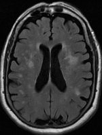
Leukoaraiosis is a particular abnormal change in appearance of white matter near the lateral ventricles. It is often seen in aged individuals, but sometimes in young adults. [1] [2] On MRI, leukoaraiosis changes appear as white matter hyperintensities (WMHs) in T2 FLAIR images. [3] [4] On CT scans, leukoaraiosis appears as hypodense periventricular white-matter lesions. [5]

Contents

Causes

The blue arrows indicate leucoaraiosis. In the left image these may well represent transependymal CSF diapedesis due to normal pressure hydrocephalus, which in turn is suggested by the narrowed superior CSF spaces and acute callosal angle. The unilateral occurrence of these alterations in right image suggests they are probably due to vascular encephalopathy. Normal pressure hydrocephalus versus atrophy.jpg
The blue arrows indicate leucoaraiosis. In the left image these may well represent transependymal CSF diapedesis due to normal pressure hydrocephalus, which in turn is suggested by the narrowed superior CSF spaces and acute callosal angle. The unilateral occurrence of these alterations in right image suggests they are probably due to vascular encephalopathy.

White matter hyperintensities can be caused by a variety of factors, including ischemia, micro-hemorrhages, gliosis, damage to small blood vessel walls, breaches of the barrier between the cerebrospinal fluid and the brain, or loss and deformation of the myelin sheath. [6] [7] Multiple small vessel infarcts in the subcortical white matter can cause the condition, often the result of chronic hypertension leading to lipohyalinosis of the small vessels. Patients may develop cognitive impairment and dementia. [8] [9]

Many patients can have leukoaraiosis without any associated clinical abnormality. However, underlying vascular mechanisms are suspected to be the cause of the imaging findings. Hypertension, smoking, diabetes, [3] hyperhomocysteinemia, and heart diseases are all risk factors for leukoaraiosis.

Leukoaraiosis has been reported to be an initial stage of Binswanger's disease but this evolution does not always happen.

Special cases

See also

References

  1. Putaala J., Kurkinen M., Tarvos V., Salonen O., Kaste M., Tatlisumak T. (2009). "Silent brain infarcts and leukoaraiosis in young adults with first-ever ischemic stroke". Neurology. 72 (21): 1823–1829. doi:10.1212/WNL.0b013e3181a711df. PMID   19470964. S2CID   593328.{{cite journal}}: CS1 maint: multiple names: authors list (link)
  2. 1 2 Aik Kah, Tan (2018). "CuRRL Syndrome: A Case Series" (PDF). Acta Scientific Ophthalmology. 1 (3): 9–13.
  3. 1 2 Habes M, Erus G, Toledo JB, Zhang T, Bryan N, Launer LJ, Rosseel Y, Janowitz D, Doshi J, Van der Auwera S, von Sarnowski B, Hegenscheid K, Hosten N, Homuth G, Völzke H, Schminke U, Hoffmann W, Grabe H, Davatzikos C (2016). "White matter hyperintensities and imaging patterns of brain ageing in the general population". Brain . 139 (Pt 4): 1164–79. doi:10.1093/brain/aww008. PMC   5006227 . PMID   26912649.
  4. Yan, Shenqiang; Wan, Jinping; Zhang, Xuting; Tong, Lusha; Zhao, Song; Sun, Jianzhong; Lin, Yuehan; Shen, Chunhong; Lou, Min (2014). "Increased Visibility of Deep Medullary Veins in Leukoaraiosis: A 3-T MRI Study". Frontiers in Aging Neuroscience. 6: 144. doi: 10.3389/fnagi.2014.00144 . PMC   4074703 . PMID   25071553.
  5. Kobari M, Meyer JS, Ichijo M, Oravez WT (1990). "Leukoaraiosis: correlation of MR and CT findings with blood flow, atrophy, and cognition". AJNR Am J Neuroradiol. 11 (2): 273–81. PMC   8334682 . PMID   2107711.
  6. Raz N, Yang Y, Dahle CL, Land S (2012). "Volume of white matter hyperintensities in healthy adults: contribution of age, vascular risk factors, and inflammation-related genetic variants". Biochimica et Biophysica Acta (BBA) - Molecular Basis of Disease . 1822 (3): 361–69. doi:10.1016/j.bbadis.2011.08.007. PMC   3245802 . PMID   21889590.
  7. Lin, Jing; Wang, Dilong; Lan, Linfang; Fan, Yuhua (2017). "Multiple Factors Involved in the Pathogenesis of White Matter Lesions". BioMed Research International. 2017 9372050. doi: 10.1155/2017/9372050 . ISSN   2314-6141. PMC   5339523 . PMID   28316994.
  8. Fauci, Anthony S.; Braunwald, Eugene; Weiner, Charles; Kasper, Dennis L.; Hauser, Stephen L.; Longo, Dan L.; Jameson, J. Larry; Loscalzo, Joseph (2008). Harrison's Principles of Internal Medicine (17th ed.). New York: McGraw-Hill. ISBN   978-0-07-149619-3.[ page needed ]
  9. Hu, He-Ying; Ou, Ya-Nan; Shen, Xue-Ning; Qu, Yi; Ma, Ya-Hui; Wang, Zuo-Teng; Dong, Qiang; Tan, Lan; Yu, Jin-Tai (January 2021). "White matter hyperintensities and risks of cognitive impairment and dementia: A systematic review and meta-analysis of 36 prospective studies". Neuroscience and Biobehavioral Reviews. 120: 16–27. doi:10.1016/j.neubiorev.2020.11.007. ISSN   1873-7528. PMID   33188821. S2CID   226301188.
  10. O'Sullivan M, Morris RG, Huckstep B, Jones DK, Williams SCR, Markus HS (2004). "Diffusion tensor MRI correlates with executive dysfunction in patients with ischaemic leukoaraiosis". J Neurol Neurosurg Psychiatry. 75 (3): 441–47. doi:10.1136/jnnp.2003.014910. PMC   1738975 . PMID   14966162.{{cite journal}}: CS1 maint: multiple names: authors list (link)
  11. Maldjian JA, Whitlow CT, Saha BN, Kota G, Vandergriff C, Davenport EM, Divers J, Freedman BI, Bowden DW. "Automated White Matter Total Lesion Volume Segmentation in Diabetes". AJNR Am J Neuroradiol. 2013 Jul 18

Further reading