Multiple sclerosis diagnosis

Last updated

Diagnosis of multiple sclerosis
Monthly multiple sclerosis anim cropped no text.gif
Animation showing dissemination of brain lesions in time and space as demonstrated by monthly MRI studies along a year
Purposediagnosis via lab test, imaging and symptoms

Current standards for diagnosing multiple sclerosis (MS) are based on the 2018 revision of McDonald criteria. They rely on MRI detection (or clinical demonstration) of demyelinating lesions in the CNS, which are distributed in space (DIS) and in time (DIT). It is also a requirement that any possible known disease that produces demyelinating lesions is ruled out before applying McDonald's criteria.[ citation needed ]

Contents

This last requirement makes MS an ill-defined entity, whose borders change every time that a new disease is set apart. Some cases previously considered MS are now considered distinct conditions, like Neuromyelitis optica or antiMOG associated encephalomyelitis. Because of the requirement of distributed lesions, a single lesion (RIS) is not considered MS. For the same reason, the prodromal stage of MS (the unknown condition that causes the lesions) would not be considered as MS if it could be found.[ citation needed ]

Sometimes the diagnosis must be retrospective, relying on gradual worsening of neurological signs/symptoms, due to the lack of understanding of the pathogenicity driving disease progression. [1] However, the only definite diagnosis of MS is post-mortem autopsy, where lesions typical of MS can be detected through histopathological techniques. [2] [3]

Overview

Multiple sclerosis is typically diagnosed based on the presenting signs and symptoms, in combination with supporting medical imaging and laboratory testing. [4] It can be difficult to confirm, especially early on, since the signs and symptoms may be similar to those of other medical problems. [5] [6]

The McDonald criteria, which focus on clinical, laboratory, and radiologic evidence of lesions at different times and in different areas, is the most commonly used method of diagnosis [7] with the Schumacher and Poser criteria being of mostly historical significance. [8] While the above criteria allow for a non-invasive diagnosis, some state that the only definitive proof is an autopsy or biopsy where lesions typical of MS are detected. [5] [2] [3]

Clinical data alone may be sufficient for a diagnosis of MS if an individual has had separate episodes of neurologic symptoms characteristic of the disease. [2] In those who seek medical attention after only one attack, other testing is needed for the diagnosis. The most commonly used diagnostic tools are neuroimaging, analysis of cerebrospinal fluid and evoked potentials. Magnetic resonance imaging of the brain and spine may show areas of demyelination (lesions or plaques). Gadolinium can be administered intravenously as a contrast agent to highlight active plaques and, by elimination, demonstrate the existence of historical lesions not associated with symptoms at the moment of the evaluation. [2] [9] Testing of cerebrospinal fluid obtained from a lumbar puncture can provide evidence of chronic inflammation in the central nervous system. The cerebrospinal fluid is tested for oligoclonal bands of IgG on electrophoresis, which are inflammation markers found in 75–85% of people with MS. [2] [10] The nervous system in MS may respond less actively to stimulation of the optic nerve and sensory nerves due to demyelination of such pathways. These brain responses can be examined using visual- and sensory-evoked potentials. [11]

Prodomal or early phase

Studies have found increased interactions with health services by people who were diagnosed with MS several years later. The range of symptoms included pain and fatigue. Studies continue on whether MS has a prodromal or early phase, and could possibly be diagnosed and treated much earlier. [12] [13] [14] [15]

DMT effectiveness

A range of disease-modifying treatments (DMT) are now available, to reduce the long-term progression of MS. Studies are increasingly indicating that early and aggressive use of DMTs is beneficial for long-term outcomes. [16] [17]

Diagnostic criteria

McDonald criteria

As of 2021, the McDonald criteria for MS are the most commonly used.

The 2017 McDonald criteria can be summarize in this table:

Clinical PresentationAdditional Data Needed
* 2 or more attacks (relapses)
* 2 or more objective clinical lesions
None; clinical evidence will suffice (additional evidence desirable but must be consistent with MS)
* 2 or more attacks
* 1 objective clinical lesion (as well as clear-cut historical evidence of a previous attack involving a lesion in a distinct anatomical location)
None.
* 2 or more attacks
* 1 objective clinical lesion
Dissemination in space, demonstrated by an additional clinical attack implication a different CNS site or by MRI.
* 1 attack
* 2 or more objective clinical lesions
Dissemination in time, demonstrated by an additional clinical attack or by MRI

OR

Demonstration of CSF-specific oligoclonal bands

* 1 attack
* 1 objective clinical lesion
(monosymptomatic presentation)
Dissemination in space demonstrated by an additional clinical attack implicating a different CNS site or by MRI.
AND
Dissemination in time demonstrated by an additional clinical attack or by MRI, OR

Demonstration of CSF-specific oligoclonal bands

Insidious neurological progression
suggestive of MS
(primary progressive MS)
One year of disease progression (retrospectively or prospectively determined) and

Two of the following:

  • One or more T2-hyperintense lesions characteristic of multiple sclerosis in one or more of the following brain regions: periventricular, cortical or juxtacortical, or infratentorial
  • Two or more T2-hyperintense lesions in the spinal cord
  • Presence of CSF-specific oligoclonal bands

Okuda Criteria

These criteria, used mainly for research in MS, define what should be considered a Radiologically Isolated Syndrome (RIS). [18] Some reports point to the possibility of predicting RIS to CIS conversion based on oligoclonal bands and neurofilament light chain. [19]

Research into diagnostic techniques

Multiple sclerosis diagnosis can only be made when there is proof of lesions disseminated in time and in space. Therefore, when damage in the CNS is big enough to be seen. It would be desirable to make it faster.

The ideal diagnosis schema would be able to determine for any given subject, if he will develop MS, at any point in his life, and when. Nevertheless, not enough is currently known about the MS underlying conditions to achieve that.

In order to get as close as possible to the ideal diagnosis status, a lot of research into multiple sclerosis biomarkers is taking place currently.[ citation needed ]

Biomarkers in MS

An active field of research is looking for biomarkers for MS that could speed-up the diagnosis doing it more accurate at the same time. While most of them are still under research, there are some of them already well stablished:

Differential diagnosis

Several conditions can mimic MS. Given the unknown pathogenesis of MS, its differential diagnosis is based in exclusion of known conditions.[ citation needed ]

Very close diseases with similar symptoms are the whole "inflammatory demyelinating diseases spectrum", specially neuromyelitis optica and anti-MOG associated encephalomyelitis.[ citation needed ]

Outside this spectrum, another important mimic is neuroborreliosis. A Borrelia-specific IgG index exists, and testing for it could make the differential diagnosis. [22]

Clinical courses

Progression of MS subtypes, 1996 revision Ms progression types.svg
Progression of MS subtypes, 1996 revision

Several phenotypes (commonly named types), or patterns of progression, have been described. Phenotypes use the past course of the disease in an attempt to predict the future course. They are important not only for prognosis but also for treatment decisions. In 1996, the United States National Multiple Sclerosis Society described four clinical courses. [23]

The original structure approved in 1996, sometimes still used, was:

  1. relapsing-remitting
  2. secondary progressive (SPMS)
  3. primary progressive (PPMS)
  4. progressive relapsing.

This set of courses was reviewed by an international panel in 2013, [24] [25] adding clinically isolated syndrome (CIS) and radiologically isolated syndrome (RIS) as phenotypes, and finally removing the "progressive relapsing" phenotype. They also added modificators for the clinical courses based on a pair of characteristics: Active/non-active and with/without progression.

The four currently accepted courses or stages are:

  1. Clinically isolated syndrome (CIS)
  2. Relapsing-remitting MS (RRMS)
  3. Primary progressive MS (PPMS)
  4. Secondary progressive MS (SPMS)

Relapsing-remitting

The relapsing-remitting subtype is characterized by unpredictable relapses followed by periods of months to years of relative quiet (remission) with no new signs of disease activity. Deficits that occur during attacks may either resolve or leave problems, the latter in about 40% of attacks and being more common the longer a person has had the disease. [4] [5] This describes the initial course of 80% of individuals with MS. [5] When deficits always resolve between attacks, this is sometimes referred to as benign MS, [26] although people will still build up some degree of disability in the long term. [5] On the other hand, the term malignant multiple sclerosis is used to describe people with MS having reached significant level of disability in a short period. [27] The relapsing-remitting subtype usually begins with a clinically isolated syndrome (CIS). In CIS, a person has an attack suggestive of demyelination, but does not fulfill the criteria for multiple sclerosis. [5] [28] 30 to 70% of persons experiencing CIS later develop MS. [28]

Secondary progressive

Nerve axon with myelin sheath Nerve.nida.jpg
Nerve axon with myelin sheath

Secondary progressive MS occurs in around 65% of those with initial relapsing-remitting MS, who eventually have progressive neurologic decline between acute attacks without any definite periods of remission. [5] [23] Occasional relapses and minor remissions may appear. [23] The most common length of time between disease onset and conversion from relapsing-remitting to secondary progressive MS is 19 years. [29]

Primary progressive

The primary progressive subtype occurs in approximately 10–20% of individuals, with no remission after the initial symptoms. [4] [30] It is characterized by progression of disability from onset, with no, or only occasional and minor, remissions and improvements. [23] The usual age of onset for the primary progressive subtype is later than of the relapsing-remitting subtype. It is similar to the age that secondary progressive usually begins in relapsing-remitting MS, around 40 years of age. [5]

Progressive relapsing

Progressive relapsing MS describes those individuals who, from onset, have a steady neurologic decline but also have clear superimposed attacks. This is the least common of all subtypes. [23]

Atypical MS

Unusual types of MS have been described; these include Devic's disease, Balo concentric sclerosis, Schilder's diffuse sclerosis, and Marburg multiple sclerosis. There is debate on whether they are MS variants or different diseases. [31] Multiple sclerosis behaves differently in children, taking more time to reach the progressive stage. [5] Nevertheless, they still reach it at a lower average age than adults usually do. [5]

History

Since the first description of multiple sclerosis (MS) by Charcot, the Neurological community has been striving to create reliable and reproducible criteria for diagnosis of MS. [32] The first attempts were made by Charcot himself, followed by Marburg and later Allison. The first criteria however were lacking in sensitivity and specificity for clinical use. [32]

The first landmark event in the history of diagnostic criteria for MS was the development of the Schumacher criteria. These were the first internationally recognized criteria for diagnosis of MS and introduced very important diagnostic concepts that are the cornerstone of MS diagnosis nowadays, such as the clinical definition of MS and the requirement of dissemination in time and space for accurate diagnosis.[ citation needed ]

Since then, other diagnostic criteria have been proposed. Among them, Poser criteria utilized several laboratory and paraclinical studies to enhance the diagnostic accuracy. McDonald criteria, which are the ones used today, successfully introduced MRI findings as surrogates for the criterion of dissemination in time and space when clinical data are lacking, thus allowing earlier diagnosis of MS. [32]

Historic diagnostic criteria

Schumacher criteria

To get a diagnosis of CDMS a patient must show the following: [33]

  1. Clinical signs of a problem in the CNS
  2. Evidence of two or more areas of CNS involvement
  3. Evidence of white matter involvement
  4. One of these: Two or more relapses (each lasting ≥ 24 hr and separated by at least 1 month) or progression (slow or stepwise)
  5. Patient should be between 10 and 50 yr old at time of examination
  6. No better explanation for patient's symptoms and signs

The last condition, no better explanation for symptoms, has been heavily criticised, but it has been preserved and it is currently included in the new McDonalds criteria in the form that "no better explanation should exist for MRI observations"[ This quote needs a citation ]

Poser criteria

Poser criteria can be summarized in this table:

Any of the five conclusions have subpossibilities. Here a table is shown with each one of them:

Clinical PresentationAdditional Data Needed
CDMS* Two or more attacks (relapses)Two clinical evidence
One clinical and one paraclinical evidence
LSDMS* At least one attack and oligoclonal bands Two attacks and one evidence (clinical or paraclinical)
One attack and two clinical evidences
One attack, one clinical and one paraclinical evidences
CPMS* At least one attackTwo attacks and one clinical evidence
One attack and two clinical evidences
One attack, one clinical and one paraclinical evidences
LSPMS* Two attacksNo more evidence is required

If none of these requirements is fulfilled, the diagnosis is "No MS", meaning that there is not enough clinical evidence to support a clinical diagnosis of MS.

Barkhof-Tintoré criteria

Barkhof criteria, [34] later modified by Tintoré [35] were an early attempt to use MRI to diagnose MS. It was developed by Frederik Barkhof. [34]

Their observations were taken into account when McDonald criteria were published, and therefore they can be considered deprecated by the latter.

Related Research Articles

<span class="mw-page-title-main">Multiple sclerosis</span> Disease that damages the myelin sheaths around nerves

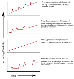
Multiple sclerosis (MS) is an autoimmune disease in which the insulating covers of nerve cells in the brain and spinal cord are damaged. This damage disrupts the ability of parts of the nervous system to transmit signals, resulting in a range of signs and symptoms, including physical, mental, and sometimes psychiatric problems. Specific symptoms can include double vision, vision loss, eye pain, muscle weakness, and loss of sensation or coordination. MS takes several forms, with new symptoms either occurring in isolated attacks or building up over time. In the relapsing forms of MS, between attacks, symptoms may disappear completely, although some permanent neurological problems often remain, especially as the disease advances. In the progressive forms of MS, bodily function slowly deteriorates and disability worsens once symptoms manifest and will steadily continue to do so if the disease is left untreated.

<span class="mw-page-title-main">Oligoclonal band</span> Marker in blood/cerebrospinal fluid testing

Oligoclonal bands (OCBs) are bands of immunoglobulins that are seen when a patient's blood serum, or cerebrospinal fluid (CSF) is analyzed. They are used in the diagnosis of various neurological and blood diseases. Oligoclonal bands are present in the CSF of more than 95% of patients with clinically definite multiple sclerosis.

Neuromyelitis optica spectrum disorders (NMOSD) are a spectrum of autoimmune diseases characterized by acute inflammation of the optic nerve and the spinal cord (myelitis). Episodes of ON and myelitis can be simultaneous or successive. A relapsing disease course is common, especially in untreated patients.

Interferon beta-1b is a cytokine in the interferon family used to treat the relapsing-remitting and secondary-progressive forms of multiple sclerosis (MS). It is approved for use after the first MS event. Closely related is interferon beta 1a, also indicated for MS, with a very similar drug profile.

<span class="mw-page-title-main">McDonald criteria</span>

The McDonald criteria are diagnostic criteria for multiple sclerosis (MS). These criteria are named after neurologist W. Ian McDonald who directed an international panel in association with the National Multiple Sclerosis Society (NMSS) of America and recommended revised diagnostic criteria for MS in April 2001. These new criteria intended to replace the Poser criteria and the older Schumacher criteria. They have undergone revisions in 2005, 2010 and 2017.

Experimental autoimmune encephalomyelitis, sometimes experimental allergic encephalomyelitis (EAE), is an animal model of brain inflammation. It is an inflammatory demyelinating disease of the central nervous system (CNS). It is mostly used with rodents and is widely studied as an animal model of the human CNS demyelinating diseases, including multiple sclerosis (MS) and acute disseminated encephalomyelitis (ADEM). EAE is also the prototype for T-cell-mediated autoimmune disease in general.

<span class="mw-page-title-main">Pathophysiology of multiple sclerosis</span>

Multiple sclerosis is an inflammatory demyelinating disease of the CNS in which activated immune cells invade the central nervous system and cause inflammation, neurodegeneration, and tissue damage. The underlying cause is currently unknown. Current research in neuropathology, neuroimmunology, neurobiology, and neuroimaging, together with clinical neurology, provide support for the notion that MS is not a single disease but rather a spectrum.

<span class="mw-page-title-main">Lesional demyelinations of the central nervous system</span>

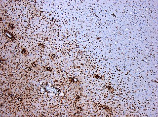
Multiple sclerosis and other demyelinating diseases of the central nervous system (CNS) produce lesions and glial scars or scleroses. They present different shapes and histological findings according to the underlying condition that produces them.

Inflammatory demyelinating diseases (IDDs), sometimes called Idiopathic (IIDDs) due to the unknown etiology of some of them, are a heterogenous group of demyelinating diseases - conditions that cause damage to myelin, the protective sheath of nerve fibers - that occur against the background of an acute or chronic inflammatory process. IDDs share characteristics with and are often grouped together under Multiple Sclerosis. They are sometimes considered different diseases from Multiple Sclerosis, but considered by others to form a spectrum differing only in terms of chronicity, severity, and clinical course.

<span class="mw-page-title-main">Balo concentric sclerosis</span> Medical condition

Baló's concentric sclerosis is a disease in which the white matter of the brain appears damaged in concentric layers, leaving the axis cylinder intact. It was described by József Mátyás Baló who initially named it "leuko-encephalitis periaxialis concentrica" from the previous definition, and it is currently considered one of the borderline forms of multiple sclerosis.

Research in multiple sclerosis may find new pathways to interact with the disease, improve function, curtail attacks, or limit the progression of the underlying disease. Many treatments already in clinical trials involve drugs that are used in other diseases or medications that have not been designed specifically for multiple sclerosis. There are also trials involving the combination of drugs that are already in use for multiple sclerosis. Finally, there are also many basic investigations that try to understand better the disease and in the future may help to find new treatments.

A clinically isolated syndrome (CIS) is a clinical situation of an individual's first neurological episode, caused by inflammation or demyelination of nerve tissue. An episode may be monofocal, in which symptoms present at a single site in the central nervous system, or multifocal, in which multiple sites exhibit symptoms. CIS with enough paraclinical evidence can be considered as a clinical stage of multiple sclerosis (MS). It can also be retrospectively diagnosed as a kind of MS when more evidence is available.

<span class="mw-page-title-main">Tumefactive multiple sclerosis</span> Medical condition

Tumefactive multiple sclerosis is a condition in which the central nervous system of a person has multiple demyelinating lesions with atypical characteristics for those of standard multiple sclerosis (MS). It is called tumefactive as the lesions are "tumor-like" and they mimic tumors clinically, radiologically and sometimes pathologically.

Diffuse myelinoclastic sclerosis, sometimes referred to as Schilder's disease, is a very infrequent neurodegenerative disease that presents clinically as pseudotumoural demyelinating lesions, making its diagnosis difficult. It usually begins in childhood, affecting children between 5 and 14 years old, but cases in adults are also possible.

Poser criteria are diagnostic criteria for multiple sclerosis (MS). They replaced the older Schumacher criteria, and are now considered obsolete as McDonald criteria have superseded them. Nevertheless, some of the concepts introduced have remained in MS research, like CDMS, and newer criteria are often calibrated against them. The criteria were unveiled in the Annals of Neurology in 1983 by a team led by Dr. Charles M. Poser.

Schumacher criteria are diagnostic criteria that were previously used for identifying multiple sclerosis (MS). Multiple sclerosis, understood as a central nervous system (CNS) condition, can be difficult to diagnose since its signs and symptoms may be similar to other medical problems. Medical organizations have created diagnostic criteria to ease and standardize the diagnostic process especially in the first stages of the disease. Schumacher criteria were the first internationally recognized criteria for diagnosis, and introduced concepts still in use, as CDMS.

<span class="mw-page-title-main">Pathology of multiple sclerosis</span> Pathologic overview

Multiple sclerosis (MS) can be pathologically defined as the presence of distributed glial scars (scleroses) in the central nervous system that must show dissemination in time (DIT) and in space (DIS) to be considered MS lesions.

MOG antibody disease (MOGAD) or MOG antibody-associated encephalomyelitis (MOG-EM) is an inflammatory demyelinating disease of the central nervous system. Serum anti-myelin oligodendrocyte glycoprotein antibodies are present in up to half of patients with an acquired demyelinating syndrome and have been described in association with a range of phenotypic presentations, including acute disseminated encephalomyelitis, optic neuritis, transverse myelitis, and neuromyelitis optica.

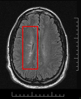
Radiologically isolated syndrome (RIS) is a clinical situation in which a person has white matter lesions suggestive of multiple sclerosis (MS), as shown on an MRI scan that was done for reasons unrelated to MS symptoms. The nerve lesions in these people show dissemination in space with an otherwise normal neurological examination and without historical accounts of typical MS symptoms.

Several biomarkers for diagnosis of multiple sclerosis, disease evolution and response to medication are under research. While most of them are still under research, there are some of them already well stablished:

References

  1. Oki S (February 2018). "Novel mechanism and biomarker of chronic progressive multiple sclerosis". Clinical and Experimental Neuroimmunology. 9 (1): 25–34. doi: 10.1111/cen3.12449 .
  2. 1 2 3 4 5 McDonald WI, Compston A, Edan G, Goodkin D, Hartung HP, Lublin FD, et al. (July 2001). "Recommended diagnostic criteria for multiple sclerosis: guidelines from the International Panel on the diagnosis of multiple sclerosis". Annals of Neurology. 50 (1): 121–127. CiteSeerX   10.1.1.466.5368 . doi:10.1002/ana.1032. PMID   11456302. S2CID   13870943.
  3. 1 2 Polman CH, Reingold SC, Edan G, Filippi M, Hartung HP, Kappos L, et al. (December 2005). "Diagnostic criteria for multiple sclerosis: 2005 revisions to the "McDonald Criteria"". Annals of Neurology. 58 (6): 840–846. CiteSeerX   10.1.1.604.2677 . doi:10.1002/ana.20703. PMID   16283615. S2CID   54512368.
  4. 1 2 3 Tsang BK, Macdonell R (December 2011). "Multiple sclerosis- diagnosis, management and prognosis". Australian Family Physician. 40 (12): 948–955. PMID   22146321.
  5. 1 2 3 4 5 6 7 8 9 10 Compston A, Coles A (October 2008). "Multiple sclerosis". Lancet. 372 (9648): 1502–1517. doi:10.1016/S0140-6736(08)61620-7. PMID   18970977. S2CID   195686659.
  6. Trojano M, Paolicelli D (November 2001). "The differential diagnosis of multiple sclerosis: classification and clinical features of relapsing and progressive neurological syndromes". Neurological Sciences. 22 (8 Suppl 2): S98-102. doi:10.1007/s100720100044. PMID   11794488. S2CID   3057096.
  7. Atlas: Multiple Sclerosis Resources in the World, 2008. World Health Organization & Multiple Sclerosis International Federation. 2008. pp. 15–16. hdl: 10665/43968 . ISBN   978-92-4-156375-8.
  8. Poser CM, Brinar VV (June 2004). "Diagnostic criteria for multiple sclerosis: an historical review". Clinical Neurology and Neurosurgery. 106 (3): 147–158. doi:10.1016/j.clineuro.2004.02.004. PMID   15177763. S2CID   23452341.
  9. Rashid W, Miller DH (February 2008). "Recent advances in neuroimaging of multiple sclerosis". Seminars in Neurology. 28 (1): 46–55. doi:10.1055/s-2007-1019127. PMID   18256986.
  10. Link H, Huang YM (November 2006). "Oligoclonal bands in multiple sclerosis cerebrospinal fluid: an update on methodology and clinical usefulness". Journal of Neuroimmunology. 180 (1–2): 17–28. doi:10.1016/j.jneuroim.2006.07.006. PMID   16945427. S2CID   22724352.
  11. Gronseth GS, Ashman EJ (May 2000). "Practice parameter: the usefulness of evoked potentials in identifying clinically silent lesions in patients with suspected multiple sclerosis (an evidence-based review): Report of the Quality Standards Subcommittee of the American Academy of Neurology". Neurology. 54 (9): 1720–1725. doi: 10.1212/wnl.54.9.1720 . PMID   10802774.
  12. Tremlett H, Okuda DT, Lebrun-Frenay C (October 2021). "The multiple sclerosis prodrome is just unspecific symptoms in radiologically isolated syndrome patients - No". Multiple Sclerosis. 27 (12): 1824–1826. doi:10.1177/13524585211035951. PMC   8521363 . PMID   34494923.
  13. Yusuf F, Wijnands JM, Kingwell E, Zhu F, Evans C, Fisk JD, et al. (February 2021). "Fatigue, sleep disorders, anaemia and pain in the multiple sclerosis prodrome". Multiple Sclerosis. 27 (2): 290–302. doi:10.1177/1352458520908163. PMID   32250183. S2CID   214810456.
  14. Disanto G, Zecca C, MacLachlan S, Sacco R, Handunnetthi L, Meier UC, et al. (June 2018). "Prodromal symptoms of multiple sclerosis in primary care". Annals of Neurology. 83 (6): 1162–1173. doi:10.1002/ana.25247. PMID   29740872. S2CID   13691058.
  15. Tremlett H, Marrie RA (January 2021). "The multiple sclerosis prodrome: Emerging evidence, challenges, and opportunities". Multiple Sclerosis. 27 (1): 6–12. doi:10.1177/1352458520914844. PMID   32228281. S2CID   214750591.
  16. Spelman T, Magyari M, Piehl F, Svenningsson A, Rasmussen PV, Kant M, et al. (October 2021). "Treatment Escalation vs Immediate Initiation of Highly Effective Treatment for Patients With Relapsing-Remitting Multiple Sclerosis: Data From 2 Different National Strategies". JAMA Neurology. 78 (10): 1197–1204. doi:10.1001/jamaneurol.2021.2738. PMC   8369379 . PMID   34398221. S2CID   237095594.
  17. Claflin SB, Campbell JA, Mason DF, Kalincik T, Simpson-Yap S, Norman R, et al. (August 2021). "The effect of national disease-modifying therapy subsidy policy on long-term disability outcomes in people with multiple sclerosis". Multiple Sclerosis. 28 (5): 831–841. doi:10.1177/13524585211035948. PMID   34387513. S2CID   236997752.
  18. Okuda DT, Mowry EM, Beheshtian A, Waubant E, Baranzini SE, Goodin DS, et al. (March 2009). "Incidental MRI anomalies suggestive of multiple sclerosis: the radiologically isolated syndrome". Neurology. 72 (9): 800–805. doi:10.1212/01.wnl.0000335764.14513.1a. PMID   19073949. S2CID   9981947.
  19. Matute-Blanch C, Villar LM, Álvarez-Cermeño JC, Rejdak K, Evdoshenko E, Makshakov G, et al. (April 2018). "Neurofilament light chain and oligoclonal bands are prognostic biomarkers in radiologically isolated syndrome". Brain. 141 (4): 1085–1093. doi: 10.1093/brain/awy021 . PMID   29452342.
  20. 1 2 Hottenrott T, Dersch R, Berger B, Rauer S, Eckenweiler M, Huzly D, Stich O (December 2015). "The intrathecal, polyspecific antiviral immune response in neurosarcoidosis, acute disseminated encephalomyelitis and autoimmune encephalitis compared to multiple sclerosis in a tertiary hospital cohort". Fluids and Barriers of the CNS. 12 (1): 27. doi: 10.1186/s12987-015-0024-8 . PMC   4677451 . PMID   26652013.
  21. Fabio Duranti; Massimo Pieri; Rossella Zenobi; Diego Centonze; Fabio Buttari; Sergio Bernardini; Mariarita Dessi (August 2015). "kFLC Index: a novel approach in early diagnosis of Multiple Sclerosis". International Journal of Scientific Research. 4 (8).
  22. Kotulska-Jóźwiak K, Pacheva I, Patrova A, Jurkiewicz EJ, Ivanov I, Kuczyński D, Geneva I (18 February 2019). "Lyme Disease or Multiple Sclerosis? Two cases with overlapping features". Journal of the International Child Neurology Association: 12. doi: 10.17724/jicna.2018.112 . S2CID   149606491.
  23. 1 2 3 4 5 Lublin FD, Reingold SC (April 1996). "Defining the clinical course of multiple sclerosis: results of an international survey. National Multiple Sclerosis Society (USA) Advisory Committee on Clinical Trials of New Agents in Multiple Sclerosis". Neurology. 46 (4): 907–911. doi:10.1212/WNL.46.4.907. PMID   8780061. S2CID   40213123.
  24. Lublin FD, Reingold SC, Cohen JA, Cutter GR, Sørensen PS, Thompson AJ, et al. (July 2014). "Defining the clinical course of multiple sclerosis: the 2013 revisions". Neurology. 83 (3): 278–286. doi:10.1212/WNL.0000000000000560. PMC   4117366 . PMID   24871874.
  25. National Multiple Sclerosis Society. "Changes in multiple sclerosis disease-course (or "type") descriptions" (PDF). Archived (PDF) from the original on 3 August 2016. Retrieved 21 August 2017. NEW COURSE ADDED: Clinically Isolated Syndrome (CIS)...COURSE ELIMINATED: Progressive Relapsing (PRMS).
  26. Pittock SJ, Rodriguez M (2008). "Benign Multiple Sclerosis: A Distinct Clinical Entity with Therapeutic Implications". Advances in multiple Sclerosis and Experimental Demyelinating Diseases. Current Topics in Microbiology and Immunology. Vol. 318. pp. 1–17. doi:10.1007/978-3-540-73677-6_1. ISBN   978-3-540-73676-9. PMID   18219812.
  27. Feinstein A (2007). The Clinical Neuropsychiatry of Multiple Sclerosis. Cambridge University Press. p. 20. ISBN   978-0-521-85234-0.
  28. 1 2 Miller D, Barkhof F, Montalban X, Thompson A, Filippi M (May 2005). "Clinically isolated syndromes suggestive of multiple sclerosis, part I: natural history, pathogenesis, diagnosis, and prognosis". The Lancet. Neurology. 4 (5): 281–288. doi:10.1016/S1474-4422(05)70071-5. PMID   15847841. S2CID   36401666.
  29. Rovaris M, Confavreux C, Furlan R, Kappos L, Comi G, Filippi M (April 2006). "Secondary progressive multiple sclerosis: current knowledge and future challenges". The Lancet. Neurology. 5 (4): 343–354. doi:10.1016/S1474-4422(06)70410-0. PMID   16545751. S2CID   39503553.
  30. Miller DH, Leary SM (October 2007). "Primary-progressive multiple sclerosis". The Lancet. Neurology. 6 (10): 903–912. doi:10.1016/S1474-4422(07)70243-0. hdl: 1871/24666 . PMID   17884680. S2CID   31389841.
  31. Stadelmann C, Brück W (November 2004). "Lessons from the neuropathology of atypical forms of multiple sclerosis". Neurological Sciences. 25 (Suppl 4): S319–S322. doi:10.1007/s10072-004-0333-1. PMID   15727225. S2CID   21212935.
  32. 1 2 3 Ntranos A, Lublin F (October 2016). "Diagnostic Criteria, Classification and Treatment Goals in Multiple Sclerosis: The Chronicles of Time and Space". Current Neurology and Neuroscience Reports. 16 (10): 90. doi:10.1007/s11910-016-0688-8. PMID   27549391. S2CID   4498332.
  33. Marriott JJ, O'Connor PW (2010). "Differential Diagnosis and Diagnostic Criteria for Multiple Sclerosis". Multiple Sclerosis 3. Blue Books of Neurology. Vol. 35. pp. 19–42. doi:10.1016/B978-1-4160-6068-0.00002-4. ISBN   978-1-4160-6068-0. S2CID   68141248.
  34. 1 2 Barkhof F, Filippi M, Miller DH, Scheltens P, Campi A, Polman CH, et al. (November 1997). "Comparison of MRI criteria at first presentation to predict conversion to clinically definite multiple sclerosis". Brain. 120 (11): 2059–2069. doi: 10.1093/brain/120.11.2059 . hdl: 1871/24709 . PMID   9397021.
  35. Tintoré M, Rovira A, Martínez MJ, Rio J, Díaz-Villoslada P, Brieva L, et al. (April 2000). "Isolated demyelinating syndromes: comparison of different MR imaging criteria to predict conversion to clinically definite multiple sclerosis". AJNR. American Journal of Neuroradiology. 21 (4): 702–706. PMC   7976636 . PMID   10782781.