The large intestine, also known as the large bowel, is the last part of the gastrointestinal tract and of the digestive system in vertebrates. Water is absorbed here and the remaining waste material is stored as feces before being removed by defecation.

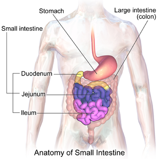
The duodenum is the first section of the small intestine in most higher vertebrates, including mammals, reptiles, and birds. In fish, the divisions of the small intestine are not as clear, and the terms anterior intestine or proximal intestine may be used instead of duodenum. In mammals the duodenum may be the principal site for iron absorption.

The lymphatic system is part of the vascular system and an important part of the immune system, comprising a large network of lymphatic vessels that carry a clear fluid called lymph directionally towards the heart. The lymphatic system was first described in the seventeenth century independently by Olaus Rudbeck and Thomas Bartholin. Unlike the circulatory system, the lymphatic system is not a closed system. The human circulatory system processes an average of 20 litres of blood per day through capillary filtration, which removes plasma while leaving the blood cells. Roughly 17 litres of the filtered plasma is reabsorbed directly into the blood vessels, while the remaining three litres remain in the interstitial fluid. One of the main functions of the lymph system is to provide an accessory return route to the blood for the surplus three litres.

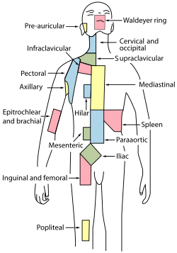
The paraaortic lymph nodes are a group of lymph nodes that lie in front of the lumbar vertebrae near the aorta. These lymph nodes receive drainage from the gastrointestinal tract and the abdominal organs.

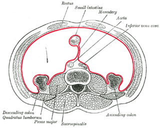
The midgut is the portion of the embryo from which most of the intestines develop. After it bends around the superior mesenteric artery, it is called the "midgut loop". It comprises the portion of the alimentary canal from the end of the foregut at the opening of the bile duct to the hindgut, about two-thirds of the way through the transverse colon.

The external iliac lymph nodes are lymph nodes, from eight to ten in number, that lie along the external iliac vessels.
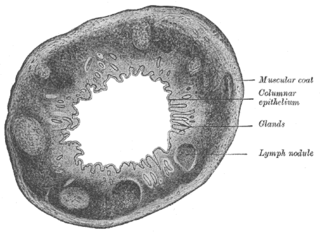
The Solitary lymphatic nodules are structures found in the small intestine and large intestine.

The ileocecal fold or ileocaecal fold is an anatomical structure in the human abdomen, located between the ileum and the cecum. It is formed by a layer of peritoneum. The upper border is fixed to the ileum, opposite its mesenteric attachment, while the lower border passes over the ileocecal junction to join the mesentery of the vermiform process, and sometimes the process itself. Behind the fold is the inferior ileocecal fossa. The structure is also called the ligament, veil, or bloodless fold of Treves after English surgeon Sir Frederick Treves.
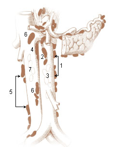
The preaortic lymph nodes lie in front of the aorta, and may be divided into celiac lymph nodes, superior mesenteric lymph nodes, and inferior mesenteric lymph nodes groups, arranged around the origins of the corresponding arteries.
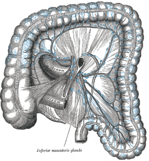
The inferior mesenteric lymph nodes consist of:

The internal iliac lymph nodes surround the internal iliac artery and its branches, and receive the lymphatics corresponding to the distribution of the branches of it, i. e., they receive lymphatics from all the pelvic viscera, from the deeper parts of the perineum, including the membranous and cavernous portions of the urethra, and from the buttock and back of the thigh. The internal iliac lymph nodes also drain the superior half of the rectum, above the pectinate line.

The popliteal lymph nodes, small in size and some six or seven in number, are embedded in the fat contained in the popliteal fossa, sometimes referred to as the 'knee pit'. One lies immediately beneath the popliteal fascia, near the terminal part of the small saphenous vein, and drains the region from which this vein derives its tributaries, such as superficial regions of the posterolateral aspect of the leg and the plantar aspect of the foot.

The superior deep cervical lymph nodes lie under the sternocleidomastoid muscle in close relation with the accessory nerve and the internal jugular vein.
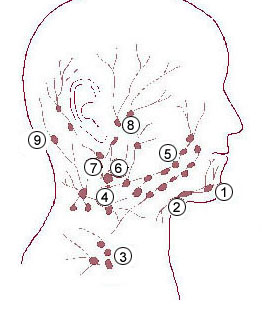
The submandibular lymph nodes, three to six in number, are placed beneath the body of the mandible in the submandibular triangle, and rest on the superficial surface of the submandibular gland.
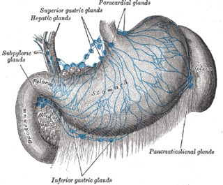
The celiac lymph nodes are associated with the branches of the celiac artery. Other lymph nodes in the abdomen are associated with the superior and inferior mesenteric arteries. The celiac lymph nodes are grouped into three sets: the gastric, hepatic and splenic lymph nodes.

The mesocolic lymph nodes are numerous, and lie between the layers of the transverse mesocolon, in close relation to the transverse colon; they are best developed in the neighborhood of the right and left colic flexures.
The superior diaphragmatic lymph nodes lie on the thoracic aspect of the diaphragm, and consist of three sets – anterior, middle, and posterior.
The public domain consists of all the creative works to which no exclusive intellectual property rights apply. Those rights may have expired, been forfeited, expressly waived, or may be inapplicable.

Gray's Anatomy is an English language textbook of human anatomy originally written by Henry Gray and illustrated by Henry Vandyke Carter. Earlier editions were called Anatomy: Descriptive and Surgical, Anatomy of the Human Body and Gray's Anatomy: Descriptive and Applied, but the book's name is commonly shortened to, and later editions are titled, Gray's Anatomy. The book is widely regarded as an extremely influential work on the subject, and has continued to be revised and republished from its initial publication in 1858 to the present day. The latest edition of the book, the 41st, was published in September 2015.