| Preaortic lymph node | |
|---|---|
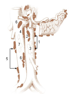
Left Lumbar Lymph Nodes (Paraaortic Lymph Nodes) 1. Lateral aortic 2. Preaortic 3. Postaortic 4. Intermediate Lumbar Right Lumbar Lymph Nodes (Paracaval Lymph Nodes) 5. Lateral caval 6. Precaval 7. Postcaval | |
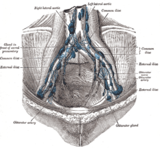
The parietal lymph glands of the pelvis. | |
| Details | |
| System | Lymphatic system |
| Source | inferior mesenteric lymph nodes |
| Drains to | primarily intestinal trunk |
| Identifiers | |
| Latin | Nodi lymphoidei praeaortici |
The preaortic lymph nodes lie in front of the aorta, and may be divided into celiac lymph nodes, superior mesenteric lymph nodes, and inferior mesenteric lymph nodes groups, arranged around the origins of the corresponding arteries.

The aorta is the main and largest artery in the human body, originating from the left ventricle of the heart and extending down to the abdomen, where it splits into two smaller arteries. The aorta distributes oxygenated blood to all parts of the body through the systemic circulation.
The celiac lymph nodes are associated with the branches of the celiac artery. Other lymph nodes in the abdomen are associated with the superior and inferior mesenteric arteries. The celiac lymph nodes are grouped into three sets: the gastric, hepatic and splenic lymph nodes.

The superior mesenteric lymph nodes may be divided into three principal groups:
Contents
The celiac lymph nodes are grouped into three sets: the gastric, hepatic and splenic lymph nodes. These groups also form their own subgroups.
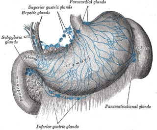
The gastric lymph nodes consist of two sets, superior and inferior.
The hepatic lymph nodes consist of the following groups:
The splenic lymph nodes are lymph nodes that accompany the lienal (splenic) artery, and are situated in relation to the posterior surface and upper border of the pancreas; one or two members of this group are found in the gastrolienal ligament.
The superior mesenteric lymph nodes are grouped into three sets: the mesenteric, ileocolic and mesocolic lymph nodes.
The mesenteric lymph nodes are one of the three principal groups of superior mesenteric lymph nodes and lie between the layers of the mesentery.
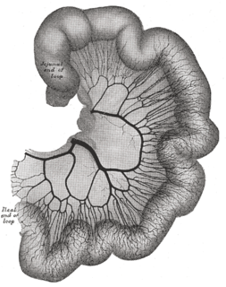
The ileocolic lymph nodes, from ten to twenty in number, form a chain around the ileocolic artery, but tend to subdivide into two groups, one near the duodenum and the other on the lower part of the trunk of the artery. Where the vessel divides into its terminal branches the chain is broken up into several groups:
The mesocolic lymph nodes are numerous, and lie between the layers of the transverse mesocolon, in close relation to the transverse colon; they are best developed in the neighborhood of the right and left colic flexures.
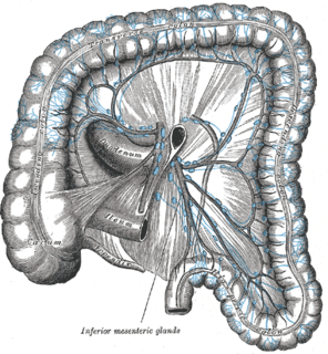
The inferior mesenteric lymph nodes have a subgroup of pararectal lymph nodes.

The pararectal lymph nodes are in contact with the muscular coat of the rectum. They drain the descending iliac and sigmoid parts of the colon and the upper part of the rectum; their efferents pass to the preaortic glands.
The preaortic lymph nodes receive a few vessels from the lateral aortic lymph nodes, but their principal afferents are derived from the organs supplied by the three arteries with which they are associated–the celiac, superior and inferior mesenteric arteries.

The celiacartery, also known as the celiac trunk, or truncus coeliacus, is the first major branch of the abdominal aorta. It is 1.25 cm in length. Branching from the aorta at thoracic vertebra 12 (T12) in humans, it is one of three anterior/ midline branches of the abdominal aorta.

In human anatomy, the superior mesenteric artery (SMA) arises from the anterior surface of the abdominal aorta, just inferior to the origin of the celiac trunk, and supplies the intestine from the lower part of the duodenum through two-thirds of the transverse colon, as well as the pancreas.

In human anatomy, the inferior mesenteric artery, often abbreviated as IMA, is the third main branch of the abdominal aorta and arises at the level of L3, supplying the large intestine from the left colic flexure to the upper part of the rectum, which includes the descending colon, the sigmoid colon, and part of the rectum. Proximally, its territory of distribution overlaps with the middle colic artery, and therefore the superior mesenteric artery. The SMA and IMA anastomose via the marginal artery of the colon and via Riolan's arcade. The territory of distribution of the IMA is more or less equivalent to the embryonic hindgut.
Some of their efferents pass to the retroaortic lymph nodes, but the majority unite to form the intestinal lymph trunk, which enters the cisterna chyli.

The retroaortic lymph nodes are placed below the cisterna chyli, on the bodies of the third and fourth lumbar vertebrae.

The intestinal trunk receives the lymph from the stomach and intestine, from the pancreas and spleen, and from the lower and front part of the liver, and empties lymph into the cisterna chyli, which in turn drains into the thoracic duct.
The cisterna chyli is a dilated sac at the lower end of the thoracic duct in most mammals into which lymph from the intestinal trunk and two lumbar lymphatic trunks flow. It receives fatty chyle from the intestines and thus acts as a conduit for the lipid products of digestion. It is the most common drainage trunk of most of the body's lymphatics. The cisterna chyli is a retro-peritoneal structure. In humans, it is located posterior to the abdominal aorta on the anterior aspect of the bodies of the first and second lumbar vertebrae. There it forms the beginning of the primary lymph vessel, the thoracic duct, which transports lymph and chyle from the abdomen via the aortic opening of the diaphragm up to the junction of left subclavian vein and internal jugular veins. In dogs, it is located to the left and often ventral to the aorta; in cats it is left and dorsal; in guinea pigs it runs to the left and drains into the left innominate vein.