
The large intestine, also known as the large bowel, is the last part of the gastrointestinal tract and of the digestive system in vertebrates. Water is absorbed here and the remaining waste material is stored as feces before being removed by defecation.
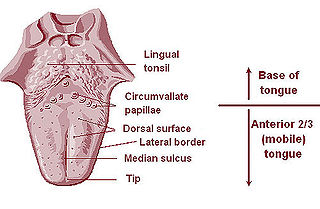
The lingual tonsils are two small mounds of lymphatic tissue located at the back of the base of the tongue, one on either side. They are composed of lymphatic tissue that functions to assist the immune system in the production of antibodies in response to invading pathogenic bacteria or viruses.

The ascending colon is the part of the colon located between the cecum and the transverse colon.
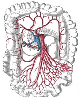
The right colic artery arises from about the middle of the concavity of the superior mesenteric artery, or from a stem common to it and the ileocolic.
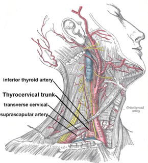
The inferior thyroid artery is an artery in the neck. It arises from the thyrocervical trunk and passes upward, in front of the vertebral artery and longus colli muscle. It then turns medially behind the carotid sheath and its contents, and also behind the sympathetic trunk, the middle cervical ganglion resting upon the vessel.
The intestinal arteries arise from the convex side of the superior mesenteric artery. They are usually from twelve to fifteen in number, and are distributed to the jejunum and ileum.

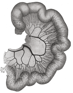
The midgut is the portion of the embryo from which most of the intestines develop. After it bends around the superior mesenteric artery, it is called the "midgut loop". It comprises the portion of the alimentary canal from the end of the foregut at the opening of the bile duct to the hindgut, about two-thirds of the way through the transverse colon.

The anterior cecal artery is a branch of the ileocolic artery which supplies the anterior region of the cecum.
The appendicular artery commonly arises from terminal branch of the ileocolic artery less commonly from the posterior cecal artery or an ileal artery. It descends behind the termination of the ileum and enters the mesoappendix of the vermiform appendix. It runs near the free margin of the mesoappendix and ends in branches which supply the appendix.

The testicular artery is a branch of the abdominal aorta that supplies blood to the testis. It is a paired artery, with one for each of the testes.
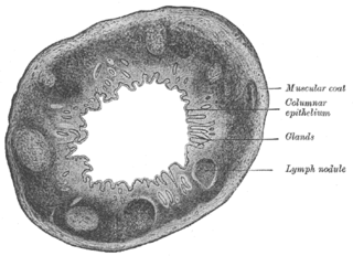
The Solitary lymphatic nodules are structures found in the small intestine and large intestine.
The colic branch of ileocolic artery is a small artery in the abdomen. The ileocolic artery of the superior mesenteric artery branches off into the ascending colic artery, the anterior and posterior cecal arteries, the appendicular artery, and the ileal branches.

The superior mesenteric lymph nodes may be divided into three principal groups:
The mesocolic lymph nodes are numerous, and lie between the layers of the transverse mesocolon, in close relation to the transverse colon; they are best developed in the neighborhood of the right and left colic flexures.
The mesenteric lymph nodes are one of the three principal groups of superior mesenteric lymph nodes and lie between the layers of the mesentery.
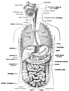
The human digestive system consists of the gastrointestinal tract plus the accessory organs of digestion. Digestion involves the breakdown of food into smaller and smaller components, until they can be absorbed and assimilated into the body. The process of digestion has many stages. The first stage is the cephalic phase of digestion which begins with gastric secretions in response to the sight and smell of food. The next stage starts in the mouth.
The public domain consists of all the creative works to which no exclusive intellectual property rights apply. Those rights may have expired, been forfeited, expressly waived, or may be inapplicable.

Gray's Anatomy is an English language textbook of human anatomy originally written by Henry Gray and illustrated by Henry Vandyke Carter. Earlier editions were called Anatomy: Descriptive and Surgical, Anatomy of the Human Body and Gray's Anatomy: Descriptive and Applied, but the book's name is commonly shortened to, and later editions are titled, Gray's Anatomy. The book is widely regarded as an extremely influential work on the subject, and has continued to be revised and republished from its initial publication in 1858 to the present day. The latest edition of the book, the 41st, was published in September 2015.


