
A hernia is the abnormal exit of tissue or an organ, such as the bowel, through the wall of the cavity in which it normally resides. Hernias come in a number of types. Most commonly they involve the abdomen, specifically the groin. Groin hernias are most commonly of the inguinal type but may also be femoral. Other hernias include hiatus, incisional, and umbilical hernias. Symptoms are present in about 66% of people with groin hernias. This may include pain or discomfort, especially with coughing, exercise or going to the bathroom. Often, it gets worse throughout the day and improves when lying down. A bulging area may appear that becomes larger when bearing down. Groin hernias occur more often on the right than left side. The main concern is strangulation, where the blood supply to part of the bowel is blocked. This usually produces severe pain and tenderness in the area. Hiatus, or hiatal, hernias often result in heartburn but may also cause chest pain or pain with eating.
The triangle is an idiophone type of musical instrument in the percussion family. It is a bar of metal, usually steel but sometimes other metals such as beryllium copper, bent into a triangle shape. The instrument is usually held by a loop of some form of thread or wire at the top curve. While the triangle theoretically has a definite pitch, it is obscured by the overtones that are produced when struck.
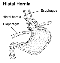
A hiatal hernia is a type of hernia in which abdominal organs slip through the diaphragm into the middle compartment of the chest. This may result in gastroesophageal reflux disease (GERD) or laryngopharyngeal reflux (LPR) with symptoms such as a taste of acid in the back of the mouth or heartburn. Other symptoms may include trouble swallowing and chest pains. Complications may include iron deficiency anemia, volvulus, or bowel obstruction.

The navel is a protruding, flat, or hollowed area on the abdomen at the attachment site of the umbilical cord. All placental mammals have a navel.

The spermatic cord is the cord-like structure in males formed by the vas deferens and surrounding tissue that runs from the deep inguinal ring down to each testicle. Its serosal covering, the tunica vaginalis, is an extension of the peritoneum that passes through the transversalis fascia. Each testicle develops in the lower thoracic and upper lumbar region and migrates into the scrotum during its descent it carries along with it vas deferens, its vessels, nerves etc. There is one on each side.

The inguinal canals are the two passages in the anterior abdominal wall of humans and animals which in males convey the spermatic cords and in females the round ligament of the uterus. The inguinal canals are larger and more prominent in males. There is one inguinal canal on each side of the midline.
An inguinal hernia is a protrusion of abdominal-cavity contents through the inguinal canal. Symptoms are present in about 66% of affected people. This may include pain or discomfort especially with coughing, exercise, or bowel movements. Often it gets worse throughout the day and improves when lying down. A bulging area may occur that becomes larger when bearing down. Inguinal hernias occur more often on the right than left side. The main concern is strangulation, where the blood supply to part of the intestine is blocked. This usually produces severe pain and tenderness of the area.

Congenital diaphragmatic hernia (CDH) is a birth defect of the diaphragm. The most common type of CDH is a Bochdalek hernia; other types include Morgagni hernia, diaphragm eventration and central tendon defects of the diaphragm. Malformation of the diaphragm allows the abdominal organs to push into the chest cavity, hindering proper lung formation.

An umbilical hernia is a health condition where the abdominal wall behind the navel is damaged. It may cause the navel to bulge outwards—the bulge consisting of abdominal fat from the greater omentum or occasionally parts of the small intestine. The bulge can often be pressed back through the hole in the abdominal wall, and may "pop out" when coughing or otherwise acting to increase intra-abdominal pressure. Treatment is surgical, and surgery may be performed for cosmetic as well as health-related reasons.

Vincent Alexander Bochdalek was a Bohemian anatomist and pathologist. His first name has also been given as Vincenc and Vincenz. Bochdalek was elected as member of the German Academy of Sciences Leopoldina.

In human anatomy, the inguinal triangle is a region of the abdominal wall. It is also known by the eponym Hesselbach's triangle, after Franz Kaspar Hesselbach.

In human anatomy, inferior epigastric artery refers to the artery that arises from the external iliac artery. It anastomoses with the superior epigastric artery. Along its course, it is accompanied by a similarly named vein, the inferior epigastric vein. These epigastric vessels form the lateral border of Hesselbach's triangle, which outlines the area through which direct inguinal hernias protrude.

The conjoint tendon is a sheath of connective tissue formed from the lower part of the common aponeurosis of the abdominal internal oblique muscle and the transversus abdominis muscle, joining the muscle to the pelvis. It forms the medial part of the posterior wall of the inguinal canal.
Femoral hernias occur just below the inguinal ligament, when abdominal contents pass through a naturally occurring weakness in the abdominal wall called the femoral canal. Femoral hernias are a relatively uncommon type, accounting for only 3% of all hernias. While femoral hernias can occur in both males and females, almost all develop in women due to the increased width of the female pelvis. Femoral hernias are more common in adults than in children. Those that do occur in children are more likely to be associated with a connective tissue disorder or with conditions that increase intra-abdominal pressure. Seventy percent of pediatric cases of femoral hernias occur in infants under the age of one.
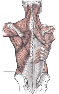
The lumbar triangle can refer to either the inferior lumbar (Petit) triangle, which lies superficially, or the superior lumbar (Grynfeltt) triangle, which is deep and superior to the inferior triangle. Of the two, the superior triangle is the more consistently found in cadavers, and is more commonly the site of herniation; however, the inferior lumbar triangle is often simply called the lumbar triangle, perhaps owing to its more superficial location and ease in demonstration.

Grynfeltt-Lesshaft hernia is a herniation of abdominal contents through the back, specifically through the superior lumbar triangle, which is defined by the quadratus lumborum muscle, twelfth rib, and internal oblique muscle.
A Richter's hernia occurs when the antimesenteric wall of the intestine protrudes through a defect in the abdominal wall. This is distinct from other types of abdominal hernias in that only one intestinal wall protrudes through the defect, such that the lumen of the intestine is incompletely contained in the defect, while the rest remains in the peritoneal cavity. If such a herniation becomes necrotic and is subsequently reduced during hernia repair, perforation and peritonitis may result. A Richter's hernia can result in strangulation and necrosis in the absence of intestinal obstruction. It is a relatively rare but dangerous type of hernia.
An obturator hernia is a rare type of hernia of the pelvic floor in which pelvic or abdominal contents protrudes through the obturator foramen. Because of differences in anatomy, it is much more common in women, especially multiparous and older women who have recently lost much weight. The diagnosis is often made intraoperatively after presenting with bowel obstruction.

Diaphragmatic rupture is a tear of the diaphragm, the muscle across the bottom of the ribcage that plays a crucial role in breathing. Most commonly, acquired diaphragmatic tears result from physical trauma. Diaphragmatic rupture can result from blunt or penetrating trauma and occurs in about 0.5% of all people with trauma.

Internal hernias occur when there is protrusion of an internal organ into a retroperitoneal fossa or a foramen in the abdominal cavity. If a loop of bowel passes through the mesenteric defect, that loop is at risk for incarceration, strangulation, or for becoming the lead point of a small bowel obstruction. Internal hernias can also trap adipose tissue (fat) and nerves. Unlike more common forms of hernias, the trapped tissue protrudes inward, rather than outward.
