
Rhinoplasty, commonly called nose job, medically called nasal reconstruction is a plastic surgery procedure for altering and reconstructing the nose. There are two types of plastic surgery used – reconstructive surgery that restores the form and functions of the nose and cosmetic surgery that changes the appearance of the nose. Reconstructive surgery seeks to resolve nasal injuries caused by various traumas including blunt, and penetrating trauma and trauma caused by blast injury. Reconstructive surgery can also treat birth defects, breathing problems, and failed primary rhinoplasties. Rhinoplasty may remove a bump, narrow nostril width, change the angle between the nose and the mouth, or address injuries, birth defects, or other problems that affect breathing, such as a deviated nasal septum or a sinus condition. Surgery only on the septum is called a septoplasty.

The ethmoid bone is an unpaired bone in the skull that separates the nasal cavity from the brain. It is located at the roof of the nose, between the two orbits. The cubical bone is lightweight due to a spongy construction. The ethmoid bone is one of the bones that make up the orbit of the eye.

The nasal cavity is a large, air-filled space above and behind the nose in the middle of the face. The nasal septum divides the cavity into two cavities, also known as fossae. Each cavity is the continuation of one of the two nostrils. The nasal cavity is the uppermost part of the respiratory system and provides the nasal passage for inhaled air from the nostrils to the nasopharynx and rest of the respiratory tract.
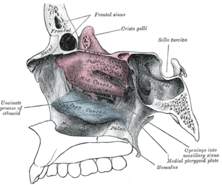
In anatomy, a nasal concha, also called a nasal turbinate or turbinal, is a long, narrow, curled shelf of bone that protrudes into the breathing passage of the nose in humans and various animals. The conchae are shaped like an elongated seashell, which gave them their name. A concha is any of the scrolled spongy bones of the nasal passages in vertebrates.
Nasal polyps (NP) are noncancerous growths within the nose or sinuses. Symptoms include trouble breathing through the nose, loss of smell, decreased taste, post nasal drip, and a runny nose. The growths are sac-like, movable, and nontender, though face pain may occasionally occur. They typically occur in both nostrils in those who are affected. Complications may include sinusitis and broadening of the nose.

Rhinoscleroma, is a chronic granulomatous bacterial disease of the nose that can sometimes infect the upper respiratory tract. It most commonly affects the nasal cavity—the nose is involved in 95–100 per cent of cases—however, it can also affect the nasopharynx, larynx, trachea, and bronchi. Slightly more females than males are affected and patients are usually 10 to 30 years of age. Rhinoscleroma is considered a tropical disease and is mostly endemic to North Africa, South Asia and Central America, less common in the United States.

A nasal septum perforation is a medical condition in which the nasal septum, the bony/cartilaginous wall dividing the nasal cavities, develops a hole or fissure.
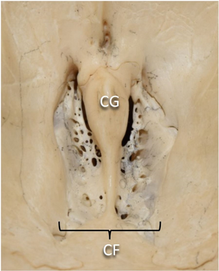
In mammalian anatomy, the cribriform plate, horizontal lamina or lamina cribrosa is part of the ethmoid bone. It is received into the ethmoidal notch of the frontal bone and roofs in the nasal cavities. It supports the olfactory bulb, and is perforated by olfactory foramina for the passage of the olfactory nerves to the roof of the nasal cavity to convey smell to the brain. The foramina at the medial part of the groove allow the passage of the nerves to the upper part of the nasal septum while the foramina at the lateral part transmit the nerves to the superior nasal concha.

Rhinorrhea, rhinorrhoea, or informally runny nose is the free discharge of a thin mucus fluid from the nose; it is a common condition. It is a common symptom of allergies or certain viral infections, such as the common cold or COVID-19. It can be a side effect of crying, exposure to cold temperatures, cocaine abuse, or drug withdrawal, such as from methadone or other opioids. Treatment for rhinorrhea is not usually undertaken, but there are a number of medical treatments and preventive techniques.

The pyramid-shaped maxillary sinus is the largest of the paranasal sinuses, located in the maxilla. It drains into the middle meatus of the nose through the semilunar hiatus. It is located to the side of the nasal cavity, and below the orbit.

The anterior ethmoidal artery is a branch of the ophthalmic artery in the orbit. It exits the orbit through the anterior ethmoidal foramen alongside the anterior ethmoidal nerve. It contributes blood supply to the ethmoid sinuses, frontal sinuses, the dura mater, lateral nasal wall, and nasal septum. It issues a meningeal branch, and nasal branches.
Chronic atrophic rhinitis, or simply atrophic rhinitis, is a chronic inflammation of the nose characterised by atrophy of nasal mucosa, including the glands, turbinate bones and the nerve elements supplying the nose. Chronic atrophic rhinitis may be primary and secondary. Special forms of chronic atrophic rhinitis are rhinitis sicca anterior and ozaena. It can also be described as the empty nose syndrome.

The nasal cycle is the unconscious alternating partial congestion and decongestion of the nasal cavities in humans and other animals. This results in greater airflow through one nostril with periodic alternation between the nostrils. It is a physiological congestion of the nasal conchae, also called the nasal turbinates, due to selective activation of one half of the autonomic nervous system by the hypothalamus. It should not be confused with pathological nasal congestion.
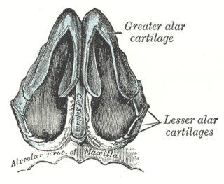
The nasal cartilages are structures within the nose that provide form and support to the nasal cavity. The nasal cartilages are made up of a flexible material called hyaline cartilage in the distal portion of the nose. There are five individual cartilages that make up the nasal cavity: septal nasal cartilage, lateral nasal cartilage, major alar cartilage, minor alar cartilage, and vomeronasal cartilage.

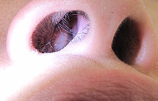
The human nose is the most protruding part of the face. It bears the nostrils and is the first organ of the respiratory system. It is also the principal organ in the olfactory system. The shape of the nose is determined by the nasal bones and the nasal cartilages, including the nasal septum which separates the nostrils and divides the nasal cavity into two. On average, the nose of a male is larger than that of a female.
A nose is a protuberance in vertebrates that houses the nostrils, or nares, which receive and expel air for respiration alongside the mouth. Behind the nose are the olfactory mucosa and the sinuses. Behind the nasal cavity, air next passes through the pharynx, shared with the digestive system, and then into the rest of the respiratory system. In humans, the nose is located centrally on the face and serves as an alternative respiratory passage especially during suckling for infants. The protruding nose that is completely separate from the mouth part is a characteristic found only in therian mammals. It has been theorized that this unique mammalian nose evolved from the anterior part of the upper jaw of the reptilian-like ancestors (synapsids).

Dacryocystocele (Dacryocystitis) or timo cyst is a benign, bluish-gray mass in the inferomedial canthus that develops within a few days or weeks after birth. The uncommon condition forms as a result as a consequence of narrowing or obstruction of the nasolacrimal duct, usually during prenatal development. Nasolacrimal duct obstruction disrupts the lacrimal drainage system, eventually creating a swelling cyst in the lacrimal sac area by the nasal cavity. The location of the cyst can cause respiratory dysfunction, compromising the airway. The obstruction ultimately leads to epiphora, an abundance of tear production.
Rhinomanometry is a form of manometry used in evaluation of the nasal cavity. Rhinomanometry is a standard diagnostic tool aiming to objectively evaluate the respiratory function of the nose. It measures pressure and flow during normal inspiration and expiration through the nose. Increased pressure during respiration is a result of increased resistance to airflow through nasal passages, while increased flow, which means the speed of airstream, is related to better patency. Nasal obstruction leads to increased values of nasal resistance. Rhinomanometry may be used to measure only one nostril at a time or both nostrils simultaneously.
Tornwaldt's disease is the inflammation or abscess of the embryonic cyst of pharyngeal bursa. It is located in the midline of the posterior wall of the nasopharynx. It is covered anteriorly by mucosa in the adenoid mass. It is bounded posteriorly by longus muscle.

Oroantral fistula (OAF) is an epithelialised oroantral communication (OAC). OAC refers to an abnormal connection between the oral cavity and antrum. The creation of an OAC is most commonly due to the extraction of a maxillary (upper) tooth closely related to the antral floor. A small OAC may heal spontaneously but a larger OAC would require surgical closure to prevent the development of persistent OAF and chronic sinusitis.
