Giant-cell arteritis

Last updated
Giant-cell arteritis
Other namesTemporal arteritis, cranial arteritis, [1] Horton disease, [2] senile arteritis, [1] granulomatous arteritis [1]
Gray508.png
The arteries of the face and scalp.
Specialty Rheumatology, emergency medicine
Symptoms Headache, pain over the temples, flu-like symptoms, double vision, difficulty opening the mouth [3]
Complications Blindness, aortic dissection, aortic aneurysm, polymyalgia rheumatica [4]
Usual onsetAge greater than 50 [4]
CausesInflammation of the small blood vessels within the walls of larger arteries [4]
Diagnostic method Based on symptoms and blood tests, confirmed by biopsy of the temporal artery [4]
Differential diagnosis Takayasu arteritis, [5] stroke, primary amyloidosis [6]
Treatment Steroids, bisphosphonates, proton pump inhibitor [4]
PrognosisLife expectancy (typically normal) [4]
Frequency~ 1 in 15,000 people a year (> 50 years old) [2]

Giant-cell arteritis (GCA), also called temporal arteritis, is an inflammatory disease of large blood vessels. [4] [7] Symptoms may include headache, pain over the temples, flu-like symptoms, double vision, and difficulty opening the mouth. [3] Complication can include blockage of the artery to the eye with resulting blindness, aortic dissection, and aortic aneurysm. [4] GCA is frequently associated with polymyalgia rheumatica. [4]

Contents

The cause is unknown. [2] The underlying mechanism involves inflammation of the small blood vessels that occur within the walls of larger arteries. [4] This mainly affects arteries around the head and neck, though some in the chest may also be affected. [4] [8] Diagnosis is suspected based on symptoms, blood tests, and medical imaging, and confirmed by biopsy of the temporal artery. [4] However, in about 10% of people the temporal artery is normal. [4]

Treatment is typically with high doses of steroids such as prednisone or prednisolone. [4] Once symptoms have resolved the dose is then decreased by about 15% per month. [4] Once a low dose is reached, the taper is slowed further over the subsequent year. [4] Other medications that may be recommended include bisphosphonates to prevent bone loss and a proton pump inhibitor to prevent stomach problems. [4]

It affects about 1 in 15,000 people over the age of 50 per year. [2] The condition typically only occurs in those over the age of 50, being most common among those in their 70s. [4] Females are more often affected than males. [4] Those of northern European descent are more commonly affected. [5] Life expectancy is typically normal. [4] The first description of the condition occurred in 1890. [1]

Signs and symptoms

Common symptoms of giant-cell arteritis include:

The inflammation may affect blood supply to the eye; blurred vision or sudden blindness may occur. In 76% of cases involving the eye, the ophthalmic artery is involved, causing arteritic anterior ischemic optic neuropathy. [13]

Giant-cell arteritis may present with atypical or overlapping features. [14] Early and accurate diagnosis is important to prevent ischemic vision loss. Therefore, this condition is considered a medical emergency. [14]

While studies vary as to the exact relapse rate of giant cell arteritis, relapse of this condition can occur. [15] It most often happens at low doses of prednisone (<20 mg/day), during the first year of treatment, and the most common signs of relapse are headache and polymyalgia rheumatica. [15]

Associated conditions

The varicella-zoster virus (VZV) antigen was found in 74% of temporal artery biopsies that were GCA-positive, suggesting that the VZV infection may trigger the inflammatory cascade. [16]

The disorder may co-exist (in about half of cases) with polymyalgia rheumatica (PMR), [12] which is characterized by sudden onset of pain and stiffness in muscles (pelvis, shoulder) of the body and is seen in the elderly. GCA and PMR are so closely linked that they are often considered to be different manifestations of the same disease process. PMR usually lacks the cranial symptoms, including headache, pain in the jaw while chewing, and vision symptoms, that are present in GCA. [17]

Giant-cell arteritis can affect the aorta and lead to aortic aneurysm and aortic dissection. [18] Up to 67% of people with GCA having evidence of an inflamed aorta, which can increase the risk of aortic aneurysm and dissection. [18] There are arguments for the routine screening of each person with GCA for this possible life-threatening complication by imaging the aorta. Screening should be done on a case-by-case basis based on the signs and symptoms of people with GCA. [18]

Mechanism

The pathological mechanism is the result of an inflammatory cascade that is triggered by an as of yet determined cause resulting in dendritic cells in the vessel wall recruiting T cells and macrophages to form granulomatous infiltrates. [18] These infiltrates erode the middle and inner layers of the arterial tunica media leading to conditions such as aneurysm and dissection. [18] Activation of T helper 17 (Th17) cells involved with interleukin (IL) 6, IL-17, IL-21 and IL-23 play a critical part; specifically, Th17 activation leads to further activation of Th17 through IL-6 in a continuous, cyclic fashion. [18] This pathway is suppressed with glucocorticoids, [19] and more recently it has been found that IL-6 inhibitors also play a suppressive role. [18]

Diagnosis

Physical exam

Laboratory tests

Biopsy

Histopathology of giant cell vasculitis in a cerebral artery. Elastica-stain. Cerebral Giant-Cell Vasculitis.jpg
Histopathology of giant cell vasculitis in a cerebral artery. Elastica-stain.

The gold standard for diagnosing temporal arteritis is biopsy, which involves removing a small part of the vessel under local anesthesia and examining it microscopically for giant cells infiltrating the tissue. [21] However, a negative result does not definitively rule out the diagnosis; since the blood vessels are involved in a patchy pattern, there may be unaffected areas on the vessel and the biopsy might have been taken from these parts. Unilateral biopsy of a 1.5–3 cm length is 85-90% sensitive (1 cm is the minimum). [22] A Characterised as intimal hyperplasia and medial granulomatous inflammation with elastic lamina fragmentation with a CD 4+ predominant T cell infiltrate, currently biopsy is only considered confirmatory for the clinical diagnosis, or one of the diagnostic criteria. [11]

Medical imaging

Radiological examination of the temporal artery with ultrasound yields a halo sign. Contrast-enhanced brain MRI and CT is generally negative in this disorder. Recent studies have shown that 3T MRI using super high resolution imaging and contrast injection can non-invasively diagnose this disorder with high specificity and sensitivity. [23]

Treatment

GCA is considered a medical emergency due to the potential of irreversible vision loss. [14] Corticosteroids, typically high-dose prednisone (1 mg/kg/day), should be started as soon as the diagnosis is suspected (even before the diagnosis is confirmed by biopsy) to prevent irreversible blindness secondary to ophthalmic artery occlusion. Steroids do not prevent the diagnosis from later being confirmed by biopsy, although certain changes in the histology may be observed towards the end of the first week of treatment and are more difficult to identify after a couple of months. [24] The dose of prednisone is lowered after 2–4 weeks, and slowly tapered over 9–12 months. Tapering may require two or more years. Oral steroids are at least as effective as intravenous steroids, [25] except in the treatment of acute visual loss where intravenous steroids appear to offer significant benefit over oral steroids. [26] Short-term side effects of prednisone are uncommon but can include mood changes, avascular necrosis, and an increased risk of infection. [27] Some of the side effects associated with long-term use include weight gain, diabetes mellitus, osteoporosis, avascular necrosis, glaucoma, cataracts, cardiovascular disease, and an increased risk of infection. [28] [29] It is unclear if adding a small amount of aspirin is beneficial or not as it has not been studied. [30] Injections of tocilizumab may also be used. [31] Tocilizumab is a humanized antibody that targets the interleukin-6 receptor, which is a key cytokine involved in the progression of GCA. [32] Tocilizumab has been found to be effective at minimizing both recurrence, and flares of GCA when used both on its own and with corticosteroids. [32] Long term use of tocilizumab requires further investigation. [32] [33] Tocilizumab may increase the risk of gastrointestinal perforation and infections, however it does not appear that there are more risks than using corticosteroids. [32] [33]

Epidemiology

Giant-cell arteritis typically only occurs in those over the age of 50; [4] particularly those in their 70s. [20] It affects about 1 in 15,000 people over the age of 50 per year. [2] It is more common in women than in men, by a ratio of 2:1, [4] and more common in those of Northern European descent, as well as in those residing further from the Equator. [5]

Terminology

The terms "giant-cell arteritis" and "temporal arteritis" are sometimes used interchangeably, because of the frequent involvement of the temporal artery. However, other large vessels such as the aorta can be involved. [34] Giant-cell arteritis is also known as "cranial arteritis" and "Horton's disease." [35] The name (giant-cell arteritis) reflects the type of inflammatory cell involved. [36]

Related Research Articles

Rheumatology is a branch of medicine devoted to the diagnosis and therapy of rheumatic diseases. Physicians who have undergone formal training in rheumatology are called rheumatologists. Rheumatologists deal mainly with immune-mediated disorders of the musculoskeletal system, soft tissues, autoimmune diseases, vasculitides, and heritable connective tissue disorders.

Erythrocyte sedimentation rate physiological quantity

The erythrocyte sedimentation rate is the rate at which red blood cells in anticoagulated whole blood descend in a standardized tube over a period of one hour. It is a common hematology test, and is a non-specific measure of inflammation. To perform the test, anticoagulated blood is traditionally placed in an upright tube, known as a Westergren tube, and the distance which the red blood cells fall is measured and reported in mm at the end of one hour.

Vasculitis vascular disease that is characterized by inflammation of the blood vessels

Vasculitis is a group of disorders that destroy blood vessels by inflammation. Both arteries and veins are affected. Lymphangitis is sometimes considered a type of vasculitis. Vasculitis is primarily caused by leukocyte migration and resultant damage.

Takayasus arteritis syndrome that involves inflammation of the aorta that carries blood from the heart to the rest of the body

Takayasu's arteritis is a form of large vessel granulomatous vasculitis with massive intimal fibrosis and vascular narrowing, most commonly affecting young or middle-age women of Asian descent, though anyone can be affected. It mainly affects the aorta and its branches, as well as the pulmonary arteries. Females are about 8–9 times more likely to be affected than males.

Arteritis inflammation of the walls of arteries, usually as a result of infection or autoimmune response.

Arteritis is the inflammation of the walls of arteries, usually as a result of infection or autoimmune response. Arteritis, a complex disorder, is still not entirely understood. Arteritis may be distinguished by its different types, based on the organ systems affected by the disease. A complication of arteritis is thrombosis, which can be fatal. Arteritis and phlebitis are forms of vasculitis.

Polymyalgia rheumatica syndrome with pain or stiffness, usually in the neck, shoulders, upper arms, and hips, but which may occur all over the body

Polymyalgia rheumatica (PMR) is a syndrome with pain or stiffness, usually in the neck, shoulders, upper arms, and hips, but which may occur all over the body. The pain can be very sudden, or can occur gradually over a period. Most people with PMR wake up in the morning with pain in their muscles; however, cases have occurred in which the person has developed the pain during the evenings or has pain and stiffness all day long.

Posterior ischemic optic neuropathy (PION) is a medical condition characterized by damage to the retrobulbar portion of the optic nerve due to inadequate blood flow (ischemia) to the optic nerve. Despite the term posterior, this form of damage to the eye's optic nerve due to poor blood flow also includes cases where the cause of inadequate blood flow to the nerve is anterior, as the condition describes a particular mechanism of visual loss as much as the location of damage in the optic nerve. In contrast, anterior ischemic optic neuropathy (AION) is distinguished from PION by the fact that AION occurs spontaneously and on one side in affected individuals with predisposing anatomic or cardiovascular risk factors.

Bruit, also called vascular murmur, is the abnormal sound generated by turbulent flow of blood in an artery due to either an area of partial obstruction or a localized high rate of blood flow through an unobstructed artery.

Giant cell multinucleated masses produced by the fusion of many cells, often associated with viral infections

A giant cell is a mass formed by the union of several distinct cells, often forming a granuloma. Although there is typically a focus on the pathological aspects of multinucleate giant cells (MGCs), they also play many important physiological roles. Osteoclasts specifically are invaluable to healthy physiological functions and are key players in the skeletal system. Osteoclasts are frequently classified and discussed separately from other MGCs which are more closely linked with human pathologies.

Retroperitoneal fibrosis or Ormond's disease is a disease featuring the proliferation of fibrous tissue in the retroperitoneum, the compartment of the body containing the kidneys, aorta, renal tract, and various other structures. It may present with lower back pain, kidney failure, hypertension, deep vein thrombosis, and other obstructive symptoms. It is named after John Kelso Ormond, who rediscovered the condition in 1948.

Arteritic anterior ischemic optic neuropathy is the cause of vision loss that occurs in temporal arteritis. Temporal arteritis is an inflammatory disease of medium-sized blood vessels that happens especially with advancing age. AAION occurs in about 15-20 percent of patients with temporal arteritis. Damage to the blood vessels supplying the optic nerves leads to insufficient blood supply (ischemia) to the nerve and subsequent optic nerve fiber death. Most cases of AAION result in nearly complete vision loss first to one eye. If the temporal arteritis is left untreated, the fellow eye will likely suffer vision loss as well within 1–2 weeks. Arteritic AION falls under the general category of anterior ischemic optic neuropathy, which also includes non-arteritic AION. AION is considered an eye emergency, immediate treatment is essential to rescue remaining vision.

Neuromuscular disease neuropathy that affect the nerves that control the voluntary muscles

Neuromuscular disease is a broad term that encompasses many diseases and ailments that impair the functioning of the muscles, either directly, being pathologies of the voluntary muscle, or indirectly, being pathologies of nerves or neuromuscular junctions.

Aortitis is the inflammation of the aortic wall. The disorder is potentially life-threatening and rare. It is reported that there are only 1–3 new cases of aortitis per year per million people in the United States and Europe. Aortitis is most common in people 10 to 40 years of age.

Kimuras disease Human disease

Kimura's disease is a benign rare chronic inflammatory disorder. Its primary symptoms are subdermal lesions in the head or neck or painless unilateral inflammation of cervical lymph nodes.

Cerebral vasculitis is vasculitis involving the brain and occasionally the spinal cord. It affects all of the vessels: very small blood vessels (capillaries), medium-size blood vessels, or large blood vessels. If blood flow in a vessel with vasculitis is reduced or stopped, the parts of the body that receive blood from that vessel begins to die. It may produce a wide range of neurological symptoms, such as headache, skin rashes, feeling very tired, joint pains, difficulty moving or coordinating part of the body, changes in sensation, and alterations in perception, thought or behavior, as well as the phenomena of a mass lesion in the brain leading to coma and herniation. Some of its signs and symptoms may resemble multiple sclerosis. 10% have associated bleeding in the brain.

Inflammatory myopathy muscle disease

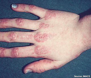
Inflammatory myopathy is disease featuring weakness and inflammation of muscles and muscle pain. The cause of much inflammatory myopathy is unknown (idiopathic), and such cases are classified according to their symptoms and signs and electromyography, MRI and laboratory findings. It can also be associated with underlying cancer. The main classes of idiopathic inflammatory myopathy are polymyositis (PM), dermatomyositis (DM), and inclusion-body myositis (IBM).

Necrotizing vasculitis also called Systemic necrotizing vasculitus (SNV) is a category of vasculitis, comprising vasculitides that present with necrosis.

Cryofibrinogenemia refers to a condition classified as a fibrinogen disorder in which the chilling of an individual's blood plasma from the normal body temperature of 37 °C to the near-freezing temperature of 4 °C causes the reversible precipitation of a complex containing fibrinogen, fibrin, fibronectin, and, occasionally, small amounts of fibrin split products, albumin, immunoglobulins and other plasma proteins. Returning this plasma to 37 °C resolubilizes the precipitate.

Inflammatory aortic aneurysm

Inflammatory aortic aneurysm (IAA), also known as Inflammatory abdominal aortic aneurysm (IAAA), is a type of abdominal aortic aneurysm (AAA) where the walls of the aneurysm become thick and inflamed. Similar to AAA, IAA occurs in the abdominal region. IAA is closely associated and believed to be a response to and extensive peri-anuerysmal fibrosis, which is the formation of excess fibrous connective tissue in an organ or tissue in a reparative or reactive process IAA accounts for 5-10% of aortic aneurysms. IAA occurs mainly in a population that is on average younger by 10 years than most AAA patients. Some common symptoms of IAA may include back pain, abdominal tenderness, fevers, weight loss or elevated Erythrocyte sedimentation rate (ESR) levels. Corticosteroids and other immunosuppressive drugs have been found to decrease symptoms and the degree of peri-aortic inflammation and fibrosis

Acute visual loss Loss of visual acuity (implying that vision was better at a certain timepoint in life).

Acute visual loss is a rapid loss of the ability to see. It is commonly caused by four conditions retinal detachment, glaucoma, macular degeneration, and giant cell arteritis.

References

  1. 1 2 3 4 Nussinovitch, Udi (2017). The Heart in Rheumatic, Autoimmune and Inflammatory Diseases: Pathophysiology, Clinical Aspects and Therapeutic Approaches. Academic Press. p. 367. ISBN   9780128032688. Archived from the original on 2017-10-22.
  2. 1 2 3 4 5 "Orphanet: Giant cell arteritis". www.orpha.net. Archived from the original on 14 September 2017. Retrieved 14 September 2017.
  3. 1 2 "Giant Cell Arteritis". National Institute of Arthritis and Musculoskeletal and Skin Diseases. 13 April 2017. Archived from the original on 22 October 2017. Retrieved 21 October 2017.
  4. 1 2 3 4 5 6 7 8 9 10 11 12 13 14 15 16 17 18 19 20 21 22 Weyand CM, Goronzy JJ (July 2014). "Clinical practice. Giant-cell arteritis and polymyalgia rheumatica". The New England Journal of Medicine. 371 (1): 50–7. doi:10.1056/NEJMcp1214825. PMC   4277693 . PMID   24988557.
  5. 1 2 3 Johnson RJ, Feehally J, Floege J (2014). Comprehensive Clinical Nephrology E-Book. Elsevier Health Sciences. p. 300. ISBN   9780323242875. Archived from the original on 2017-10-22.
  6. Ferri, Fred F. (2010). Ferri's Differential Diagnosis E-Book: A Practical Guide to the Differential Diagnosis of Symptoms, Signs, and Clinical Disorders. Elsevier Health Sciences. p. 195. ISBN   978-0323081634. Archived from the original on 2017-10-22.
  7. Jennette JC, Falk RJ, Bacon PA, Basu N, Cid MC, Ferrario F, et al. (January 2013). "2012 revised International Chapel Hill Consensus Conference Nomenclature of Vasculitides". Arthritis and Rheumatism. 65 (1): 1–11. doi:10.1002/art.37715. PMID   23045170.
  8. "Giant Cell Arteritis". National Institute of Arthritis and Musculoskeletal and Skin Diseases. 13 April 2017. Archived from the original on 22 October 2017. Retrieved 21 October 2017.
  9. Moutray TN, Williams MA, Best JL (August 2008). "Suspected giant cell arteritis: a study of referrals for temporal artery biopsy". Canadian Journal of Ophthalmology. Journal Canadien d'Ophtalmologie. 43 (4): 445–8. doi:10.3129/i08-070. PMID   18711459.
  10. Sainuddin S, Saeed NR (December 2008). "Acute bilateral tongue necrosis--a case report". The British Journal of Oral & Maxillofacial Surgery. 46 (8): 671–2. doi:10.1016/j.bjoms.2008.03.027. PMID   18499311.
  11. 1 2 Zadik Y, Findler M, Maly A, Rushinek H, Czerninski R (January 2011). "A 78-year-old woman with bilateral tongue necrosis". Oral Surgery, Oral Medicine, Oral Pathology, Oral Radiology, and Endodontics. 111 (1): 15–9. doi:10.1016/j.tripleo.2010.09.001. PMID   21176820.
  12. 1 2 Hunder, Gene G. "Polymyalgia rheumatica and giant cell (temporal) arteritis". uptodate.com. Wolters Kluwer. Archived from the original on 25 September 2015. Retrieved 23 September 2015.
  13. Hayreh (April 3, 2003). "Ocular Manifestations of GCA". University of Iowa Health Care. Archived from the original on 2007-10-25. Retrieved 2007-10-15.
  14. 1 2 3 Rana AQ, Saeed U, Khan OA, Qureshi AR, Paul D (October 2014). "Giant cell arteritis or tension-type headache?: A differential diagnostic dilemma". Journal of Neurosciences in Rural Practice. 5 (4): 409–11. doi:10.4103/0976-3147.140005. PMC   4173245 . PMID   25288850.
  15. 1 2 "UpToDate". www.uptodate.com. Retrieved 2019-11-25.
  16. Gilden D, White T, Khmeleva N, Heintzman A, Choe A, Boyer PJ, et al. (May 2015). "Prevalence and distribution of VZV in temporal arteries of patients with giant cell arteritis". Neurology. 84 (19): 1948–55. doi:10.1212/WNL.0000000000001409. PMC   4433460 . PMID   25695965.
  17. "Giant cell arteritis". BMJ Best Practice. Retrieved 2019-11-25.
  18. 1 2 3 4 5 6 7 Chen, John; Warrington, Kenneth; Garrity, James; Prasad, Sashank (2017). "Is Routine Imaging of the Aorta Warranted in Patients With Giant Cell Arteritis?". Journal of Neuro-Ophthalmology. 37 (3): 314–319. doi:10.1097/WNO.0000000000000538. ISSN   1070-8022. PMID   28614098.
  19. Weyand, Cornelia M.; Goronzy, Jörg J. (2014-07-03). Solomon, Caren G. (ed.). "Giant-Cell Arteritis and Polymyalgia Rheumatica". New England Journal of Medicine. 371 (1): 50–57. doi:10.1056/NEJMcp1214825. ISSN   0028-4793. PMC   4277693 . PMID   24988557.
  20. 1 2 3 Mackie, SL; Dejaco, C; Appenzeller, S; Camellino, D; Duftner, C; Gonzalez-Chiappe, S; Mahr, A; Mukhtyar, C; Reynolds, G; de Souza, AWS; Brouwer, E; Bukhari, M; Buttgereit, F; Byrne, D; Cid, MC; Cimmino, M; Direskeneli, H; Gilbert, K; Kermani, TA; Khan, A; Lanyon, P; Luqmani, R; Mallen, C; Mason, JC; Matteson, EL; Merkel, PA; Mollan, S; Neill, L; Sullivan, EO; Sandovici, M; Schmidt, WA; Watts, R; Whitlock, M; Yacyshyn, E; Ytterberg, S; Dasgupta, B (1 March 2020). "British Society for Rheumatology guideline on diagnosis and treatment of giant cell arteritis". Rheumatology (Oxford, England). 59 (3): e1–e23. doi:10.1093/rheumatology/kez672. PMID   31970405.
  21. Cahais J, Houdart R, Lupinacci RM, Valverde A (June 2017). "Operative technique: Superficial temporal artery biopsy". Journal of Visceral Surgery. 154 (3): 203–207. doi:10.1016/j.jviscsurg.2017.05.001. PMID   28601496.
  22. Ypsilantis E, Courtney ED, Chopra N, Karthikesalingam A, Eltayab M, Katsoulas N, et al. (November 2011). "Importance of specimen length during temporal artery biopsy". The British Journal of Surgery. 98 (11): 1556–60. doi:10.1002/bjs.7595. PMID   21706476.
  23. Bley TA, Uhl M, Carew J, Markl M, Schmidt D, Peter HH, et al. (October 2007). "Diagnostic value of high-resolution MR imaging in giant cell arteritis". AJNR. American Journal of Neuroradiology. 28 (9): 1722–7. doi: 10.3174/ajnr.A0638 . PMID   17885247.
  24. Font RL, Prabhakaran VC (February 2007). "Histological parameters helpful in recognising steroid-treated temporal arteritis: an analysis of 35 cases". The British Journal of Ophthalmology. 91 (2): 204–9. doi:10.1136/bjo.2006.101725. PMC   1857614 . PMID   16987903.
  25. "BestBets: Steroids and Temporal Arteritis". Archived from the original on 2009-02-27.
  26. Chan CC, Paine M, O'Day J (September 2001). "Steroid management in giant cell arteritis". The British Journal of Ophthalmology. 85 (9): 1061–4. doi:10.1136/bjo.85.9.1061. PMC   1724128 . PMID   11520757.
  27. Richards, Robert N. (March 2008). "Side Effects of Short-Term Oral Corticosteroids". Journal of Cutaneous Medicine and Surgery. 12 (2): 77–81. doi:10.2310/7750.2008.07029. ISSN   1203-4754. PMID   18346404.
  28. Youssef, Jameel; Novosad, Shannon A.; Winthrop, Kevin L. (2016). "Infection Risk and Safety of Corticosteroid Use". Rheumatic Disease Clinics of North America. 42 (1): 157–176. doi:10.1016/j.rdc.2015.08.004. ISSN   0889-857X. PMC   4751577 . PMID   26611557.
  29. Oray, Merih; Samra, Khawla Abu; Ebrahimiadib, Nazanin; Meese, Halea; Foster, C. Stephen (2016-04-02). "Long-term side effects of glucocorticoids". Expert Opinion on Drug Safety. 15 (4): 457–465. doi:10.1517/14740338.2016.1140743. ISSN   1474-0338. PMID   26789102.
  30. Mollan SP, Sharrack N, Burdon MA, Denniston AK (August 2014). "Aspirin as adjunctive treatment for giant cell arteritis". The Cochrane Database of Systematic Reviews (8): CD010453. doi:10.1002/14651858.CD010453.pub2. PMID   25087045.
  31. "Press Announcements - FDA approves first drug to specifically treat giant cell arteritis". www.fda.gov. Retrieved 10 February 2018.
  32. 1 2 3 4 Mariano, Vincent J.; Frishman, William H. (2018). "Tocilizumab in Giant Cell Arteritis". Cardiology in Review. 26 (6): 321–330. doi:10.1097/CRD.0000000000000204. ISSN   1061-5377. PMID   29570475.
  33. 1 2 Rinden, T.; Miller, E.; Nasr, R. (2019-07-01). "Giant cell arteritis: An updated review of an old disease". Cleveland Clinic Journal of Medicine. 86 (7): 465–472. doi: 10.3949/ccjm.86a.18103 . ISSN   0891-1150. PMID   31291180.
  34. Walter MA, Melzer RA, Graf M, Tyndall A, Müller-Brand J, Nitzsche EU (May 2005). "[18F]FDG-PET of giant-cell aortitis". Rheumatology. 44 (5): 690–1. doi: 10.1093/rheumatology/keh551 . PMID   15728420.
  35. James WD, Elston DM, Berger TG, Andrews GC (2006). Andrews' Diseases of the Skin: clinical Dermatology. Saunders Elsevier. p. 840. ISBN   978-0-7216-2921-6. OCLC   663444979.
  36. " giant cell arteritis " at Dorland's Medical Dictionary
Classification
D
External resources