Polymyalgia rheumatica

Last updated

Polymyalgia rheumatica
Polymyalgia rheumatica man.svg
In polymyalgia rheumatica, pain is usually located in the shoulders and hips.
Specialty Rheumatology
Symptoms Shoulder, neck and hip pain [1]
Usual onsetAge greater than 50
Diagnostic method Elevated inflammatory markers, CRP and ESR
Differential diagnosis Myositis, giant cell arteritis
Medication Corticosteroids

Polymyalgia rheumatica (PMR) is a systemic inflammatory disease characterized by pain or stiffness, usually in the neck, shoulders, upper arms, and hips, but which may occur all over the body. Almost all cases occur in people age 50 or older. Pain and stiffness of PMR is worst in the morning and improves throughout the day, but these symptoms frequently persist to some extent throughout the day and into the evening. [2]

Contents

People who have polymyalgia rheumatica may also have temporal arteritis (giant cell arteritis), an inflammation of blood vessels in the face which can cause blindness if not treated quickly. [3] The pain and stiffness can result in a lowered quality of life, and can lead to depression. [1] The exact cause of PMR, including whether or not it may be an autoimmune disease, is unclear. [4] Persons of Northern European descent are at greater risk. [5] There is no definitive laboratory test, but C-reactive protein (CRP) and erythrocyte sedimentation rate (ESR) can be useful, as non-specific markers of systemic inflammation. [2]

PMR is usually treated with corticosteroids taken by mouth. [6] Most people need to continue the corticosteroid treatment for two to three years. [7] PMR sometimes goes away on its own in a year or two, but medications and self-care measures (e.g., eating the recommended amount of fruits and vegetables) can improve the rate of recovery. [8]

PMR was first established as a distinct disease in 1966 by a case report [9] on 11 patients at Mount Sinai Hospital in New York City. [10] It takes its name from the Greek word Πολυμυαλγία polymyalgia, which means "pain in many muscles".

Signs and symptoms

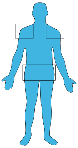
A wide range of symptoms can indicate if a person has polymyalgia rheumatica. The classic symptoms include: [2] [11]

Temporal arteritis

About 20% of people who are diagnosed with polymyalgia rheumatica also have temporal arteritis (also called giant cell arteritis), and about 50% of people with temporal arteritis have polymyalgia rheumatica. [2] Some symptoms of temporal arteritis include headaches, scalp tenderness, jaw or facial soreness, distorted vision, or aching in the limbs caused by decreased blood flow, and fatigue. [13]

Causes

The pathophysiology of polymyalgia rheumatica is not well-understood. Evidence shows that there is likely a combined genetic and environmental pathophysiology behind the disease, but concrete identification of the causes, including whether or not polymyalgia rheumatica is an autoimmune disease, remains elusive. [4] It is, at the very least, an immune-mediated disease, with both innate and adaptive immune system elements being known to play a role. [2] [4] Infectious diseases have historically been hypothesized as a likely trigger for disease in genetically susceptible people. Individual studies have sometimes found correlations between specific pathogens and development of the disease, but broader analysis fails to find significant correlations. Proposed causative pathogens, none of which have been proven, include: [4]

The immune cell involvement in polymyalgia rheumatica includes the activation of dendritic cells and monocytes/macrophages, leading to inflammation in the synovium and bursae of the shoulder and hip girdles which is primarily mediated by the innate immune system. There is an altered balance between Th17 and Treg cells, with increased IL-6 levels driving Th17 cell activation. Disturbed B cell distribution and function are also observed, with a decrease in circulating B cells that recover after steroid treatment. Additionally, systemic activation of circulating monocytes is associated with increased IL-6 and IL-1 beta production. Associations of uncertain significance with multiple types of TNF have also been found. [4]

Despite the severe pain associated with the condition in multiple muscle groups, as well as the signs of systemic inflammation, muscle biopsies have found no signs of localized inflammation in muscle tissue in patients with PMR. Electromyography studies also typically turn up normal. The only locations known definitively to be inflamed in PMR are the synovial membranes and bursae of joints. [4]

Persons having the HLA-DR4 type of human leucocyte antigen appear to have a higher risk of PMR. [14]

Diagnosis

No specific test exists to diagnose polymyalgia rheumatica; many other diseases can cause inflammation and pain in muscles, but a few tests can help narrow down the cause of the pain. Limitation in shoulder motion or swelling of the joints in the wrists or hands, are noted by the doctor. [15]

One blood test usually performed is the erythrocyte sedimentation rate (ESR) which measures how fast the patient's red blood cells settle in a test tube. The faster the red blood cells settle, the higher the ESR value (measured in mm/hour), which suggests that inflammation may be present. Many conditions can cause an elevated ESR, so this test alone is not proof that a person has polymyalgia rheumatica. [16] [17]

Another test that checks the level of C-reactive protein (CRP) in the blood may also be conducted. CRP is produced by the liver in response to an injury or infection, and people with polymyalgia rheumatica usually have high levels. [16] [17] However, like the ESR, this test is also not very specific. [18]

Polymyalgia rheumatica is sometimes associated with temporal arteritis, a condition requiring more aggressive therapy. To test for this additional disorder, a biopsy sample may be taken from the temporal artery. [16]

Treatment

Prednisone is the drug of choice for PMR, [19] and treatment duration is frequently greater than one year. [15] If the patient does not experience dramatic improvement after three days of 10–20 mg oral prednisone per day, the diagnosis should be reconsidered. [20] Sometimes relief of symptoms occurs in only several hours. [19]

Nonsteroidal anti-inflammatory drugs (NSAIDs) such as ibuprofen are ineffective in the initial treatment of PMR, [21] but they may be used in conjunction with the maintenance dose of corticosteroid. [22]

Along with medical treatment, patients are encouraged to exercise and eat healthily, helping to maintain a strong immune system and build strong muscles and bones. [23] A diet of fruits, vegetables, whole grains, and low-fat meat and dairy products, avoiding foods with high levels of refined sugars and salt is recommended. [24] Research in the UK has also suggested that people with polymyalgia rheumatica would benefit from a falls assessment when first diagnosed, and regular treatment reviews. [25] [26]

Epidemiology

No circumstances are certain as to which an individual will get polymyalgia rheumatica, but a few factors show a relationship with the disorder:

See also

Related Research Articles

<span class="mw-page-title-main">Giant cell arteritis</span> Inflammatory disease of large blood vessels

Giant cell arteritis (GCA), also called temporal arteritis, is an inflammatory autoimmune disease of large blood vessels. Symptoms may include headache, pain over the temples, flu-like symptoms, double vision, and difficulty opening the mouth. Complications can include blockage of the artery to the eye with resulting blindness, as well as aortic dissection, and aortic aneurysm. GCA is frequently associated with polymyalgia rheumatica.

<span class="mw-page-title-main">Crohn's disease</span> Type of inflammatory bowel disease

Crohn's disease is a chronic inflammatory bowel disease characterized by recurrent episodes of intestinal inflammation, primarily manifesting as diarrhea and abdominal pain. Unlike ulcerative colitis, inflammation can occur anywhere in the gastrointestinal tract, though it most frequently affects the ileum and colon, involving all layers of the intestinal wall. Symptoms may be non-specific and progress gradually, often delaying diagnosis. About one-third of patients have colonic disease, another third have ileocolic disease, and the remaining third have isolated ileal disease. Systemic symptoms such as chronic fatigue, weight loss, and low-grade fevers are common. Organs such as the skin and joints can also be affected. Complications can include bowel obstructions, fistulas, nutrition problems, and an increased risk of intestinal cancers.

<span class="mw-page-title-main">Myalgia</span> Painful sensations in muscle tissue

Myalgia or muscle pain is a painful sensation evolving from muscle tissue. It is a symptom of many diseases. The most common cause of acute myalgia is the overuse of a muscle or group of muscles; another likely cause is viral infection, especially when there has been no injury.

Rheumatology is a branch of medicine devoted to the diagnosis and management of disorders whose common feature is inflammation in the bones, muscles, joints, and internal organs. Rheumatology covers more than 100 different complex diseases, collectively known as rheumatic diseases, which includes many forms of arthritis as well as lupus and Sjögren's syndrome. Doctors who have undergone formal training in rheumatology are called rheumatologists.

<span class="mw-page-title-main">Erythrocyte sedimentation rate</span> Physiological quantity

The erythrocyte sedimentation rate is the rate at which red blood cells in anticoagulated whole blood descend in a standardized tube over a period of one hour. It is a common hematology test, and is a non-specific measure of inflammation. To perform the test, anticoagulated blood is traditionally placed in an upright tube, known as a Westergren tube, and the distance which the red blood cells fall is measured and reported in millimetres at the end of one hour.

<span class="mw-page-title-main">Prednisone</span> Steroid medication

Prednisone is a glucocorticoid medication mostly used to suppress the immune system and decrease inflammation in conditions such as asthma, COPD, and rheumatologic diseases. It is also used to treat high blood calcium due to cancer and adrenal insufficiency along with other steroids. It is taken by mouth.

<span class="mw-page-title-main">Vasculitis</span> Medical disorders that destroy blood vessels by inflammation

Vasculitis is a group of disorders that destroy blood vessels by inflammation. Both arteries and veins are affected. Lymphangitis is sometimes considered a type of vasculitis. Vasculitis is primarily caused by leukocyte migration and resultant damage. Although both occur in vasculitis, inflammation of veins (phlebitis) or arteries (arteritis) on their own are separate entities.

<span class="mw-page-title-main">Polyarteritis nodosa</span> Systemic necrotizing inflammation of medium-sized muscular arteries

Polyarteritis nodosa (PAN) is a systemic necrotizing inflammation of blood vessels (vasculitis) affecting medium-sized muscular arteries, typically involving the arteries of the kidneys and other internal organs but generally sparing the lungs' circulation. Small aneurysms are strung like the beads of a rosary, therefore making this "rosary sign" an important diagnostic feature of the vasculitis. PAN is sometimes associated with infection by the hepatitis B or hepatitis C virus. The condition may be present in infants.

<span class="mw-page-title-main">Arteritis</span> Medical condition

Arteritis is a vascular disorder characterized by inflammation of the walls of arteries, usually as a result of infection or autoimmune responses. Arteritis, a complex disorder, is still not entirely understood. Arteritis may be distinguished by its different types, based on the organ systems affected by the disease. A complication of arteritis is thrombosis, which can be fatal. Arteritis and phlebitis are forms of vasculitis.

<span class="mw-page-title-main">Ear pain</span> Pain in the ear

Ear pain, also known as earache or otalgia, is pain in the ear. Primary ear pain is pain that originates from the ear. Secondary ear pain is a type of referred pain, meaning that the source of the pain differs from the location where the pain is felt.

Posterior ischemic optic neuropathy (PION) is a medical condition characterized by damage to the retrobulbar portion of the optic nerve due to inadequate blood flow (ischemia) to the optic nerve. Despite the term posterior, this form of damage to the eye's optic nerve due to poor blood flow also includes cases where the cause of inadequate blood flow to the nerve is anterior, as the condition describes a particular mechanism of visual loss as much as the location of damage in the optic nerve. In contrast, anterior ischemic optic neuropathy (AION) is distinguished from PION by the fact that AION occurs spontaneously and on one side in affected individuals with predisposing anatomic or cardiovascular risk factors.

<span class="mw-page-title-main">Giant cell</span> Mass resulting from the fusion of many cells

A giant cell is a mass formed by the union of several distinct cells, often forming a granuloma.

<span class="mw-page-title-main">Myositis</span> Inflammation of skeletal muscle

Myositis is a rarely-encountered medical condition characterized by inflammation affecting the muscles. The manifestations of this condition may include skin issues, muscle weakness, and the potential involvement of other organs. Additionally, systemic symptoms like weight loss, fatigue, and low-grade fever can manifest in individuals with myositis.

Arteritic anterior ischemic optic neuropathy is vision loss that occurs in giant cell arteritis. Temporal arteritis is an inflammatory disease of medium-sized blood vessels that happens especially with advancing age. AAION occurs in about 15-20 percent of patients with temporal arteritis. Damage to the blood vessels supplying the optic nerves leads to insufficient blood supply (ischemia) to the nerve and subsequent optic nerve fiber death. Most cases of AAION result in nearly complete vision loss first to one eye. If the temporal arteritis is left untreated, the affected eye will likely suffer vision loss as well within 1–2 weeks. Arteritic AION (AAION) falls under the general category of anterior ischemic optic neuropathy (AION), which also includes non-arteritic AION (NAION). AAION is considered an eye emergency, immediate treatment is essential to rescue remaining vision.

<span class="mw-page-title-main">Neuromuscular disease</span> Medical condition

A neuromuscular disease is any disease affecting the peripheral nervous system (PNS), the neuromuscular junctions, or skeletal muscles, all of which are components of the motor unit. Damage to any of these structures can cause muscle atrophy and weakness. Issues with sensation can also occur.

Aortitis is the inflammation of the aortic wall. The disorder is potentially life-threatening and rare. It is reported that there are only 1–3 new cases of aortitis per year per million people in the United States and Europe. Aortitis is most common in people 10 to 40 years of age.

AA amyloidosis is a form of amyloidosis, a disease characterized by the abnormal deposition of fibers of insoluble protein in the extracellular space of various tissues and organs. In AA amyloidosis, the deposited protein is serum amyloid A protein (SAA), an acute-phase protein which is normally soluble and whose plasma concentration is highest during inflammation.

<span class="mw-page-title-main">Systemic vasculitis</span> Medical condition

Necrotizing vasculitis, also called systemic necrotizing vasculitis, is a general term for the inflammation of veins and arteries that develops into necrosis and narrows the vessels.

Autoimmunity refers to a pathological immune response of the body's immune system against itself. Autoimmune disease is widely recognized to be significantly more common in women than in men, and often presents differently between the sexes. The reasons for these disparities are still under investigation, but may in part involve the presence of an additional X chromosome in women, as well as the higher presence of female sex hormones such as estrogen. The risk, incidence, and character of autoimmune disease in women may also be associated with female-specific physiological changes, such as hormonal shifts during menses, pregnancy, and menopause.

<span class="mw-page-title-main">Acute visual loss</span> Loss of visual acuity associated with illness or aging

Acute visual loss is a rapid loss of the ability to see. It is caused by many ocular conditions like retinal detachment, glaucoma, macular degeneration, and giant cell arteritis, etc.

References

  1. 1 2 "Polymyalgia Rheumatica". National Institute of Arthritis and Musculoskeletal and Skin Diseases. April 11, 2017. Retrieved February 10, 2021.
  2. 1 2 3 4 5 6 7 8 9 10 Espígol-Frigolé, Georgina; Dejaco, Christian; Mackie, Sarah L; Salvarani, Carlo; Matteson, Eric L; Cid, Maria C (October 2023). "Polymyalgia rheumatica". The Lancet. 402 (10411): 1459–1472. doi:10.1016/S0140-6736(23)01310-7.
  3. Schmidt J, Warrington KJ (August 1, 2011). "Polymyalgia rheumatica and giant cell arteritis in older patients: diagnosis and pharmacological management". Drugs & Aging. 28 (8): 651–66. doi:10.2165/11592500-000000000-00000. PMID   21812500. S2CID   44787949.
  4. 1 2 3 4 5 6 Guggino, G.; Ferrante, A.; Macaluso, F.; Triolo, G.; Ciccia, F. (March 27, 2018). "Pathogenesis of polymyalgia rheumatica". Reumatismo. 70 (1): 10–17. doi:10.4081/reumatismo.2018.1048. ISSN   2240-2683.
  5. Cimmino MA (1997). "Genetic and environmental factors in polymyalgia rheumatica". Annals of the Rheumatic Diseases. 56 (10): 576–577. doi:10.1136/ard.56.10.576. PMC   1752263 . PMID   9389216.
  6. Dejaco C, Singh YP (October 2015). "2015 Recommendations for the management of polymyalgia rheumatica: a European League Against Rheumatism/American College of Rheumatology collaborative initiative". Annals of the Rheumatic Diseases. 74 (10): 1799–807. doi: 10.1136/annrheumdis-2015-207492 . hdl: 2445/115060 . PMID   26359488.
  7. "Polymyalgia Rheumatica treatments and drugs". MayoClinic. December 4, 2010. Retrieved January 19, 2012.
  8. "Polymyalgia Rheumatica definition". MayoClinic. May 17, 2008. Archived from the original on June 23, 2008. Retrieved January 19, 2012.
  9. Davison S, Spiera H, Plotz CM (February 1966). "Polymyalgia rheumatica". Arthritis and Rheumatism. 9 (1): 18–23. doi:10.1002/art.1780090103. PMID   4952416.
  10. Plotz, Charles; Docken, William (May 2013). "Letters: More on the History of Polymyalgia Rheumatica and Giant Cell Arteritis". The Rheumatologist. ACR/ARHP. Archived from the original on February 12, 2015. Retrieved June 1, 2014.
  11. "Polymyalgia rheumatica". NHS. October 20, 2017. Retrieved July 23, 2021.
  12. "Polymyalgia Rheumatica symptoms". MayoClinic. December 4, 2010. Retrieved January 19, 2012.
  13. 1 2 3 4 Gelfand JL (November 18, 2007). "Polymyalgia Rheumatica and Temporal Arteritis". WebMD . Retrieved June 10, 2008.
  14. Page 255 in: Elizabeth D Agabegi; Agabegi, Steven S. (2008). Step-Up to Medicine (Step-Up Series) . Hagerstwon, MD: Lippincott Williams & Wilkins. ISBN   978-0-7817-7153-5.
  15. 1 2 Shiel Jr WC (March 13, 2008). "Polymyalgia Rheumatica (PMR) & Giant Cell Arteritis (Temporal Arteritis)". MedicineNet. Retrieved June 10, 2008.
  16. 1 2 3 "Polymyalgia Rheumatica tests and diagnosis". MayoClinic. December 4, 2010. Retrieved January 19, 2012.
  17. 1 2 "Polymyalgia Rheumatica tests and diagnosis". MayoClinic. May 17, 2008. Archived from the original on June 23, 2008. Retrieved January 19, 2012.
  18. Buttgereit, Frank; Dejaco, Christian; Matteson, Eric L.; Dasgupta, Bhaskar (June 14, 2016). "Polymyalgia Rheumatica and Giant Cell Arteritis: A Systematic Review". JAMA. 315 (22): 2442. doi:10.1001/jama.2016.5444. ISSN   0098-7484.
  19. 1 2 Hernández-Rodríguez J, Cid MC, López-Soto A, Espigol-Frigolé G, Bosch X (November 2009). "Treatment of polymyalgia rheumatica: a systematic review". Archives of Internal Medicine. 169 (20): 1839–50. doi: 10.1001/archinternmed.2009.352 . PMID   19901135.
  20. McPhee SJ, Papadakis MA (2010). Current Medical Diagnosis and Treatment . p.  767. ISBN   978-0071624442.
  21. 1 2 Docken WP (August 2009). "Polymyalgia rheumatica". American College of Rheumatology. Retrieved January 20, 2012.
  22. "Polymyalgia rheumatica". MDGuidelines. Retrieved January 20, 2012.
  23. "Polymyalgia Rheumatica lifestyle and home remedies". MayoClinic. May 17, 2008. Archived from the original on June 23, 2008. Retrieved January 19, 2012.
  24. "Polymyalgia Rheumatica lifestyle and home remedies". MayoClinic. December 4, 2010. Retrieved January 19, 2012.
  25. "Polymyalgia rheumatica: treatment reviews are needed". NIHR Evidence. June 21, 2022. doi:10.3310/nihrevidence_51304. S2CID   251774691 . Retrieved August 5, 2022.
  26. Singh Sokal, Balamrit; Hilder, Samantha L.; Paskins, Zoe; Mallen, Christian D; Muller, Sara (2021). "Fragility fractures and prescriptions of medications for osteoporosis in patients with polymyalgia rheumatica: results from the PMR Cohort Study". Rheumatology Advances in Practice. 5 (3): rkab094. doi:10.1093/rap/rkab094. PMC   8712242 . PMID   34988356.
  27. 1 2 "Polymyalgia Rheumatica risk factors". MayoClinic. December 4, 2010. Retrieved January 19, 2012.