Interphalangeal joints of the foot

Last updated
Interphalangeal joints of foot
DIP, PIP, IP and MTP joints of the foot.png
The MTP, IP, PIP, and DIP joints of the foot:
  • MetaTarsoPhalangeal
  • InterPhalangeal (big toe only has one joint)
  • Proximal InterPhalangeal
  • Distal InterPhalangeal
Articulationes interphalangeae pedis-la.svg
Bones of the foot. Interphalangeal joints highlighted (orange background area)
Details
Identifiers
Latin articulationes interphalangeae pedis
MeSH D014033
TA98 A03.6.10.901
TA2 1968
FMA 71357
Anatomical terminology

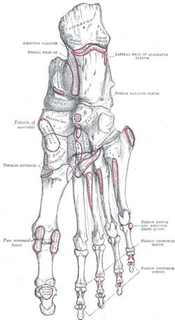
The interphalangeal joints of the foot are between the phalanx bones of the toes in the feet.

Contents

Since the great toe only has two phalanx bones (proximal and distal phalanges), it only has one interphalangeal joint, which is often abbreviated as the "IP joint". The rest of the toes each have three phalanx bones (proximal, middle, and distal phalanges), so they have two interphalangeal joints: the proximal interphalangeal joint between the proximal and middle phalanges (abbreviated "PIP joint") and the distal interphalangeal joint between the middle and distal phalanges (abbreviated "DIP joint").

All interphalangeal joints are ginglymoid (hinge) joints, and each has a plantar (underside) and two collateral ligaments. In the arrangement of these ligaments, extensor tendons supply the places of dorsal ligaments, which is similar to that in the metatarsophalangeal articulations.

Toe bones or phalanges of the foot. Note the big toe has no middle phalanx.
People vary; sometimes the smallest toe also has none (not shown). [1]

   Distal phalanges of the foot
   Middle phalanges of the foot
   Proximal phalanges of the foot

Movements

The only movements permitted in the joints of the digits are flexion and extension; these movements are more extensive between the first and second phalanges than between the second and third. The flexor hallucis longus and flexor digitorum longus flex the interphalangeal joint of the big toe and lateral four toes, respectively. The tendons of both of these muscles cross as they reach their distal attachments. In other words, the flexor hallucis longus arises laterally, while the flexor digitorum longus arises medially.

The amount of flexion is very considerable, but extension is limited by the plantar and collateral ligaments.

Bones of the right foot, bottom (plantar) surface. Joints are not labelled, but region is visible. Gray269.png
Bones of the right foot, bottom (plantar) surface. Joints are not labelled, but region is visible.

See also

Related Research Articles

<span class="mw-page-title-main">Foot</span> Anatomical structure found in vertebrates

The foot is an anatomical structure found in many vertebrates. It is the terminal portion of a limb which bears weight and allows locomotion. In many animals with feet, the foot is a separate organ at the terminal part of the leg made up of one or more segments or bones, generally including claws and/or nails.

<span class="mw-page-title-main">Human leg</span> Lower extremity or limb of the human body (foot, lower leg, thigh and hip)

The human leg is the entire lower limb of the human body, including the foot, thigh or sometimes even the hip or buttock region. The major bones of the leg are the femur, tibia, and adjacent fibula. The thigh is between the hip and knee, while the calf (rear) and shin (front) are between the knee and foot.

<span class="mw-page-title-main">Toe</span> Digit of a foot

Toes are the digits of the foot of a tetrapod. Animal species such as cats that walk on their toes are described as being digitigrade. Humans, and other animals that walk on the soles of their feet, are described as being plantigrade; unguligrade animals are those that walk on hooves at the tips of their toes.

<span class="mw-page-title-main">Extensor hallucis longus muscle</span> Thin muscle, situated between the tibialis anterior and the extensor digitorum longus

The extensor hallucis longus muscle is a thin skeletal muscle, situated between the tibialis anterior and the extensor digitorum longus. It extends the big toe and dorsiflects the foot. It also assists with foot eversion and inversion.

<span class="mw-page-title-main">Extensor digitorum muscle</span> Muscle of the posterior forearm

The extensor digitorum muscle is a muscle of the posterior forearm present in humans and other animals. It extends the medial four digits of the hand. Extensor digitorum is innervated by the posterior interosseous nerve, which is a branch of the radial nerve.

<span class="mw-page-title-main">Dorsal interossei of the foot</span> Four muscles situated between the metatarsal bones

In human anatomy, the dorsal interossei of the foot are four muscles situated between the metatarsal bones.

<span class="mw-page-title-main">Flexor hallucis longus muscle</span> One of the three deep muscles in the lower leg

The flexor hallucis longus muscle (FHL) attaches to the plantar surface of phalanx of the great toe and is responsible for flexing that toe. The FHL is one of the three deep muscles of the posterior compartment of the leg, the others being the flexor digitorum longus and the tibialis posterior. The tibialis posterior is the most powerful of these deep muscles. All three muscles are innervated by the tibial nerve which comprises half of the sciatic nerve.

<span class="mw-page-title-main">Flexor digitorum longus muscle</span> Muscle located on the tibial side of the leg

The flexor digitorum longus muscle is situated on the tibial side of the leg. At its origin it is thin and pointed, but it gradually increases in size as it descends. It serves to flex the second, third, fourth, and fifth toes.

<span class="mw-page-title-main">Extensor digitorum longus muscle</span> Pennate muscle, situated at the lateral part of the front of the leg

The extensor digitorum longus is a pennate muscle, situated at the lateral part of the front of the leg.

<span class="mw-page-title-main">Extensor digitorum brevis muscle</span> Muscle on the upper surface of the foot that helps extend digits 2 through 4

The extensor digitorum brevis muscle is a muscle on the upper surface of the foot that helps extend digits 2 through 4.

<span class="mw-page-title-main">Adductor hallucis muscle</span> Muscle responsible for adducting the big toe

The Adductor hallucis arises by two heads—oblique and transverse and is responsible for adducting the big toe. It has two heads, both are innervated by the lateral plantar nerve.

<span class="mw-page-title-main">Flexor hallucis brevis muscle</span> Muscle in sole of the foot that leads to the big toe

Flexor hallucis brevis muscle is a muscle of the foot that flexes the big toe.

<span class="mw-page-title-main">Flexor digitorum brevis muscle</span> Lies in the middle of the sole of the foot

The flexor digitorum brevis is a muscle which lies in the middle of the sole of the foot, immediately above the central part of the plantar aponeurosis, with which it is firmly united.

<span class="mw-page-title-main">Quadratus plantae muscle</span> Muscles of the sole of the foot

The quadratus plantae is separated from the muscles of the first layer by the lateral plantar vessels and nerve. It acts to aid in flexing the 2nd to 5th toes and is one of the few muscles in the foot with no homolog in the hand.

<span class="mw-page-title-main">Lumbricals of the foot</span> Four small skeletal muscles

The lumbricals are four small skeletal muscles, accessory to the tendons of the flexor digitorum longus muscle. They are numbered from the medial side of the foot.

<span class="mw-page-title-main">Dorsal interossei of the hand</span> Muscles between the metacarpals

In human anatomy, the dorsal interossei (DI) are four muscles in the back of the hand that act to abduct (spread) the index, middle, and ring fingers away from hand's midline and assist in flexion at the metacarpophalangeal joints and extension at the interphalangeal joints of the index, middle and ring fingers.

<span class="mw-page-title-main">Interphalangeal joints of the hand</span> Hinge joints between finger phalanges

The interphalangeal joints of the hand are the hinge joints between the phalanges of the fingers that provide flexion towards the palm of the hand.

<span class="mw-page-title-main">Sole (foot)</span> Bottom part of foot

The sole is the bottom of the foot.

In the human foot, the plantar or volar plates are fibrocartilaginous structures found in the metatarsophalangeal (MTP) and interphalangeal (IP) joints. The anatomy and composition of the plantar plates are similar to the palmar plates in the metacarpophalangeal (MCP) and interphalangeal joints in the hand; the proximal origin is thin but the distal insertion is stout. Due to the weight-bearing nature of the human foot, the plantar plates are exposed to extension forces not present in the human hand.

<span class="mw-page-title-main">Extrinsic extensor muscles of the hand</span>

The extrinsic extensor muscles of the hand are located in the back of the forearm and have long tendons connecting them to bones in the hand, where they exert their action. Extrinsic denotes their location outside the hand. Extensor denotes their action which is to extend, or open flat, joints in the hand. They include the extensor carpi radialis longus (ECRL), extensor carpi radialis brevis (ECRB), extensor digitorum (ED), extensor digiti minimi (EDM), extensor carpi ulnaris (ECU), abductor pollicis longus (APL), extensor pollicis brevis (EPB), extensor pollicis longus (EPL), and extensor indicis (EI).

References

  1. Hatch, RL; Hacking, S (15 December 2003). "Evaluation and management of toe fractures". American Family Physician. 68 (12): 2413–8. PMID   14705761.

PD-icon.svgThis article incorporates text in the public domain from page 359 of the 20th edition of Gray's Anatomy (1918)