Periaortic lymph nodes

Last updated
Periaortic lymph nodes
Lymph node regions.svg
Lymph nodes. (Paraaortic labeled in center in blue.)
Illu lymph chain07.jpg
Details
System Lymphatic system
Anatomical terminology

The periaortic lymph nodes (also known as lumbar) are a group of lymph nodes that lie in front of the lumbar vertebrae near the aorta. These lymph nodes receive drainage from the gastrointestinal tract and the abdominal organs.

Contents

The periaortic lymph nodes are different from the paraaortic lymph nodes. The periaortic group is the general group, that is subdivided into: preaortic, paraaortic, and retroaortic groups. The paraaortic group is synonymous with the lateral aortic group.

Divisions

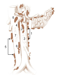
The periaortic lymph node group is divided into three subgroups: preaortic, paraaortic, and retroaortic:

The right paraaortic nodes, are situated partly in front of the inferior vena cava near the termination of the renal vein, and partly behind it on the origin of the psoas major, and on the right crus of diaphragm.

The left paraaortic nodes form a chain on the left side of the abdominal aorta in front of the origin of the psoas major and on the left crus of the diaphragm.

The paraaortic and retroaortic nodes receive:

Most of the efferent vessels from the paraaortic nodes converge to form the right and left lumbar trunks which join the cisterna chyli, but some enter the preaortic and retroaortic lymph nodes, and others pierce the crura of the diaphragm to join the lower end of the thoracic duct.

Dissection

The lateral aortic lymph nodes, typically 15 to 20 on each side, are the ones usually chosen for dissection or biopsy in the treatment or diagnosis of cancer.

A dissection usually includes the region from the bifurcation of the aorta to the superior mesenteric artery or the renal veins.

Additional images

Bibliography

See also

Related Research Articles

<span class="mw-page-title-main">Ureter</span> Tubes used in the urinary system in most animals

The ureters are tubes made of smooth muscle that propel urine from the kidneys to the urinary bladder. In a human adult, the ureters are usually 20–30 cm (8–12 in) long and around 3–4 mm (0.12–0.16 in) in diameter. The ureter is lined by urothelial cells, a type of transitional epithelium, and has an additional smooth muscle layer that assists with peristalsis in its lowest third.

Articles related to anatomy include:

<span class="mw-page-title-main">Thoracic duct</span> Lymphatic vessel

In human anatomy, the thoracic duct is the larger of the two lymph ducts of the lymphatic system. The thoracic duct usually begins from the upper aspect of the cisterna chyli, passing out of the abdomen through the aortic hiatus into first the posterior mediastinum and then the superior mediastinum, extending as high up as the root of the neck before descending to drain into the systemic (blood) circulation at the venous angle.

<span class="mw-page-title-main">Thoracic diaphragm</span> Sheet of internal skeletal muscle

The thoracic diaphragm, or simply the diaphragm, is a sheet of internal skeletal muscle in humans and other mammals that extends across the bottom of the thoracic cavity. The diaphragm is the most important muscle of respiration, and separates the thoracic cavity, containing the heart and lungs, from the abdominal cavity: as the diaphragm contracts, the volume of the thoracic cavity increases, creating a negative pressure there, which draws air into the lungs. Its high oxygen consumption is noted by the many mitochondria and capillaries present; more than in any other skeletal muscle.

<span class="mw-page-title-main">Mesentery</span> Contiguous fold of tissues that supports the intestines

The mesentery is an organ that attaches the intestines to the posterior abdominal wall and is formed by the double fold of peritoneum. It helps in storing fat and allowing blood vessels, lymphatics, and nerves to supply the intestines, among other functions.

<span class="mw-page-title-main">Abdominal aorta</span> Largest artery in the abdomen

In human anatomy, the abdominal aorta is the largest artery in the abdominal cavity. As part of the aorta, it is a direct continuation of the descending aorta.

<span class="mw-page-title-main">Inguinal lymph nodes</span> Lymph nodes in the human groin

Inguinal lymph nodes are lymph nodes in the groin. They are situated in the femoral triangle of the inguinal region. They are subdivided into two groups: the superficial inguinal lymph nodes and deep inguinal lymph nodes.

<span class="mw-page-title-main">Inferior mesenteric artery</span>

In human anatomy, the inferior mesenteric artery (IMA) is the third main branch of the abdominal aorta and arises at the level of L3, supplying the large intestine from the distal transverse colon to the upper part of the anal canal. The regions supplied by the IMA are the descending colon, the sigmoid colon, and part of the rectum.

<span class="mw-page-title-main">Cisterna chyli</span> Component of the lymphoid system

The cisterna chyli or receptaculum chyli is a dilated sac at the lower end of the thoracic duct in most mammals into which lymph from the intestinal trunk and two lumbar lymphatic trunks flow. It receives fatty chyle from the intestines and thus acts as a conduit for the lipid products of digestion. It is the most common drainage trunk of most of the body's lymphatics. The cisterna chyli is a retroperitoneal structure.

<span class="mw-page-title-main">Internal iliac artery</span> Main artery of the pelvis

The internal iliac artery is the main artery of the pelvis.

<span class="mw-page-title-main">Median sacral artery</span>

The median sacral artery is a small artery that arises posterior to the abdominal aorta and superior to its bifurcation.

<span class="mw-page-title-main">Lumbar arteries</span> Arteries within the lower back

The lumbar arteries are arteries located in the lower back or lumbar region. The lumbar arteries are in parallel with the intercostals.

<span class="mw-page-title-main">Aortic hiatus</span> Anatomical detail

The aortic hiatus is a midline opening in the posterior part of the diaphragm giving passage to the descending aorta as well as the thoracic duct, and variably the azygos and hemiazygos veins. It is the lowest and most posterior of the large apertures.

<span class="mw-page-title-main">Crus of diaphragm</span> Anatomical structure

The crus of diaphragm, refers to one of two tendinous structures that extends below the diaphragm to the vertebral column. There is a right crus and a left crus, which together form a tether for muscular contraction. They take their name from their leg-shaped appearance – crus meaning leg in Latin.

<span class="mw-page-title-main">Axillary lymph nodes</span> Lymph nodes in the human armpit

The axillary lymph nodes or armpit lymph nodes are lymph nodes in the human armpit. Between 20 and 49 in number, they drain lymph vessels from the lateral quadrants of the breast, the superficial lymph vessels from thin walls of the chest and the abdomen above the level of the navel, and the vessels from the upper limb. They are divided in several groups according to their location in the armpit. These lymph nodes are clinically significant in breast cancer, and metastases from the breast to the axillary lymph nodes are considered in the staging of the disease.

<span class="mw-page-title-main">Common iliac lymph nodes</span> Lymph nodes found in the pelvis

The common iliac lymph nodes, four to six in number, are grouped behind and on the sides of the common iliac artery, one or two being placed below the bifurcation of the aorta, in front of the fifth lumbar vertebra.

<span class="mw-page-title-main">Lumbar lymph trunk</span>

The lumbar trunks are formed by the union of the efferent vessels from the lateral aortic lymph nodes.

<span class="mw-page-title-main">Outline of human anatomy</span> Overview of and topical guide to human anatomy

The following outline is provided as an overview of and topical guide to human anatomy:

The superior diaphragmatic lymph nodes lie on the thoracic aspect of the diaphragm, and consist of three sets – anterior, middle, and posterior.