Protein disulfide-isomerase | |
---|---|
Identifiers | |
Symbol | ? |
InterPro | IPR005792 |
Protein disulfide-isomerase | |||||||||
---|---|---|---|---|---|---|---|---|---|
Identifiers | |||||||||
EC no. | 5.3.4.1 | ||||||||
CAS no. | 37318-49-3 | ||||||||
Databases | |||||||||
IntEnz | IntEnz view | ||||||||
BRENDA | BRENDA entry | ||||||||
ExPASy | NiceZyme view | ||||||||
KEGG | KEGG entry | ||||||||
MetaCyc | metabolic pathway | ||||||||
PRIAM | profile | ||||||||
PDB structures | RCSB PDB PDBe PDBsum | ||||||||
Gene Ontology | AmiGO / QuickGO | ||||||||
|
protein disulfide isomerase family A, member 2 | |||||||
---|---|---|---|---|---|---|---|
Identifiers | |||||||
Symbol | PDIA2 | ||||||
Alt. symbols | PDIP | ||||||
NCBI gene | 64714 | ||||||
HGNC | 14180 | ||||||
OMIM | 608012 | ||||||
RefSeq | NM_006849 | ||||||
UniProt | Q13087 | ||||||
Other data | |||||||
Locus | Chr. 16 p13.3 | ||||||
|
protein disulfide isomerase family A, member 3 | |||||||
---|---|---|---|---|---|---|---|
Identifiers | |||||||
Symbol | PDIA3 | ||||||
Alt. symbols | GRP58 | ||||||
NCBI gene | 2923 | ||||||
HGNC | 4606 | ||||||
OMIM | 602046 | ||||||
RefSeq | NM_005313 | ||||||
UniProt | P30101 | ||||||
Other data | |||||||
Locus | Chr. 15 q15 | ||||||
|
protein disulfide isomerase family A, member 4 | |||||||
---|---|---|---|---|---|---|---|
Identifiers | |||||||
Symbol | PDIA4 | ||||||
NCBI gene | 9601 | ||||||
HGNC | 30167 | ||||||
RefSeq | NM_004911 | ||||||
UniProt | P13667 | ||||||
Other data | |||||||
Locus | Chr. 7 q35 | ||||||
|
protein disulfide isomerase family A, member 5 | |||||||
---|---|---|---|---|---|---|---|
Identifiers | |||||||
Symbol | PDIA5 | ||||||
NCBI gene | 10954 | ||||||
HGNC | 24811 | ||||||
RefSeq | NM_006810 | ||||||
UniProt | Q14554 | ||||||
Other data | |||||||
EC number | 5.3.4.1 | ||||||
Locus | Chr. 3 q21.1 | ||||||
|
protein disulfide isomerase family A, member 6 | |||||||
---|---|---|---|---|---|---|---|
Identifiers | |||||||
Symbol | PDIA6 | ||||||
Alt. symbols | TXNDC7 | ||||||
NCBI gene | 10130 | ||||||
HGNC | 30168 | ||||||
RefSeq | NM_005742 | ||||||
UniProt | Q15084 | ||||||
Other data | |||||||
Locus | Chr. 2 p25.1 | ||||||
|
Protein disulfide isomerase (EC 5.3.4.1), or PDI, is an enzyme in the endoplasmic reticulum (ER) in eukaryotes and the periplasm of bacteria that catalyzes the formation and breakage of disulfide bonds between cysteine residues within proteins as they fold. [1] [2] [3] This allows proteins to quickly find the correct arrangement of disulfide bonds in their fully folded state, and therefore the enzyme acts to catalyze protein folding.
Protein disulfide-isomerase has two catalytic thioredoxin-like domains (active sites), each containing the canonical CGHC motif, and two non catalytic domains. [4] [5] [6] This structure is similar to the structure of enzymes responsible for oxidative folding in the intermembrane space of the mitochondria; an example of this is mitochondrial IMS import and assembly (Mia40), which has 2 catalytic domains that contain a CX9C, which is similar to the CGHC domain of PDI. [7] Bacterial DsbA, responsible for oxidative folding, also has a thioredoxin CXXC domain. [8]
PDI displays oxidoreductase and isomerase properties, both of which depend on the type of substrate that binds to protein disulfide-isomerase and changes in protein disulfide-isomerase's redox state. [4] These types of activities allow for oxidative folding of proteins. Oxidative folding involves the oxidation of reduced cysteine residues of nascent proteins; upon oxidation of these cysteine residues, disulfide bridges are formed, which stabilizes proteins and allows for native structures (namely tertiary and quaternary structures). [4]
PDI is specifically responsible for folding proteins in the ER. [6] In an unfolded protein, a cysteine residue forms a mixed disulfide with a cysteine residue in an active site (CGHC motif) of protein disulfide-isomerase. A second cysteine residue then forms a stable disulfide bridge within the substrate, leaving protein disulfide-isomerase's two active-site cysteine residues in a reduced state. [4]
Afterwards, PDI can be regenerated to its oxidized form in the endoplasmic reticulum by transferring electrons to reoxidizing proteins such ER oxidoreductin 1 (Ero 1), VKOR (vitamin K epoxide reductase), glutathione peroxidase (Gpx7/8), and PrxIV (peroxiredoxin IV). [4] [9] [10] [6] Ero1 is thought to be the main reoxidizing protein of PDI, and the pathway of reoxidation of PDI for Ero1 is more understood than that of other proteins. [10] Ero1 accepts electrons from PDI and donates these electrons to oxygen molecules in the ER, which leads to the formation of hydrogen peroxide. [10]
The reduced (dithiol) form of protein disulfide-isomerase is able to catalyze a reduction of a misformed disulfide bridge of a substrate through either reductase activity or isomerase activity. [11] For the reductase method, a misfolded substrate disulfide bond is converted to a pair of reduced cysteine residues by the transfer of electrons from glutathione and NADPH. Afterwards, normal folding occurs with oxidative disulfide bond formation between the correct pairs of substrate cysteine residues, leading to a properly folded protein. For the isomerase method, intramolecular rearrangement of substrate functional groups is catalyzed near the N terminus of each active site. [4] Therefore, protein disulfide-isomerase is capable of catalyzing the post-translational modification disulfide exchange.
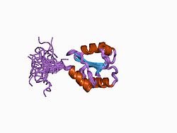
In the chloroplasts of the unicellular algae Chlamydomonas reinhardtii the protein disulfide-isomerase RB60 serves as a redox sensor component of an mRNA-binding protein complex implicated in the photoregulation of the translation of psbA, the RNA encoding for the photosystem II core protein D1. Protein disulfide-isomerase has also been suggested to play a role in the formation of regulatory disulfide bonds in chloroplasts. [12]
Protein disulfide-isomerase helps load antigenic peptides into MHC class I molecules. These molecules (MHC I) are related to the peptide presentation by antigen-presenting cells in the immune response.
Protein disulfide-isomerase has been found to be involved in the breaking of bonds on the HIV gp120 protein during HIV infection of CD4 positive cells, and is required for HIV infection of lymphocytes and monocytes. [13] Some studies have shown it to be available for HIV infection on the surface of the cell clustered around the CD4 protein. Yet conflicting studies have shown that it is not available on the cell surface, but instead is found in significant amounts in the blood plasma.
Another major function of protein disulfide-isomerase relates to its activity as a chaperone; its b' domain aids in the binding of misfolded protein for subsequent degradation. [4] This is regulated by three ER membrane proteins, Protein Kinase RNA-like endoplasmic reticulum kinase (PERK), inositol-requiring kinase 1 (IRE1), and activating transcription factor 6 (ATF6). [4] [14] They respond to high levels of misfolded proteins in the ER through intracellular signaling cascades that can activate PDI's chaperone activity. [4] These signals can also inactivate translation of these misfolded proteins, because the cascade travels from the ER to the nucleus. [4]
Insulin turbidity assay: protein disulfide-isomerase breaks the two disulfide bonds between two insulin (a and b) chains that results in precipitation of b chain. This precipitation can be monitored at 650 nm, which is indirectly used to monitor protein disulfide-isomerase activity. [15] Sensitivity of this assay is in micromolar range.
ScRNase assay: protein disulfide-isomerase converts scrambled (inactive) RNase into native (active) RNase that further acts on its substrate. [16] The sensitivity is in micromolar range.
Di-E-GSSG assay: This is the fluorometric assay that can detect picomolar quantities of protein disulfide-isomerase and therefore is the most sensitive assay to date for detecting protein disulfide-isomerase activity. [17] Di-E-GSSG has two eosin molecules attached to oxidized glutathione (GSSG). The proximity of eosin molecules leads to the quenching of its fluorescence. However, upon breakage of disulfide bond by protein disulfide-isomerase, fluorescence increases 70-fold.
Redox dysregulation leads to increases in nitrosative stress in the endoplasmic reticulum. Such adverse changes in the normal cellular environment of susceptible cells, such as neurons, leads to nonfunctioning thiol-containing enzymes. [14] More specifically, protein disulfide-isomerase can no longer fix misfolded proteins once its thiol group in its active site has a nitric monoxide group attached to it; as a result, accumulation of misfolded proteins occurs in neurons, which has been associated with the development of neurodegenerative diseases such as Alzheimer's disease and Parkinson's disease. [4] [14]
Due to the role of protein disulfide-isomerase in a number of disease states, small molecule inhibitors of protein disulfide-isomerase have been developed. These molecules can either target the active site of protein disulfide-isomerase irreversibly [18] or reversibly. [19]
It has been shown that protein disulfide-isomerase activity is inhibited by red wine and grape juice, which could be the explanation for the French paradox. [20]
Human genes encoding protein disulfide isomerases include: [3] [21] [22]
The endoplasmic reticulum (ER) is a part of a transportation system of the eukaryotic cell, and has many other important functions such as protein folding. It is a type of organelle made up of two subunits – rough endoplasmic reticulum (RER), and smooth endoplasmic reticulum (SER). The endoplasmic reticulum is found in most eukaryotic cells and forms an interconnected network of flattened, membrane-enclosed sacs known as cisternae, and tubular structures in the SER. The membranes of the ER are continuous with the outer nuclear membrane. The endoplasmic reticulum is not found in red blood cells, or spermatozoa.
In chemistry, a disulfide is a compound containing a R−S−S−R′ functional group or the S2−
2 anion. The linkage is also called an SS-bond or sometimes a disulfide bridge and usually derived from two thiol groups.
Thioredoxin reductases are enzymes that reduce thioredoxin (Trx). Two classes of thioredoxin reductase have been identified: one class in bacteria and some eukaryotes and one in animals. In bacteria TrxR also catalyzes the reduction of glutaredoxin like proteins known as NrdH. Both classes are flavoproteins which function as homodimers. Each monomer contains a FAD prosthetic group, a NADPH binding domain, and an active site containing a redox-active disulfide bond.
Thioredoxin is a class of small redox proteins known to be present in all organisms. It plays a role in many important biological processes, including redox signaling. In humans, thioredoxins are encoded by TXN and TXN2 genes. Loss-of-function mutation of either of the two human thioredoxin genes is lethal at the four-cell stage of the developing embryo. Although not entirely understood, thioredoxin is linked to medicine through their response to reactive oxygen species (ROS). In plants, thioredoxins regulate a spectrum of critical functions, ranging from photosynthesis to growth, flowering and the development and germination of seeds. Thioredoxins play a role in cell-to-cell communication.
Oxidative protein folding is a process that is responsible for the formation of disulfide bonds between cysteine residues in proteins. The driving force behind this process is a redox reaction, in which electrons pass between several proteins and finally to a terminal electron acceptor.
Tissue transglutaminase is a 78-kDa, calcium-dependent enzyme of the protein-glutamine γ-glutamyltransferases family. Like other transglutaminases, it crosslinks proteins between an ε-amino group of a lysine residue and a γ-carboxamide group of glutamine residue, creating an inter- or intramolecular bond that is highly resistant to proteolysis. Aside from its crosslinking function, tTG catalyzes other types of reactions including deamidation, GTP-binding/hydrolyzing, and isopeptidase activities. Unlike other members of the transglutaminase family, tTG can be found both in the intracellular and the extracellular spaces of various types of tissues and is found in many different organs including the heart, the liver, and the small intestine. Intracellular tTG is abundant in the cytosol but smaller amounts can also be found in the nucleus and the mitochondria. Intracellular tTG is thought to play an important role in apoptosis. In the extracellular space, tTG binds to proteins of the extracellular matrix (ECM), binding particularly tightly to fibronectin. Extracellular tTG has been linked to cell adhesion, ECM stabilization, wound healing, receptor signaling, cellular proliferation, and cellular motility.
ER oxidoreductin 1 (Ero1) is an oxidoreductase enzyme that catalyses the formation and isomerization of protein disulfide bonds in the endoplasmic reticulum (ER) of eukaryotes. ER Oxidoreductin 1 (Ero1) is a conserved, luminal, glycoprotein that is tightly associated with the ER membrane, and is essential for the oxidation of protein dithiols. Since disulfide bond formation is an oxidative process, the major pathway of its catalysis has evolved to utilise oxidoreductases, which become reduced during the thiol-disulfide exchange reactions that oxidise the cysteine thiol groups of nascent polypeptides. Ero1 is required for the introduction of oxidising equivalents into the ER and their direct transfer to protein disulfide isomerase (PDI), thereby ensuring the correct folding and assembly of proteins that contain disulfide bonds in their native state.
Endoplasmic-reticulum-associated protein degradation (ERAD) designates a cellular pathway which targets misfolded proteins of the endoplasmic reticulum for ubiquitination and subsequent degradation by a protein-degrading complex, called the proteasome.
Glutaredoxins are small redox enzymes of approximately one hundred amino-acid residues that use glutathione as a cofactor. In humans this oxidation repair enzyme is also known to participate in many cellular functions, including redox signaling and regulation of glucose metabolism. Glutaredoxins are oxidized by substrates, and reduced non-enzymatically by glutathione. In contrast to thioredoxins, which are reduced by thioredoxin reductase, no oxidoreductase exists that specifically reduces glutaredoxins. Instead, glutaredoxins are reduced by the oxidation of glutathione. Reduced glutathione is then regenerated by glutathione reductase. Together these components compose the glutathione system.
The unfolded protein response (UPR) is a cellular stress response related to the endoplasmic reticulum (ER) stress. It has been found to be conserved between mammalian species, as well as yeast and worm organisms.
Protein disulfide-isomerase A3 (PDIA3), also known as glucose-regulated protein, 58-kD (GRP58), is an isomerase enzyme encoded by the autosomal gene PDIA3 in humans. This protein localizes to the endoplasmic reticulum (ER) and interacts with lectin chaperones calreticulin and calnexin (CNX) to modulate folding of newly synthesized glycoproteins. It is thought that complexes of lectins and this protein mediate protein folding by promoting formation of disulfide bonds in their glycoprotein substrates.
Binding immunoglobulin protein (BiPS) also known as 78 kDa glucose-regulated protein (GRP-78) or heat shock 70 kDa protein 5 (HSPA5) is a protein that in humans is encoded by the HSPA5 gene.
Protein disulfide-isomerase, also known as the beta-subunit of prolyl 4-hydroxylase (P4HB), is an enzyme that in humans encoded by the P4HB gene. The human P4HB gene is localized in chromosome 17q25. Unlike other prolyl 4-hydroxylase family proteins, this protein is multifunctional and acts as an oxidoreductase for disulfide formation, breakage, and isomerization. The activity of P4HB is tightly regulated. Both dimer dissociation and substrate binding are likely to enhance its enzymatic activity during the catalysis process.
Endoplasmic reticulum protein 29 (ERp29) is a chaperone protein that in humans is encoded by the ERP29 gene.
ERO1-like protein alpha is a protein that in humans is encoded by the ERO1L gene.
Glutaredoxin 2 (GLRX2) is an enzyme that in humans encoded by the GLRX2 gene. GLRX2, also known as GRX2, is a glutaredoxin family protein and a thiol-disulfide oxidoreductase that maintains cellular thiol homeostasis. This gene consists of four exons and three introns, spanned 10 kilobase pairs, and localized to chromosome 1q31.2–31.3.
Thioredoxin domain-containing protein 5 is a protein that in humans is encoded by the TXNDC5 gene.
DsbA is a bacterial thiol disulfide oxidoreductase (TDOR). DsbA is a key component of the Dsb family of enzymes. DsbA catalyzes intrachain disulfide bond formation as peptides emerge into the cell's periplasm.
Thioredoxins are small disulfide-containing redox proteins that have been found in all the kingdoms of living organisms. Thioredoxin serves as a general protein disulfide oxidoreductase. It interacts with a broad range of proteins by a redox mechanism based on reversible oxidation of 2 cysteine thiol groups to a disulfide, accompanied by the transfer of 2 electrons and 2 protons. The net result is the covalent interconversion of a disulfide and a dithiol.
Protein disulfide isomerase family A member 2 is a protein that in humans is encoded by the PDIA2 gene.