7-Dehydrocholesterol

Last updated
7-Dehydrocholesterol
7-Dehydrocholesterol.svg
7-Dehydrocholesterol molecule ball.png
Names
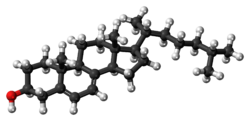
IUPAC name
Cholesta-5,7-dien-3β-ol
Systematic IUPAC name
(1R,3aR,7S,9aR,9bS,11aR)-9a,11a-Dimethyl-1-[(2R)-6-methylheptan-2-yl]-2,3,3a,6,7,8,9,9a,9b,10,11,11a-dodecahydro-1H-cyclopenta[a]phenanthren-7-ol
Identifiers
3D model (JSmol)
ChEBI
ChemSpider
ECHA InfoCard 100.006.456 OOjs UI icon edit-ltr-progressive.svg
MeSH 7-dehydrocholesterol
PubChem CID
UNII
  • InChI=1S/C27H44O/c1-18(2)7-6-8-19(3)23-11-12-24-22-10-9-20-17-21(28)13-15-26(20,4)25(22)14-16-27(23,24)5/h9-10,18-19,21,23-25,28H,6-8,11-17H2,1-5H3/t19-,21+,23-,24+,25+,26+,27-/m1/s1-dehydrocholesterol X mark.svgN
    Key: UCTLRSWJYQTBFZ-DDPQNLDTSA-N Yes check.svgY
  • InChI=1/C27H44O/c1-18(2)7-6-8-19(3)23-11-12-24-22-10-9-20-17-21(28)13-15-26(20,4)25(22)14-16-27(23,24)5/h9-10,18-19,21,23-25,28H,6-8,11-17H2,1-5H3/t19-,21+,23-,24+,25+,26+,27-/m1/s1
    Key: UCTLRSWJYQTBFZ-DDPQNLDTBZ
  • O[C@@H]4C/C3=C/C=C1\[C@H](CC[C@]2([C@H]1CC[C@@H]2[C@H](C)CCCC(C)C)C)[C@@]3(C)CC4
Properties
C27H44O
Molar mass 384.638
Except where otherwise noted, data are given for materials in their standard state (at 25 °C [77 °F], 100 kPa).
X mark.svgN  verify  (what is  Yes check.svgYX mark.svgN ?)
The epidermal strata of the skin. Skinlayers.png
The epidermal strata of the skin.

7-Dehydrocholesterol (7-DHC) is a zoosterol that functions in the serum as a cholesterol precursor, and is photochemically converted to vitamin D3 in the skin, therefore functioning as provitamin-D3. The presence of this compound in human skin enables humans to manufacture vitamin D3 (cholecalciferol). Upon exposure to ultraviolet UV-B rays in the sun light, 7-DHC is converted into vitamin D3 via previtamin D3 as an intermediate isomer. It is also found in the milk of several mammalian species. [1] [2] Lanolin, a waxy substance that is naturally secreted by wool-bearing mammals, contains 7-DHC which is converted into vitamin D by sunlight and then ingested during grooming as a nutrient. In insects 7-dehydrocholesterol is a precursor for the hormone ecdysone, required for reaching adulthood. [3] It was discovered by Nobel-laureate organic chemist Adolf Windaus.

Contents

Biosynthesis

It is synthesized from lathosterol by the enzyme lathosterol oxidase (lathosterol 5-desaturase). This is the next-to-last step of cholesterol biosynthesis. [4] Defective synthesis results in the human inherited disorder lathosterolosis resembling Smith–Lemli–Opitz syndrome. [4] Mice where this gene has been deleted lose the ability to increase vitamin D3 in the blood following UV exposure of the skin. [5]

Location

The skin consists of two primary layers: an inner layer, the dermis, comprising largely connective tissue, and an outer, thinner epidermis. The thickness of the epidermis ranges from 0.08 mm to greater than 0.6 mm (from 0.003 to 0.024 inches). [6] The epidermis comprises five strata; from outer to inner, they are the stratum corneum, stratum lucidum, stratum granulosum, stratum spinosum, and stratum basale. The highest concentrations of 7-dehydrocholesterol are found in the epidermal layer of skin—specifically in the stratum basale and stratum spinosum. [7] The production of pre-vitamin D3 is, therefore, greatest in these two layers.

Radiation

Synthesis of pre-vitamin D3 in the skin involves UVB radiation, which effectively penetrates only the epidermal layers of skin. 7-Dehydrocholesterol absorbs UV light most effectively at wavelengths between 295 and 300 nm and, thus, the production of vitamin D3 will occur primarily at those wavelengths. [8] The two most important factors that govern the generation of pre-vitamin D3 are the quantity (intensity) and quality (appropriate wavelength) of the UVB irradiation reaching the 7-dehydrocholesterol deep in the stratum basale and stratum spinosum. [7] Light-emitting diodes (LEDs) can be used to produce the radiation. [9]

Another important consideration is the quantity of 7-dehydrocholesterol present in the skin. Under normal circumstances, ample quantities of 7-dehydrocholesterol (about 2550 μg/cm2 of skin) are available in the stratum spinosum and stratum basale of human skin to meet the body's vitamin D requirements. 7-DHC insufficiency has been proposed as an alternate cause for Vitamin D deficiency. [10]

Sources

7-DHC can be produced by animals and plants via different pathways. It is not produced by fungi in significant amounts. It is made by some algae, but the pathway is poorly understood. [11] It can also be produced by some bacteria. [10]

Industrially, 7-DHC generally comes from lanolin, and is used to produce vitamin D3 by UV exposure. [12] Lichen (Cladonia rangiferina) is used to produce vegan D3. [13] [14]

7-DHC is used for vitamin D3 synthesis via lanosterol in land animals, via cycloartenol in plants, and in algae together with another provitamin D ergosterol for D2. In fungi solely ergosterol is used for synthesis of D2 via lanosterol. [15]

Interactive pathway map

Click on genes, proteins and metabolites below to link to respective articles. [§ 1]

[[File:
VitaminDSynthesis WP1531.png Go to articleGo to articleGo to articleGo to articlego to articleGo to articleGo to articleGo to articlego to articlego to articlego to articlego to articleGo to articleGo to articlego to articleGo to articlego to articlego to articlego to articleGo to articlego to article
[[
]]
[[
]]
[[
]]
[[
]]
[[
]]
[[
]]
[[
]]
[[
]]
[[
]]
[[
]]
[[
]]
[[
]]
[[
]]
[[
]]
[[
]]
[[
]]
[[
]]
[[
]]
[[
]]
[[
]]
[[
]]
[[
]]
VitaminDSynthesis WP1531.png Go to articleGo to articleGo to articleGo to articlego to articleGo to articleGo to articleGo to articlego to articlego to articlego to articlego to articleGo to articleGo to articlego to articleGo to articlego to articlego to articlego to articleGo to articlego to article
|alt=Vitamin D Synthesis Pathway (view / edit)]]
Vitamin D Synthesis Pathway (view / edit)
  1. The interactive pathway map can be edited at WikiPathways: "VitaminDSynthesis_WP1531".

See also

Related Research Articles

<span class="mw-page-title-main">Integumentary system</span> Skin and other protective organs

The integumentary system is the set of organs forming the outermost layer of an animal's body. It comprises the skin and its appendages, which act as a physical barrier between the external environment and the internal environment that it serves to protect and maintain the body of the animal. Mainly it is the body's outer skin.

<span class="mw-page-title-main">Keratinocyte</span> Primary type of cell found in the epidermis

Keratinocytes are the primary type of cell found in the epidermis, the outermost layer of the skin. In humans, they constitute 90% of epidermal skin cells. Basal cells in the basal layer of the skin are sometimes referred to as basal keratinocytes. Keratinocytes form a barrier against environmental damage by heat, UV radiation, water loss, pathogenic bacteria, fungi, parasites, and viruses. A number of structural proteins, enzymes, lipids, and antimicrobial peptides contribute to maintain the important barrier function of the skin. Keratinocytes differentiate from epidermal stem cells in the lower part of the epidermis and migrate towards the surface, finally becoming corneocytes and eventually be shed off, which happens every 40 to 56 days in humans.

<span class="mw-page-title-main">Epidermis</span> Outermost of the three layers that make up the skin

The epidermis is the outermost of the three layers that comprise the skin, the inner layers being the dermis and hypodermis. The epidermis layer provides a barrier to infection from environmental pathogens and regulates the amount of water released from the body into the atmosphere through transepidermal water loss.

<span class="mw-page-title-main">Cholecalciferol</span> Vitamin D3, a chemical compound

Cholecalciferol, also known as vitamin D3 and colecalciferol, is a type of vitamin D that is made by the skin when exposed to sunlight; it is found in some foods and can be taken as a dietary supplement.

<span class="mw-page-title-main">Ergocalciferol</span> Vitamin D2, a chemical compound

Ergocalciferol, also known as vitamin D2 and nonspecifically calciferol, is a type of vitamin D found in food and used as a dietary supplement. As a supplement it is used to prevent and treat vitamin D deficiency. This includes vitamin D deficiency due to poor absorption by the intestines or liver disease. It may also be used for low blood calcium due to hypoparathyroidism. It is used by mouth or injection into a muscle.

<span class="mw-page-title-main">Ergosterol</span> Chemical compound

Ergosterol (ergosta-5,7,22-trien-3β-ol) is a sterol found in cell membranes of fungi and protozoa, serving many of the same functions that cholesterol serves in animal cells. Because many fungi and protozoa cannot survive without ergosterol, the enzymes that synthesize it have become important targets for drug discovery. In human nutrition, ergosterol is a provitamin form of vitamin D2; exposure to ultraviolet (UV) light causes a chemical reaction that produces vitamin D2.

<span class="mw-page-title-main">Stratum basale</span> Deepest layer of the five layers of the epidermis

The stratum basale is the deepest layer of the five layers of the epidermis, the external covering of skin in mammals.

A provitamin is a substance that may be converted within the body to a vitamin. The term previtamin is a synonym.

<span class="mw-page-title-main">Stratum spinosum</span>

The stratum spinosum is a layer of the epidermis found between the stratum granulosum and stratum basale. This layer is composed of polyhedral keratinocytes. These are joined with desmosomes. Their spiny appearance is due to shrinking of the microfilaments between desmosomes that occurs when stained with H&E. Keratinization begins in the stratum spinosum, although the actual keratinocytes begin in the stratum basale. They have large pale-staining nuclei as they are active in synthesizing fibrilar proteins, known as cytokeratin, which build up within the cells aggregating together forming tonofibrils. The tonofibrils go on to form the desmosomes, which allow for strong connections to form between adjacent keratinocytes. The stratum spinosum also contains Langerhans cells, which functions as a macrophage by engulfing bacteria, foreign particles, and damaged cells that occur in this layer.

<span class="mw-page-title-main">Spinous cell</span>

Spinous cells, or prickle cells, are keratin producing epidermal cells owing their prickly appearance to their numerous intracellular connections. They make up the stratum spinosum of the epidermis and provide a continuous net-like layer of protection for underlying tissue. They are susceptible to mutations caused by sunlight and can become malignant.

UV-induced apoptosis UV-induced apoptosis is an adequate (physiological) reaction of a cell damaged by UV radiation (UVR) in a sufficiently large (lethal) dose and it prevents the disordered destruction of UV damaged cells by help necrosis. Cell elimination by apoptosis occurs when UV-induced cell damage which cannot be repaired by the intracellular repair system exceeds at it certain limit. Through apoptosis, the cells are self-disassembled into compartments with their subsequent utilization. The first time sign of the beginning of the apoptosis system is working in a UV damaged cell is the activation of restriction enzymes, which divide cell DNA into fragments convenient for utilization. But too large a dose of UVR can lead to breakdown (inactivation) of the energy-dependent mechanism of apoptosis. In this case, cell destruction occurs randomly, not orderly, and during a significantly longer time interval. UV-irradiated cells do not change their appearance for a long time [1, 6], as a result of which the researchers may make the erroneous conclusion that “revealed an unexpected response to a dose at which a higher dose of UV increased the viability of keratinocytes” [2]. The fact that UV-induced apoptosis at high doses of UVR begins to be replaced by necrosis was established in 2000 [3]. For keratinocytes, the proportion of cells that have elimination by help apoptosis, with an increase in UVR dose can reach to achieve 45%, but with a further increase in the dose of UVR, destruction of damaged cells by help necrosis and the part of cells that eliminated by apoptosis begins to decrease [4, 11]. In the dose range of UVR from “lethal” to “super-lethal”, “pro-inflammatory” apoptosis can be manifested, which was experimentally discovered in 2003 [5]. This may be the result of partial damage to the apoptosis mechanism by UV radiation [1]. If at moderate doses “pure” apoptosis does not cause an inflammatory reaction, then at sufficiently large doses, an inflammatory reaction arises due to pro-inflammatory apoptosis, which leads to the appearance of “fast” erythema for UV irradiated skin keratinocytes. Kinetic of “fast” erythema is much faster by the time of development of UV erythema caused by necrosis of UV damaged keratinocytes [6]. The most erythemogenic is UVB the spectral range of UVR, since radiation in this range is less absorbed by the outer layers of the skin, which allows UVB radiation, in contrast to UVC, to reach more deep layers skin and act on keratinocytes of the deep-lying basal layer of the epidermis of the skin. The ability to induce apoptosis for UVB and UVC radiation is due to the fact that the DNA of the nucleus [7] and / or mitochondria [8] of the cell absorbs UVR well in the UVC and UVB spectral range. Keratinocytes of the skin are in a state of programmed apoptosis, during which the keratinocytes of the basal layer are removed from it and during the transition through all layers of the epidermis within 28 days turn into flakes of the outer stratum corneum, which are subsequently desquamated. It is clear that the keratinocyte response to UV exposure will depend on what phase of programmed apoptosis the keratinocyte experienced UV exposure, and this is the main reason for the difference of the UV effect for UVC and UVB on the skin. There are also differences in the initiation of mitochondrial (internal) and caspase-dependent (external) apoptosis for the UVC and UVB spectral ranges [9]. Sunburn cells (SBS) are the keratinocytes in the process of UV-induced apoptosis. The appearance of SBC may be not associated with an inflammatory reaction, but the role of UV-induced apoptosis of skin keratinocytes in the development of UV erythema of the skin has been established, which allowed the development of a patent-protected METHOD FOR QUANTITATIVE ASSESSMENT OF APOPTOSIS SYSTEM [10], in which “the brightest lamp of skin display "(photoerythema) is used to diagnose the state of the body systems involved in the elimination of UV-induced damage. Such systems include the immune system, the intracellular repair system, the microcirculation system and not only.

<span class="mw-page-title-main">Human skin</span> Outer covering of the body

The human skin is the outer covering of the body and is the largest organ of the integumentary system. The skin has up to seven layers of ectodermal tissue guarding muscles, bones, ligaments and internal organs. Human skin is similar to most of the other mammals' skin, and it is very similar to pig skin. Though nearly all human skin is covered with hair follicles, it can appear hairless. There are two general types of skin, hairy and glabrous skin (hairless). The adjective cutaneous literally means "of the skin".

Previtamin D<sub>3</sub> Chemical compound

Previtamin D3 is an intermediate in the production of cholecalciferol (vitamin D3).

<span class="mw-page-title-main">Light skin</span> Human skin color

Light skin is a human skin color that has a base level of eumelanin pigmentation that has adapted to environments of low UV radiation. Light skin is most commonly found amongst the native populations of Europe, Central Asia, and Northeast Asia as measured through skin reflectance. People with light skin pigmentation are often referred to as "white" although these usages can be ambiguous in some countries where they are used to refer specifically to certain ethnic groups or populations.

UV-B lamps are lamps that emit a spectrum of ultraviolet light with wavelengths ranging from 290–320 nanometers. This spectrum is also commonly called the biological spectrum due to the human body's sensitivity to light of such a wavelength. UV-B light does not tan the skin very much, compared to the UV-A lamps that are used in tanning beds.

<span class="mw-page-title-main">Vitamin D</span> Group of fat-soluble secosteroids

Vitamin D is a group of fat-soluble secosteroids responsible for increasing intestinal absorption of calcium, magnesium, and phosphate, and for many other biological effects. In humans, the most important compounds in this group are vitamin D3 (cholecalciferol) and vitamin D2 (ergocalciferol).

<span class="mw-page-title-main">Health effects of sunlight exposure</span>

Exposing skin to the ultraviolet radiation in sunlight has both positive and negative health effects. On the positive side, exposure allows for the synthesis of vitamin D3. Vitamin D has been suggested as having a wide range of positive health effects, which include strengthening bones and possibly inhibiting the growth of some cancers. A dietary supplement can also supply vitamin D, but there are also benefits to exposure not obtainable through Vitamin D supplementation. Long-term sun exposure is associated with reduced all-cause mortality and reduced mortality risk from cardiovascular disease (CVD), some forms of cancer, and non-CVD/noncancer related disease, with indications in these studies that Vitamin D is not the mediator. Supplementation offers limited bioavailability and no synthesis of subdermal nitric oxide. UV exposure also has positive effects for endorphin levels, and possibly for protection against multiple sclerosis. Abundant visible light to the eyes gives health benefits through its association with the timing of melatonin synthesis, maintenance of normal and robust circadian rhythms, and reduced risk of seasonal affective disorder.

Skin sloughing is the process of shedding dead surface cells from the skin. It is most associated with cosmetic skin maintenance via exfoliation, but can also occur biologically or for medical reasons.

<span class="mw-page-title-main">Michael F. Holick</span> American physician–scientist

Michael F. Holick is an American adult endocrinologist, specializing in vitamin D, such as the identification of both calcidiol, the major circulating form of vitamin D, and calcitriol, the active form of vitamin D. His work has been the basis for diagnostic tests and therapies for vitamin D-related diseases. He is a professor of medicine at the Boston University Medical Center and editor-in-chief of the journal Clinical Laboratory.

<span class="mw-page-title-main">C-5 sterol desaturase</span> Class of enzymes

C-5 sterol desaturase is an enzyme that is highly conserved among eukaryotes and catalyzes the dehydrogenation of a C-5(6) bond in a sterol intermediate compound as a step in the biosynthesis of major sterols. The precise structure of the enzyme's substrate varies by species. For example, the human C-5 sterol desaturase oxidizes lathosterol, while its ortholog ERG3 in the yeast Saccharomyces cerevisiae oxidizes episterol.

References

  1. "7-dehydrocholesterol". The American Heritage Stedman's Medical Dictionary. Houghton Mifflin Company. 21 January 2007.
  2. "7-dehydrocholesterol". Answers.com. Archived from the original on 25 October 2012.
  3. Young E (2012). "Thanks to one gene, this fly needs a cactus to escape Neverland". Not Exactly Rocket Science. Archived from the original on 2012-09-30. Retrieved 2012-09-28.
  4. 1 2 Krakowiak PA, Wassif CA, Kratz L, Cozma D, Kovárová M, Harris G, Grinberg A, Yang Y, Hunter AG, Tsokos M, Kelley RI, Porter FD (July 2003). "Lathosterolosis: an inborn error of human and murine cholesterol synthesis due to lathosterol 5-desaturase deficiency". Human Molecular Genetics. 12 (13): 1631–41. doi: 10.1093/hmg/ddg172 . PMID   12812989.
  5. Makarova AM, Pasta S, Watson G, Shackleton C, Epstein EH (July 2017). "Attenuation of UVR-induced vitamin D3 synthesis in a mouse model deleted for keratinocyte lathosterol 5-desaturase". The Journal of Steroid Biochemistry and Molecular Biology. 171: 187–194. doi:10.1016/j.jsbmb.2017.03.017. PMID   28330720. S2CID   206502190.
  6. Martini F, Timmons MJ, Tallitsch RB (2006). Human Anatomy. Pearson/Benjamin-Cummings Publishers. p. 89. ISBN   0-8053-7211-3.
  7. 1 2 Norman AW (June 1998). "Sunlight, season, skin pigmentation, vitamin D, and 25-hydroxyvitamin D: integral components of the vitamin D endocrine system". The American Journal of Clinical Nutrition. 67 (6): 1108–1110. doi: 10.1093/ajcn/67.6.1108 . PMID   9625080.
  8. MacLaughlin JA, Anderson RR, Holick MF (May 1982). "Spectral character of sunlight modulates photosynthesis of previtamin D3 and its photoisomers in human skin". Science. 216 (4549): 1001–3. doi:10.1126/science.6281884. PMID   6281884.
  9. Kalajian TA, Aldoukhi A, Veronikis AJ, Persons K, Holick MF (September 2017). "Ultraviolet B Light Emitting Diodes (LEDs) Are More Efficient and Effective in Producing Vitamin D3 in Human Skin Compared to Natural Sunlight". Scientific Reports. 7 (1): 11489. doi:10.1038/s41598-017-11362-2. PMC   5597604 . PMID   28904394.
  10. 1 2 Gokhale S, Bhaduri A (December 2019). "Provitamin D3 modulation through prebiotics supplementation: simulation based assessment". Scientific Reports. 9 (1): 19267. Bibcode:2019NatSR...919267G. doi: 10.1038/s41598-019-55699-2 . PMC   6917722 . PMID   31848400.
  11. Jäpelt RB, Jakobsen J (2013). "Vitamin D in plants: a review of occurrence, analysis, and biosynthesis". Frontiers in Plant Science. 4: 136. doi: 10.3389/fpls.2013.00136 . PMC   3651966 . PMID   23717318.
  12. Holick MF (November 2005). "The vitamin D epidemic and its health consequences". The Journal of Nutrition. 135 (11): 2739S–2748S. doi: 10.1093/jn/135.11.2739S . PMID   16251641. [Vitamin D3] is produced commercially by extracting 7-dehydrocholesterol from wool fat, followed by UVB irradiation and purification [...] [Vitamin D2] is commercially made by irradiating and then purifying the ergosterol extracted from yeast
  13. "Vitamin D". The Vegan Society.
  14. Gangwar, Gourvendra (1 July 2023). "Formulation of Lichen Based Pill a Natural Source of Vitamin D3 with a High Absorption Rate by Ambrosiya Neo-Medicine Pvt. Ltd". International Journal of Biomedical Investigation: 1.
  15. Göring, Horst (November 2018). "Vitamin D in Nature: A Product of Synthesis and/or Degradation of Cell Membrane Components". Biokhimiya (Moscow). 83 (11): 1350–1357. doi:10.1134/S0006297918110056 . Retrieved December 2, 2023.