Coprinellus micaceus

Last updated

Contents

Coprinellus micaceus
Coprinellus micaceus Glimmer-Tintling.jpg
Mushrooms near Erbach an der Donau, Germany
Scientific classification OOjs UI icon edit-ltr.svg
Domain: Eukaryota
Kingdom: Fungi
Division: Basidiomycota
Class: Agaricomycetes
Order: Agaricales
Family: Psathyrellaceae
Genus: Coprinellus
Species:
C. micaceus
Binomial name
Coprinellus micaceus
(Bull.:Fr.) Vilgalys, Hopple & Jacq.Johnson (2001)
Synonyms [1]
  • Agaricus micaceusBull. (1786)
  • Coprinus micaceus(Bull.) Fr. (1838)
Coprinellus micaceus
Information icon.svg
Gills icon.png Gills on hymenium
Conical cap icon.svgCampanulate cap icon.svg Cap is conical or campanulate
Adnexed gills icon2.svg Hymenium is adnexed
Bare stipe icon.svg Stipe is bare
Transparent spore print icon.svg
Spore print is black
Saprotrophic fungus.svgEcology is saprotrophic
Mycomorphbox Edible.pngEdibility is edible

Coprinellus micaceus, commonly known as the mica cap, glistening inky cap, or shiny cap, is a common species of mushroom-forming fungus in the family Psathyrellaceae with a cosmopolitan distribution. The fruit bodies of the saprobe typically grow in clusters on or near rotting hardwood tree stumps or underground tree roots. Depending on their stage of development, the tawny-brown mushroom caps may range in shape from oval to bell-shaped to convex, and reach diameters up to 3 cm (1+14 in). The caps, marked with fine radial or linear grooves that extend nearly to the center, rest atop whitish stipes up to 10 cm (4 in) long. In young specimens, the entire cap surface is coated with a fine layer of reflective mica-like cells. Although small and with thin flesh, the mushrooms are usually bountiful, as they typically grow in dense clusters. A few hours after collection, the gills will begin to slowly dissolve into a black, inky, spore-laden liquid—an enzymatic process called autodigestion or deliquescence. The fruit bodies are edible before the gills blacken and dissolve, and cooking will stop the autodigestion process.

The microscopic characteristics and cytogenetics of C. micaceus are well known, and it has been used frequently as a model organism to study cell division and meiosis in basidiomycetes. Chemical analysis of the fruit bodies has revealed the presence of antibacterial and enzyme-inhibiting compounds. Formerly known as Coprinus micaceus, the species was transferred to Coprinellus in 2001 as phylogenetic analyses provided the impetus for a reorganization of the many species formerly grouped together in the genus Coprinus . Based on external appearance, C. micaceus is virtually indistinguishable from C. truncorum , and it has been suggested that many reported collections of the former may be of the latter.

History and taxonomy

Agaricus micaceus, illustrated by Bulliard in 1786 Agaricus micaceus Bulliard.png
Agaricus micaceus, illustrated by Bulliard in 1786

Coprinellus micaceus was illustrated in a woodcut by the 16th-century botanist Carolus Clusius in what is arguably the first published monograph on fungi, the 1601 Rariorum plantarum historia (History of rare plants), in an appendix, [2] [3] Clusius erroneously believed the species to be poisonous, and classified it as a genus of Fungi perniciales (harmful fungi). The species was first described scientifically by French botanist Jean Baptiste François Pierre Bulliard in 1786 as Agaricus micaceus in his work Herbier de la France. [4] In 1801, Christian Hendrik Persoon grouped together all of the gilled fungi that auto-digested (deliquesced) during spore discharge into the section Coprinus of the genus Agaricus. [5] Elias Magnus Fries later raised Persoon's section Coprinus to genus rank in his Epicrisis Systematis Mycologici, and the species became known as Coprinus micaceus. [6] It was the type species of subsection Exannulati in section Micacei of the genus Coprinus, a grouping of related taxa with veils made of sphaerocysts (round swollen cells usually formed in clusters) exclusively or with thin-filamentous connective hyphae intermixed. [7] Molecular studies published in the 1990s [8] [9] demonstrated that many of the coprinoid (Coprinus-like) mushrooms were in fact unrelated to each other. This culminated in a 2001 revision of the genus Coprinus , which was split into four genera; C. micaeus was transferred to Coprinellus . [10]

Due partly to their ready availability and the ease with which they may be grown in the laboratory, C. micaceus and other coprinoid mushrooms were common subjects in cytological studies of the 19th and 20th centuries. The German botanist Johann Heinrich Friedrich Link reported his observations of the structure of the hymenium (the fertile spore-bearing surface) in 1809, [11] but misinterpreted what he had seen. Link thought that microscopic structures known today as basidia were thecae, comparable in form to the asci of the Ascomycetes, and that each theca contained four series of spores. His inaccurate drawings of the hymenium of C. micaceus were copied in subsequent mycological publications by other authors, and it was not until microscopy had advanced that mycologists were able to determine the true nature of the basidia, when nearly three decades later in 1837 Joseph-Henri Léveillé and August Corda independently published correct descriptions of the structure of the hymenium. [3] In 1924, A. H. Reginald Buller published a comprehensive description and analysis of the processes of spore production and release in the third volume of his Researches on Fungi. [12]

The specific epithet micaceus is derived from the Latin word mica, for "crumb, grain of salt" and the suffix -aceus, "like, similar"; [13] the modern application of "mica" to a very different substance comes from the influence of micare, "glitter". [14] The mushroom is commonly known as the "shiny cap", [15] the "mica cap" or the "glistening inky cap", all in reference to the mealy particles found on the cap that glisten like mica. [16]

Description

Coprinellus.micaceus2.-.lindsey.jpg
Gills of young...
Coprinellus micaceus 82670.jpg
...and old fruit bodies

The cap is initially 1–2.5 cm (12–1 in) in diameter, oval to cylindrical, but expands to become campanulate (bell-shaped), sometimes with an umbo (a central nipple-like protrusion); finally it flattens somewhat, becoming convex. When expanded, the cap diameter reaches .8–5 cm (14–2 in) with the margin torn into rays and turned upwards slightly. The color is yellow-brown or tan often with a darker center, then pale yellow or buff from the margin inwards. [17] The cap margin is prominently grooved almost all the way to the center; the grooves mark the positions of the longer gills on the underside of the cap. When young, the cap surface is covered with white or whitish shiny particles, remnants of the universal veil that covers immature specimens. [18] The particles are loosely attached and easily washed away, so that older specimens are often smooth. [19] Coprinellus micaceus is hygrophanous, meaning it assumes different colors depending on its state of hydration. [20]

The gills are crowded together closely, and have an adnexed (narrow) attachment to the stipe. [21] Initially white, they change color to dark brown then eventually black as the spores mature. Expansion of the cap causes the gills to split open down their median planes, tearing the cap margin into rays. The process of spore discharge and autodigestion begin at the bottom of the gills before the upper parts of the gills have become completely blackened. [22] The brittle stipe is hollow, and measures 3–10 cm (1+14–4 in) long by 2–5 mm (116316 in) thick and is roughly the same diameter throughout the length of the stipe. It is generally white but may discolor to pale dirty cream from the base up. [17] The stipe surface is at first velvety with a very fine whitish powder, but this eventually wears off, leaving it more or less smooth. Stipes may have a rudimentary ring at the base, another universal veil remnant. [16] The spore print is dark brown or black. [23] The flesh is thin, fragile, white in the stipe, and brownish in the cap. [24] Its odor and taste are not distinctive. [25] Individual fruit bodies take an average of five to seven days to fully mature. [26]

Microscopic characteristics

Spores are mitriform (shield-shaped). Coprinellus sp. 68717.jpg
Spores are mitriform (shield-shaped).

The spores of C. micaceus are reddish-brown or black, [17] with dimensions of 7–10 by 4.5–6  μm. Generally, they are lentiform (shaped like a biconvex lens), but viewed from the side they appear more almond-shaped or spindle-shaped, while in front view they appear oval or mitriform (roughly the shape of a miter—a peaked cap). Spores have a germ pore, a flattened area in the center of the spore surface through which a germ tube may emerge. [23] The spore-bearing cells (the basidia) are four-spored, club-shaped, and measure 10–15 by 4–7 μm. [27] Studies have shown that the basidia develop in four discrete generations. The first generation basidia are the most protuberant and extend out the greatest distance from the surface of the hymenium. Subsequent generations of basidia have shorter and less protuberant bodies. When a living gill is viewed with a microscope, the four sets of basidia can be seen distinctly. Arthur Buller coined the term inaequihymeniiferous to describe this mode of hymenial development. The purpose of the staggered basidia sizes is to facilitate the release of spores from the hymenium. There are four zones of spore discharge that correspond to the four sets of basidia, and basidia that have released all of their spores quickly begin to autodigest. The staggered setup minimizes the chance of spores colliding with neighboring basidia during release. [28]

Cystidia that are located along the edge of the cap (called cheilocystidia) are spherical, and 30–120 by 20–74 μm. The facial cystidia (called pleurocystidia) are club-shaped or elongated ellipses, up to 130–155 μm in length. The pleurocystidia protrude from the face of the gill and act as guards, preventing adjacent gills from touching each other, and also ensuring that the basidia and spores have sufficient room for development. [29] C. micaceus may also have scattered caulocystidia (cystidia on the stipe) that are 60–100 by 5–10 μm, but their presence is variable and cannot reliably be used for identification. [27] Both De Bary and Buller, in their investigations into the structure of the cystidia, concluded that there is a central mass of cytoplasm formed where numerous thin plates of cytoplasm meet at the center of the cell. De Bary believed that the plates were filamentous branching processes, [30] but Buller thought that they were formed in a process similar to the walls of foam bubbles and that the central mass was able to slowly change form and position by altering the relative volumes of the vacuoles enclosed by the numerous thin cytoplasmic walls. In older cells, the cytoplasm may be limited to the periphery of the cell, with one huge vacuole occupying the cell center. [31]

The globular cells that make up the mica-resembling scales on the cap are colorless, smooth-walled, and range in size from about 25–65 μm, although most are between 40 and 50 μm. [22] Buller explained the "glitter" of these cells as follows: "The sparkling of the meal-cells, as well as of the cystidia on the edges and faces of the gills, is simply due to light which strikes them from without and is refracted and reflected to the eye in the same manner as from the minute drops of water one so often sees at the tips of grass leaves on English lawns early in the morning after a dewy night." [32]

In 1914, Michael Levine was the first to report successfully cultivating C. micaceus from spores in the laboratory. In his experiments, fruit bodies appeared roughly 40 to 60 days after initially inoculating the growth media (agar supplemented with soil, horse dung, or cornmeal) with spores. [33] Like other coprinoid species, C. micaceus undergoes synchronous meiosis. The chromosomes are readily discernible with light microscopy, and all of the meiotic stages are well-defined. These features have made the species a useful tool in laboratory investigations of Basidiomycete cytogenetics. [34] [35] The chromosome number of C. micaceus is n=12. [36]

Edibility

Coprinellus micaceus is an edible species, [21] [37] and cooking inactivates the enzymes that cause autodigestion or deliquescence—a process that can begin as soon as one hour after collection. [38] It is considered good for omelettes, [24] and as a flavor for sauces, [15] although it is "a very delicate species easily spoiled by overcooking". [39] The flavor is so delicate that it is easy to overpower and hide with almost anything. The fungus also appeals to fruit flies of the genus Drosophila , who frequently use the fruit bodies as hosts for larvae production. [40] [41]

A study of the mineral contents of various edible mushrooms found that C. micaceus contained the highest concentration of potassium in the 34 species tested, close to half a gram of potassium per kilogram of mushroom. [42] Because the species can bioaccumulate detrimental heavy metals like lead and cadmium, it has been advised to restrict consumption of specimens collected from roadsides or other collection sites that may be exposed to or contain pollutants. [43]

Similar species

The related species Coprinellus disseminatus invariably grows in large clusters on wood. Coprinus disseminatus.JPG
The related species Coprinellus disseminatus invariably grows in large clusters on wood.

The edible Coprinellus bisporus is nearly identical but lacks the yellowish cap granules and only has two spores per basidium. The scaly inky cap (Coprinus variegatus = Coprinus quadrifidus ) has a grayish-brown cap with dull white to brownish scales; its odor is disagreeable. The trooping crumble cap ( Coprinellus disseminatus , edible) has smaller, yellow-brown to grey-brown caps and white gills that turn black but do not dissolve away; it always grows in large clusters on rotting wood (sometimes buried wood). [44] Coprinopsis atramentaria is a larger, [45] gray species that grows in dense clusters on stumps or on the ground from buried wood, lacks glistening particles on the cap, and the cap and gills dissolve at maturity. Coprinellus radians develops singly or in clumps on wood, from a tufted mat of coarse yellow-orange mycelium. [46] Coprinellus truncorum is also covered with glistening granules and is said to be almost indistinguishable from C. micaceus in the field; microscopy is needed to tell the difference, as C. truncorum has ellipsoid spores with a rounded germ pore, compared to the shield-shaped (mitriform) spores with truncated germ pores of C. micaceus. [47] One study suggests that compared to C. truncorum, C. micaceus is browner in the center of the cap (rather than grayish) and has a greater tendency to grow in clusters; more molecular evidence is required to determine if the two taxa are genetically identical. [27] C. flocculosus is another similar species. [17]

Ecology, habitat and distribution

Coprinellus micaceus is a saprotrophic species, deriving nutrients from dead and decomposing organic matter, and grows in and around stumps or logs of broad-leaved trees or attached to buried wood. It prefers feeding on bark, particularly the secondary phloem, rather than the wood. [48] In the scheme of the succession of fungal species involved in the decomposition of wood, C. micaceus is a late stage colonizer, and prefers to feed on wood that has already decomposed sufficiently to have reached "a friable softened consistency". [49] A 2010 study suggests that the fungus can also live as an endophyte, inhabiting the woody tissue of healthy trees without causing disease symptoms. [50] The fungus is also associated with disturbed or developed ground, such as the sides of roads and paths, gardens, building sites and the edges of parking lots; [51] it has also been noted for growing indoors on rotting wood in humid environments. [16] In one instance it was discovered about 120 m (400 ft) underground in an abandoned coal mine, growing on wooden gangways and props used to support the roof. [52]

A cluster of fruit bodies at the base of a tree in Wayne National Forest, Ohio, USA Coprinellus micaceus 41717.jpg
A cluster of fruit bodies at the base of a tree in Wayne National Forest, Ohio, USA

Fruit bodies are commonly found growing in dense clusters, but can also be found growing singly or in small clumps, especially in forested areas. [20] In North America, C. micaceus is one of the first edible mushrooms to appear in the spring, [25] and fruits from April to September. In Europe, it fruits from May to December. [39] Although it can grow at any time of the year, it is more prevalent during the spring and fall, coinciding with the higher humidity resulting from spring and autumn rains. [46] A study of air quality conducted in the city of Santiago de Compostela in the Iberian Peninsula, concluded that most "Coprinus" spores present in the atmosphere belonged to C. micaceus, and that the number of spores went up with increased humidity and rainfall, but decreased with greater temperatures. [53] The species is known for reappearing with successive fruitings at the same location. In one case, a total of 38 lb (17.2 kg) of fresh mushrooms were collected from one elm stump in 10 successive crops over a spring and summer. [54] [55]

Coprinellus micaceus has a cosmopolitan distribution, [24] and has been collected in northern Africa, [56] South Africa, [57] Europe (including Turkey [58] ), North America (as far north as Alaska), [59] the Hawaiian islands, [27] [47] South America, [27] India, [60] [61] Australia, [39] New Zealand, [62] and Japan. [63] Phylogenetic analysis of rDNA sequences from specimens collected in southeastern Asia and Hawaii show that the Hawaiian species form a distinct clade with little genetic diversity compared to Asian populations; this suggests that the Hawaiian populations have been introduced relatively recently and have not had much time to develop genetic variation. [64] One study suggests that in South Africa, where C. micaceus is rare, it has been frequently confused with the similar-appearing C. truncorum , a more common species in that region. A similar inference has been raised about North American species. [59] [65]

Bioactive compounds

Research into the natural product chemistry of Coprinellus micaceus has revealed the presence of several chemical compounds unique to the species. Micaceol is a sterol with "modest" antibacterial activity against the pathogens Corynebacterium xerosis and Staphylococcus aureus . The compound (Z,Z)-4-oxo-2,5-heptadienedioic acid has inhibitory activity against glutathione S-transferase, an enzyme that has been implicated in the resistance of cancer cells against chemotherapeutic agents, especially alkylating drugs. [66] [67] A 2003 study did not find any antibacterial activity in this species. [68] A 1962 publication reported the presence of the biologically active indole compound tryptamine in C. micaceus, although the concentration was not determined. [69] The fruit bodies additionally produce a variety of pigment compounds known as melanins—complex chemical polymers that contribute to the formation of soil humus after the fruit bodies have disintegrated. [70] C. micaceus has been found to be devoid of the toxin coprine, the disulfiram-mimicking chemical found in Coprinopsis atramentaria that causes illness when consumed simultaneously with alcohol. [71]

Related Research Articles

<span class="mw-page-title-main">Agaricaceae</span> Family of fungi

The Agaricaceae are a family of basidiomycete fungi and include the genus Agaricus, as well as basidiomycetes previously classified in the families Tulostomataceae, Lepiotaceae, and Lycoperdaceae.

<i>Coprinus</i> Genus of fungi

Coprinus is a small genus of mushroom-forming fungi consisting of Coprinus comatus—the shaggy ink cap (British) or shaggy mane (American)—and several of its close relatives. Until 2001, Coprinus was a large genus consisting of all agaric species in which the lamellae autodigested to release their spores. The black ink-like liquid this creates gave these species their common name "ink cap" (British) or "inky cap" (American).

<i>Coprinopsis atramentaria</i> Species of fungus

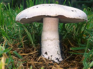
Coprinopsis atramentaria, commonly known as the common ink cap, tippler's bane, or inky cap, is an edible mushroom found in Europe and North America. Previously known as Coprinus atramentarius, it is the second best known ink cap and previous member of the genus Coprinus after C. comatus. It is a widespread and common fungus found throughout the northern hemisphere. Clumps of mushrooms arise after rain from spring to autumn, commonly in urban and disturbed habitats such as vacant lots and lawns, as well as grassy areas. The grey-brown cap is initially bell-shaped before opening, after which it flattens and disintegrates. The flesh is thin and the taste mild. It can be eaten, but due to the presence of coprine within the mushroom, it is poisonous when consumed with alcohol, as it heightens the body's sensitivity to ethanol in a similar manner to the anti-alcoholism drug disulfiram.

<i>Neolentinus ponderosus</i> Species of fungus

Neolentinus ponderosus, commonly known as the giant sawgill, or ponderous lentinus, is a species of fungus in the family Gloeophyllaceae. Found in western North America, it was originally described in 1965 as a species of Lentinus by American mycologist Orson K. Miller.

<i>Coprinopsis lagopus</i> Species of fungus

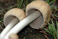
Coprinopsis lagopus is a species of fungus in the family Psathyrellaceae. Until 2001, the species was known as Coprinus lagopus; advances in the understanding of phylogenetic relationships between the various coprinoid species led to a major reorganization of that genus. It is a delicate and short-lived fungus, the fruit bodies lasting only a few hours before dissolving into a black ink – a process called deliquescence. The vague resemblance of the young fruit body to the paw of a white rabbit has earned this species the common name harefoot mushroom.

<i>Macrolepiota clelandii</i> Species of mushroom-forming fungus

Macrolepiota clelandii, commonly known as the slender parasol or graceful parasol, is a species of mushroom-forming fungus in the family Agaricaceae. The species is found in Australia and New Zealand, where it fruits singly or in small groups on the ground in eucalypt woodlands, parks, and roadsides. It is a tall mushroom up to roughly 20 cm (8 in), with a broad cap covered with distinctive rings of dark brown scales. The whitish gills on the cap underside are closely spaced and free from attachment to the slender stipe, which has a loose ring on its upper half, and a bulbous base. The edibility of the mushroom is not known with certainty, but closely related parasol mushrooms are edible and some are very sought after.

<i>Tulosesus amphithallus</i> Species of fungus

Tulosesus amphithallus is a species of mushroom producing fungus in the family Psathyrellaceae.

<i>Mycena adscendens</i> Species of fungus

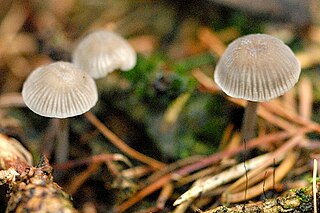
Mycena adscendens, commonly known as the frosty bonnet, is a species of fungus in the family Mycenaceae. The fungus produces small white fruit bodies (mushrooms) with caps up to 7.5 mm (0.3 in) in diameter that appear to be dusted with sugar-like granules. Caps are supported by thin, hollow stems up to 20 mm (0.8 in) long, which are set on a disc-like base. Its distribution includes Europe, Turkey and the Pacific coast of the United States. The fruit bodies grow on fallen twigs and other woody debris on the forest floor, including fallen hazel nuts. The variety carpophila is known from Japan. There are several small white Mycena species that are similar in appearance to M. adscendens, some of which can be reliably distinguished only by examining microscopic characteristics.

<i>Mycena nargan</i> Species of fungus in the family Mycenaceae

Mycena nargan, commonly known as the Nargan's bonnet, is a species of fungus in the family Mycenaceae, and the sole member of the section Nargan in the genus Mycena. Reported as a new species in 1995, it is known predominantly from Southern Australia. The saprobic fungus produces mushrooms that grow on well-decayed wood, often on the underside of wood lying in litter. The dark chestnut-coloured caps are covered with white, easily removed scales, and reach diameters of up to 2 cm (0.8 in) wide. The pale, slender stems are up to 5 cm (2.0 in) long and have white scales at the base. On the underside of the cap, the cream-coloured gills are widely spaced and bluntly attached to the stem. The edibility of the mushroom is unknown.

<i>Mycena overholtsii</i> Species of fungus

Mycena overholtsii, commonly known as the snowbank fairy helmet or fuzzy foot, is a species of fungus in the family Mycenaceae. The mushrooms produced by the fungus are relatively large for the genus Mycena, with convex grayish caps up to 5 cm (2 in) in diameter and stems up to 15 cm (6 in) long. The gills on the underside of the cap are whitish to pale gray, and initially closely spaced before becoming well-spaced at maturity after the cap enlarges. The mushrooms are characterized by the dense covering of white "hairs" on the base of the stem. M. overholtsii is an example of a snowbank fungus, growing on well-decayed conifer logs near snowbanks, during or just after snowmelt. Formerly known only from high-elevation areas of western North America, particularly the Rocky Mountain and Cascade regions, it was reported for the first time in Japan in 2010. The edibility of the mushroom is unknown. M. overholtsii can be distinguished from other comparable species by differences in location, or spore size.

<i>Amanita onusta</i> Species of fungus

Amanita onusta, commonly known as the loaded Lepidella, the gunpowder Lepidella or the gunpowder amanita, is a species of fungus in the mushroom family Amanitaceae. It is characterized by its small to medium-sized fruit bodies that have white to pale gray caps crowded with roughly conical, pyramidal, or irregular gray warts. The stipe is whitish-gray with woolly or wart-like veil remnants, and at the base is a spindle- or turnip-shaped base that is rooted somewhat deeply in the soil.

<i>Tulosesus impatiens</i> Species of fungus

Tulosesus impatiens is a species of fungus in the family Psathyrellaceae. First described in 1821, it has been classified variously in the genera Psathyrella, Pseudocoprinus, Coprinarius, and Coprinus, before molecular phylogenetics reaffirmed it as a Coprinellus species in 2001. The fungus is found in North America and Europe, where the mushrooms grow on the ground in deciduous forests. The fruit bodies have buff caps that are up to 4 cm (1.6 in) in diameter, held by slender whitish stems that can be up to 10 cm (3.9 in) tall. Several other Coprinopsis species that resemble C. impatiens may be distinguished by differences in appearance, habit, or spore morphology.

<i>Coprinopsis variegata</i> Species of fungus

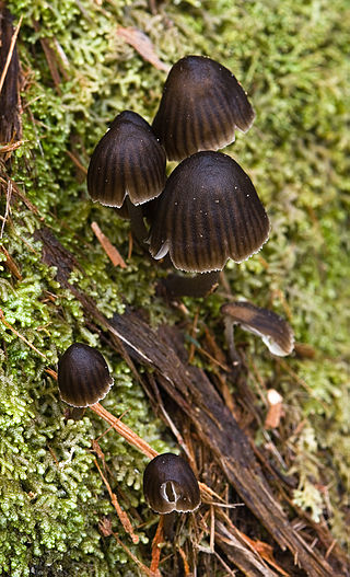
Coprinopsis variegata, commonly known as the scaly ink cap or the feltscale inky cap, is a species of fungus in the family Psathyrellaceae. Distributed in eastern North America, it has a medium-sized, bell-shaped to flattened cap up to 7.5 cm (3.0 in) in diameter, with felt-like, patchy scales. The gills, initially white, turn black in maturity and eventually dissolve into a black "ink". Fruit bodies grow in clusters or groups on leaf litter or rotted hardwood, although the wood may be buried, giving the appearance of growing in the soil. The fungus is found in the United States, in areas east of the Great Plains. Coprinus ebulbosus and Coprinus quadrifidus are names assigned by Charles Horton Peck to what he believed were species distinct from C. variegata; they were later shown to represent the same species, and are now synonyms. The mushroom is not recommended for consumption, and has been shown to cause allergic reactions in susceptible individuals.

<i>Mycena stylobates</i> Species of fungus

Mycena stylobates, commonly known as the bulbous bonnet, is a species of inedible mushroom in the family Mycenaceae. Found in North America and Europe, it produces small whitish to gray fruit bodies with bell-shaped caps that are up to 15 mm (0.6 in) in diameter. The distinguishing characteristic of the mushroom is the fragile stipe, which is seated on a flat disk marked with distinct grooves, and fringed with a row of bristles. The mushrooms grow in small troops on leaves and other debris of deciduous and coniferous trees. The mushroom's spores are white in deposit, smooth, and ellipsoid-shaped with dimensions of 6–10 by 3.5–4.5 μm. In the development of the fruit body, the preliminary stipe and cap structures appear at the same time within the primordium, and hyphae originating from the stipe form a cover over the developing structures. The mycelia of the mushroom is believed to have bioluminescent properties.

<i>Volvariella surrecta</i> Species of fungus

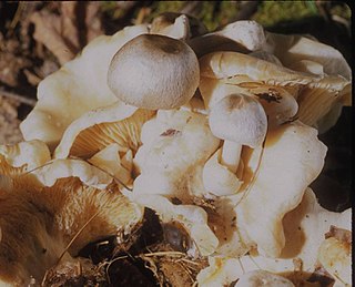
Volvariella surrecta, commonly known as the piggyback rosegill, is an agaric fungus in the family Pluteaceae. Although rare, the species is widely distributed, having been reported from Asia, North America, Northern Africa, Europe, and New Zealand. The fungus grows as a parasite on the fruit bodies of other gilled mushrooms, usually Clitocybe nebularis. V. surrecta mushrooms have white or greyish silky-hairy caps up to 8 cm (3.1 in) in diameter, and white gills that turns pink in maturity. The stipe, also white, is up to 9 cm (3.5 in) long, and has a sack-like volva at its base.

<i>Tricholoma vaccinum</i> Fungus of the agaric genus Tricholoma

Tricholoma vaccinum, commonly known as the russet scaly tricholoma, the scaly knight, or the fuzztop, is a fungus of the agaric genus Tricholoma. It produces medium-sized fruit bodies (mushrooms) that have a distinctive hairy reddish-brown cap with a shaggy margin when young. The cap, which can reach a diameter of up to 6.5 cm (2.6 in) wide, breaks up into flattened scales in maturity. It has cream-buff to pinkish gills with brown spots. Its fibrous, hollow stipe is white above and reddish brown below, and measures 4 to 7.5 cm long. Although young fruit bodies have a partial veil, it does not leave a ring on the stipe.

<i>Parasola auricoma</i> Species of fungus

Parasola auricoma is a species of agaric fungus in the family Psathyrellaceae. First described scientifically in 1886, the species is found in Europe, Japan, and North America. The mushroom was reported in February 2019 in Colombia, in the city of Bogota by the mycologist Juan Camilo Rodriguez Martinez. The small, umbrella-shaped fruit bodies (mushrooms) of the fungus grow in grass or woodchips and are short-lived, usually collapsing with age in a few hours. The caps are up to 6 cm (2.4 in) wide, initially elliptical before flattening out, and colored reddish-brown to greyish, depending on their age and hydration. They are pleated with radial grooves extending from the center to the edge of the cap. The slender, whitish stems are up to 12 cm (4.7 in) long and a few millimeters thick. Microscopically, P. auricoma is characterized by the presence of setae in its cap cuticle. This characteristic, in addition to the relatively large, ellipsoid spores can be used to distinguish it from other morphologically similar Parasola species.

<i>Agrocybe putaminum</i> Species of fungus

Agrocybe putaminum, commonly known as the mulch fieldcap, is a species of agaric fungus in the family Strophariaceae in the Agrocybe sororia complex. Described as new to science in 1913, it is found in Asia, Australia, Europe, and western North America, where it grows in parks, gardens, and roadsides in woodchip mulch. Fruitbodies of the fungus have a dull brownish-orange cap with a matte texture, a grooved stipe, and a bitter, mealy taste.

<i>Strobilurus tenacellus</i> Species of fungus

Strobilurus tenacellus, commonly known as the pinecone cap, is a species of agaric fungus in the family Physalacriaceae. It is found in Asia and Europe, where it grows on the fallen cones of pine and spruce trees. The fruit bodies (mushrooms) are small, with convex to flat, reddish to brownish caps up to 15 mm (0.6 in) in diameter, set atop thin cylindrical stems up to 4–7.5 cm (1.6–3.0 in) long with a rooting base. A characteristic microscopic feature of the mushroom is the sharp, thin-walled cystidia found on the stipe, gills, and cap. The mushrooms, sometimes described as edible, are too small to be of culinary interest. The fungus releases compounds called strobilurins that suppress the growth and development of other fungi. Derivatives of these compounds are used as an important class of agricultural fungicides.

<i>Tubaria punicea</i> Species of fungus

Tubaria punicea is a rare species of agaric fungus in the family Tubariaceae. It is found on the west coast of North America, where it grows on the bases and in hollows of madrone.
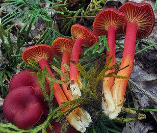
References

  1. "Species synonymy – Coprinellus micaceus". Index Fungorum. CAB International. 2008. Archived from the original on 2011-06-10. Retrieved 2010-04-14.
  2. Fungorum in Pannoniis observatorum brevis historia (Brief history of fungi observed in Pannonia [Hungary]), genus XVI, p. cclxxxii.
  3. 1 2 Buller, 1924, p. 331.
  4. Bulliard JBF (1786). Herbier de la France [Guide to the Herbs of France] (in French). Vol. 6. pp. 241–88, plate 246. Click on page 11.
  5. Persoon CH (1801). Synopsis Methodica Fungorum (Göttingen) (in Latin). Vol. 1. p. 403.
  6. Fries EM (1838). Epicrisis Systematis Mycologici (in Latin). Uppsala, Sweden: W.G. Farlow. p. 247.
  7. Singer R. (1986). The Agaricales in Modern Taxonomy (4th ed.). Königstein im Taunus, Germany: Koeltz Scientific Books. p. 522. ISBN   3-87429-254-1.
  8. Hopple JS, Vilgalys R (1994). "Phylogenetic relationships among coprinoid taxa and allies based on data from restriction site mapping of nuclear rDNA" (PDF). Mycologia . 86 (1): 96–107. doi:10.2307/3760723. JSTOR   3760723. Archived from the original (PDF) on 2012-02-27. Retrieved 2009-10-13.
  9. Park DS, Go SJ, Kim YS, Seok SJ, Song JK, Yeo YS, Ryu JC, Sung JM (1999). "Genetic relationships of Coprinus spp. on the basis of sequences in ITS II region". Korean Journal of Mycology (in Korean). 27 (1): 27–31. ISSN   0253-651X.
  10. Redhead SA, Vilgalys R, Moncalvo J-M, Johnson J, Hopple JS Jr (2001). "Coprinus Pers. and the disposition of Coprinus species sensu lato". Taxon. 50 (1): 203–41. doi:10.2307/1224525. JSTOR   1224525.
  11. Link HF (1809). "Nova plantarum genera e classe Lichenum, Algarum, Fungorum". Schrader's Neues Journal für die Botanik. 3: 10–15.
  12. Buller, 1924, pp. 328–56.
  13. Stearn WT (1973). Botanical Latin (2nd annot. and rev. ed.). Newton Abbot: David & Charles. pp. 379, 463.
  14. Oxford English Dictionary Online. Oxford: Oxford University Press. 2010. Archived from the original on 2006-06-25. Retrieved 2010-03-12. ss. vv. "mica", "micaceous".
  15. 1 2 Ammirati JF, McKenny M, Stuntz DE (1987). The New Savory Wild Mushroom. Seattle, Washington: University of Washington Press. p. 156. ISBN   0-295-96480-4.
  16. 1 2 3 Arora, 1986, p. 348 Retrieved 2010-04-16.
  17. 1 2 3 4 Davis, R. Michael; Sommer, Robert; Menge, John A. (2012). Field Guide to Mushrooms of Western North America. Berkeley: University of California Press. pp. 205–206. ISBN   978-0-520-95360-4. OCLC   797915861.
  18. Evenson VS (1997). Mushrooms of Colorado and the Southern Rocky Mountains. Westcliffe Publishers. p. 131. ISBN   978-1-56579-192-3.
  19. Buller, 1924, p. 335.
  20. 1 2 McKnight VB, McKnight KH (1987). A Field Guide to Mushrooms, North America. Boston: Houghton Mifflin. p. 34. ISBN   0-395-91090-0.
  21. 1 2 Orr DB, Orr RT (1979). Mushrooms of Western North America. Berkeley, California: University of California Press. p. 176. ISBN   0-520-03656-5.
  22. 1 2 Buller, 1924, p. 337.
  23. 1 2 Orton PD, Watling R (1979). Coprinaceae: Coprinus. Edinburgh, Scotland: Royal Botanic Garden. pp. 54–55. ISBN   0-11-491565-2.
  24. 1 2 3 Schalkwijk-Barendsen HME. (1991). Mushrooms of Western Canada. Edmonton, Canada: Lone Pine Publishing. p.  334. ISBN   0-919433-47-2.
  25. 1 2 Roody WC (2003). Mushrooms of West Virginia and the Central Appalachians. Lexington, Kentucky: University Press of Kentucky. p. 156. ISBN   0-8131-9039-8.
  26. Pady SM (1941). "Notes on Coprinus micaceus growing in an unusual habitat". Mycologia. 33 (4): 411–14. doi:10.2307/3754895. JSTOR   3754895.
  27. 1 2 3 4 5 Keirle MR, Hemmes DE, Desjardin DE (2004). "Agaricales of the Hawaiian Islands. 8. Agaricaceae: Coprinus and Podaxis; Psathyrellaceae: Coprinopsis, Coprinellus and Parasola" (PDF). Fungal Diversity. 15 (4): 33–124.
  28. Buller, 1924, pp. 351–53.
  29. Buller, 1924, pp. 329, 342.
  30. De Bary A. (1873). "On cystidia". Grevillea. 1: 181–83.
  31. Buller, 1924, pp. 248–50.
  32. Buller, 1924, p. 340.
  33. Levine M. (1914). "The origin and development of the lamellae of Coprinus micaceus". American Journal of Botany. 1 (7): 343–56. doi:10.2307/2435139. JSTOR   2435139.
  34. Bos CJ (1996). Fungal Genetics: Principles and Practice. New York: Marcel Dekker. pp. 152–53. ISBN   0-8247-9544-X.
  35. Moore D, Chiu S-W (1996). Patterns in Fungal Development. Cambridge, UK: Cambridge University Press. p. 127. ISBN   0-521-56047-0.
  36. Lu BC, Raju NB (1970). "Meiosis in Coprinus. II. Chromosome pairing and the lampbrush diplotene stage of meiotic prophase". Chromosoma (Berlin). 29 (3): 305–16. doi:10.1007/BF00325945. PMID   5445348. S2CID   12287280.
  37. Russell B. (2006). Field Guide to Wild Mushrooms of Pennsylvania and the Mid-Atlantic. University Park, Pennsylvania: Pennsylvania State University Press. p. 29. ISBN   978-0-271-02891-0.
  38. Smith AH (1975). A Field Guide to Western Mushrooms. Ann Arbor, Michigan: University of Michigan Press. p. 229. ISBN   0-472-85599-9.
  39. 1 2 3 Dickinson C, Lucas J (1982). VNR Color Dictionary of Mushrooms. New York: Van Nostrand Reinhold. p. 78. ISBN   978-0-442-21998-7.
  40. Kimura MT (1980). "Evolution of food preferences in fungus-feeding Drosophila: an ecological study". Evolution. 34 (5): 1009–18. doi:10.2307/2408009. JSTOR   2408009. PMID   28581130.
  41. Toda MJ, Kimura MT, Tuno N (1999). "Coexistence mechanisms of mycophagous drosophilids on multispecies fungal hosts: aggregation and resource partitioning". Journal of Animal Ecology. 68 (4): 794–803. doi: 10.1046/j.1365-2656.1999.00328.x . JSTOR   2647329.
  42. Dursun N, Ozcan MM, Ozturk C (2006). "Mineral contents of 34 species of edible mushrooms growing wild in Turkey". Journal of the Science of Food and Agriculture. 86 (7): 1087–94. Bibcode:2006JSFA...86.1087D. doi:10.1002/jsfa.2462.
  43. Das N. (2007). "Heavy metal levels in wild edible mushroom samples from Nayagram Block of Midnapore District, West Bengal". Indian Forester. 133 (2): 171–78.
  44. Sundberg W, Bessette A (1987). Mushrooms: A Quick Reference Guide to Mushrooms of North America (Macmillan Field Guides). New York: Collier Books. p. 134. ISBN   0-02-063690-3.
  45. Trudell, Steve; Ammirati, Joe (2009). Mushrooms of the Pacific Northwest. Timber Press Field Guides. Portland, OR: Timber Press. p. 196. ISBN   978-0-88192-935-5.
  46. 1 2 Healy RA, Huffman DR, Tiffany LH, Knaphaus G (2008). Mushrooms and Other Fungi of the Midcontinental United States (Bur Oak Guide). Iowa City, Iowa: University of Iowa Press. p. 63. ISBN   978-1-58729-627-7.
  47. 1 2 Hemmes DE, Desjardin D (2002). Mushrooms of Hawai'i: An Identification Guide. Berkeley, California: Ten Speed Press. p. 127. ISBN   1-58008-339-0.
  48. Buller, 1924, p. 333.
  49. Peiris D, Dunn WB, Brown M, Kell DB, Roy I, Hedger JN (2008). "Metabolite profiles of interacting mycelial fronts differ for pairings of the wood decay basidiomycete fungus, Stereum hirsutum with its competitors Coprinus micaceus and Coprinus disseminatus". Metabolomics. 4 (1): 52–62. doi:10.1007/s11306-007-0100-4. S2CID   22052503.
  50. de Errasti A, Carmarán CC, Novas MV (2010). "Diversity and significance of fungal endophytes from living stems of naturalized trees from Argentina". Fungal Diversity. 40 (1): 29–40. doi:10.1007/s13225-009-0012-x. S2CID   30981477.
  51. Arora, 1986, p. 48. Retrieved 2010-04-13.
  52. Atkinson GF (1898). "Society for Plant Morphology and Physiology. 2. Studies on some mycelium and fungi from a coal mine". Botanical Gazette. 25 (2): 106–18. doi:10.1086/327641. JSTOR   2464466. S2CID   224843835.
  53. Aira MJ, Rodriguez-Rajo FJ, Castro M, Jato V (2009). "Characterization of Coprinus spores in the NW of the Iberian Peninsula. Identification and count in aerobiological samples". Cryptogamie, Mycologie. 30 (1): 57–66.
  54. Christensen CM (1981). Edible Mushrooms. Minnesota, Minneapolis: University of Minnesota Press. p. 80. ISBN   978-0-8166-1049-5.
  55. Stewart FC (1926). "The mica ink-cap or glistening Coprinus". New York (Geneva) Agricultural Experiment Station Bulletin. 535: 1–30.
  56. Melchers LE (1931). "A check list of plant diseases and fungi occurring in Egypt". Transactions of the Kansas Academy of Science. 34: 41–106. doi:10.2307/3624470. JSTOR   3624470.
  57. Reid DA, Eicker A (1999). "South African fungi 10: New species, new records and some new observations". Mycotaxon. 73: 169–97.
  58. Gezer K, Ekici FT, Turkoglu A (2008). "Macrofungi of Karci Mountain (Denizli, Turkey)". Turkish Journal of Botany. 32 (1): 91–6. ISSN   1300-008X.
  59. 1 2 Watling R, Miller OK (1971). "8 species of Coprinus of Yukon Territory and adjacent Alaska". Canadian Journal of Botany. 49 (9): 1687–90. doi:10.1139/b71-237.
  60. Kaul TN, Kachroo JL (1974). "Common edible mushrooms of Jammu and Kashmir India". Journal of the Bombay Natural History Society. 71 (1): 26–31.
  61. Manjula R, Surjit S, Dutta BB, Krishnendu A (2005). "Some additions to the Coprinaceae of Sikkim Himalaya". Journal of Mycopathological Research. 43 (1): 101–03. ISSN   0971-3719.
  62. Crowe A. (1983). A Field Guide to the Native Edible Plants of New Zealand : including Those Plants Eaten by the Maori. Collins. p. 135. ISBN   0-00-216983-5.
  63. Uljé, K. "Coprinus micaceus". Kees Uljé Coprinus site. Retrieved 2010-04-14.
  64. Ko KS. (2001). "Phylogeographic divergences of nuclear ITS sequences in Coprinus species sensu lato". Mycological Research. 105 (12): 1519–26. doi:10.1017/S0953756201005184.
  65. Kuo M. (February 2008). "Coprinellus micaceus". MushroomExpert.Com. Retrieved 2011-03-11.
  66. Zahid S, Udenigwe CC, Ata A, Eze MO, Segstro EP, Holloway P (2006). "New bioactive natural products from Coprinus micaceus". Natural Product Research. 20 (14): 1283–89. doi:10.1080/14786410601101829. PMID   17393652. S2CID   7494031.
  67. Ata A. (2009). "Novel bioactive natural products from marine and terrestrial sources". In Şener B (ed.). Innovations in Chemical Biology. Dordrecht, The Netherlands: Springer Netherlands. pp. 51–60. doi:10.1007/978-1-4020-6955-0. ISBN   978-1-4020-6954-3.
  68. Efremenkova OV, Ershova EY, Tolstych IV, Zenkova VA, Dudnik YV (2003). "Antimicrobial activity of medicinal mushrooms from the genus Coprinus (Fr.) S. F. Gray (Agaricomycetideae)". International Journal of Medicinal Mushrooms. 5 (1): 37–41. doi:10.1615/InterJMedicMush.v5.i1.
  69. Worthen LR, Stessel GJ, Youngken HW (1962). "The occurrence of indole compounds in Coprinus species". Economic Botany. 16 (4): 315–18. doi:10.1007/BF02860190. S2CID   22240178.
  70. Saiz-Jimenez C. (1983). "The chemical nature of the melanins from Coprinus spp". Soil Science. 136 (2): 65–74. Bibcode:1983SoilS.136...65S. doi:10.1097/00010694-198308000-00001. hdl: 10261/76183 . S2CID   26206774.
  71. Brossi A. (1991). The Alkaloids: Chemistry and Pharmacology. Vol. 40. Amsterdam, the Netherlands: Elsevier. p. 298. ISBN   0-12-469540-X.

Cited books