Formate dehydrogenase

Last updated
Formate dehydrogenase N, transmembrane
1kqg.jpg
Formate dehydrogenase-N hetero9mer, E.Coli
Identifiers
SymbolForm-deh_trans
Pfam PF09163
InterPro IPR015246
SCOP2 1kqf / SCOPe / SUPFAM
OPM superfamily 3
OPM protein 1kqf
Available protein structures:
Pfam   structures / ECOD  
PDB RCSB PDB; PDBe; PDBj
PDBsum structure summary
PDB 1kqf B:246-289 1kqg B:246-289

Formate dehydrogenases are a set of enzymes that catalyse the oxidation of formate to carbon dioxide, donating the electrons to a second substrate, such as NAD+ in formate:NAD+ oxidoreductase (EC 1.17.1.9) or to a cytochrome in formate:ferricytochrome-b1 oxidoreductase (EC 1.2.2.1). [1] This family of enzymes has attracted attention as inspiration or guidance on methods for the carbon dioxide fixation, relevant to global warming. [2]

Contents

Function

NAD-dependent formate dehydrogenases are important in methylotrophic yeast and bacteria, being vital in the catabolism of C1 compounds such as methanol. [3] The cytochrome-dependent enzymes are more important in anaerobic metabolism in prokaryotes. [4] For example, in E. coli , the formate:ferricytochrome-b1 oxidoreductase is an intrinsic membrane protein with two subunits and is involved in anaerobic nitrate respiration. [5] [6]

NAD-dependent reaction

Formate + NAD+ CO2 + NADH + H+

Cytochrome-dependent reaction

Formate + 2 ferricytochrome b1 CO2 + 2 ferrocytochrome b1 + 2 H+

Molybdopterin, molybdenum and selenium dependence

The metal-dependent Fdh's feature Mo or W at their active sites. These active sites resemble the motif seen in DMSO reductase, with two molybdopterin cofactors bound to Mo/W in a bidentate fashion. The fifth and sixth ligands are sulfide and either cysteinate or selenocysteinate. [7]

The mechanism of action appears to involve 2e redox of the metal centers, induced by hydride transfer from formate and release of carbon dioxide:

S=MVI(Scys)(SR)4 + HCO2 ⇌ HS−MIV(Scys)(SR)4 + CO2
S=MVI(Secys)(SR)4 + HCO2 ⇌ HS−MIV(Secys)(SR)4 + CO2

In this scheme, (SR)4 represents the four thiolate-like ligands provided by the two dithiolene cofactors, the molybdopterins. The dithiolene and cysteinyl/selenocysteinyl ligands are redox-innocent. In terms of the molecular details, the mechanism remains uncertain, despite numerous investigations. Most mechanisms assume that formate does not coordinate to Mo/W, in contrast to typical Mo/W oxo-transferases (e.g., DMSO reductase). A popular mechanistic proposal entails transfer of H- from formate to the Mo/WVI=S group. [8]

Formate Dehydrogenase (PDB 1KQF, 1.6 A resolution, from E. coli); overall view of the electron transport chain showing the [Fe4S4] clusters in the periplasmic alpha and beta subunits, and the cytoplasmic gamma subunit showing the Fe(heme b)P and the Fe-(heme b)C menoquinone binding site where an HQNO ligand is bound close the Fe(heme b)C. Atom colours: Fe = orange, S = yellow, C = grey, O = red, N = blue. Formate Dehydrogenase 1kqf overall protein 1 (KC).png
Formate Dehydrogenase (PDB 1KQF, 1.6 A resolution, from E. coli); overall view of the electron transport chain showing the [Fe4S4] clusters in the periplasmic alpha and beta subunits, and the cytoplasmic gamma subunit showing the Fe(heme b)P and the Fe-(heme b)C menoquinone binding site where an HQNO ligand is bound close the Fe(heme b)C. Atom colours: Fe = orange, S = yellow, C = grey, O = red, N = blue.

Transmembrane domain

Formate dehydrogenase consists of two transmembrane domains; three α-helices of the β-subunit and four transmembrane helices from the gamma-subunit.

The β-subunit of formate dehydrogenase is present in the periplasm with a single transmembrane α-helix spanning the membrane by anchoring the β-subunit to the inner-membrane surface. The β-subunit has two subdomains, where each subdomain has two [4Fe-4S] ferredoxin clusters. The judicious alignment of the [4Fe-4S] clusters in a chain through the subunit have low separation distances, which allow rapid electron flow through [4Fe-4S]-1, [4Fe-4S]-4, [4Fe-4S]-2, and [4Fe-4S]-3 to the periplasmic heme b in the γ-subunit. The electron flow is then directed across the membrane to a cytoplasmic heme b in the γ-subunit .

The γ-subunit of formate dehydrogenase is a membrane-bound cytochrome b consisting of four transmembrane helices and two heme b groups which produce a four-helix bundle which aids in heme binding. The heme b cofactors bound to the gamma subunit allow for the hopping of electrons through the subunit. The transmembrane helices maintain both heme b groups, while only three provide the heme ligands thereby anchoring Fe-heme. The periplasmic heme b group accepts electrons from [4Fe-4S]-3 clusters of the  β-subunit’s periplasmic domain. The cytoplasmic heme b group accepts electrons from the periplasmic heme b group, where electron flow is then directed towards the menaquinone (vitamin K) reduction site, present in the transmembrane domain of the gamma subunit. The menaquinone reduction site in the γ-subunit, accepts electrons through the binding of a histidine ligand of the cytoplasmic heme b. [9]

Menaquinone binding site alongside proposed water proton pathway Colored electron tunneling F-DHN.jpg
Menaquinone binding site alongside proposed water proton pathway

See also

Additional reading

Related Research Articles

<span class="mw-page-title-main">Oxidative phosphorylation</span> Metabolic pathway

Oxidative phosphorylation or electron transport-linked phosphorylation or terminal oxidation is the metabolic pathway in which cells use enzymes to oxidize nutrients, thereby releasing chemical energy in order to produce adenosine triphosphate (ATP). In eukaryotes, this takes place inside mitochondria. Almost all aerobic organisms carry out oxidative phosphorylation. This pathway is so pervasive because it releases more energy than alternative fermentation processes such as anaerobic glycolysis.

<span class="mw-page-title-main">Coenzyme Q – cytochrome c reductase</span> Class of enzymes

The coenzyme Q : cytochrome c – oxidoreductase, sometimes called the cytochrome bc1 complex, and at other times complex III, is the third complex in the electron transport chain, playing a critical role in biochemical generation of ATP. Complex III is a multisubunit transmembrane protein encoded by both the mitochondrial and the nuclear genomes. Complex III is present in the mitochondria of all animals and all aerobic eukaryotes and the inner membranes of most eubacteria. Mutations in Complex III cause exercise intolerance as well as multisystem disorders. The bc1 complex contains 11 subunits, 3 respiratory subunits, 2 core proteins and 6 low-molecular weight proteins.

<span class="mw-page-title-main">Succinate dehydrogenase</span> Enzyme

Succinate dehydrogenase (SDH) or succinate-coenzyme Q reductase (SQR) or respiratory complex II is an enzyme complex, found in many bacterial cells and in the inner mitochondrial membrane of eukaryotes. It is the only enzyme that participates in both the citric acid cycle and the electron transport chain. Histochemical analysis showing high succinate dehydrogenase in muscle demonstrates high mitochondrial content and high oxidative potential.

Ferredoxins are iron–sulfur proteins that mediate electron transfer in a range of metabolic reactions. The term "ferredoxin" was coined by D.C. Wharton of the DuPont Co. and applied to the "iron protein" first purified in 1962 by Mortenson, Valentine, and Carnahan from the anaerobic bacterium Clostridium pasteurianum.

Cytochrome b<sub>6</sub>f complex Enzyme

The cytochrome b6f complex (plastoquinol/plastocyanin reductase or plastoquinol/plastocyanin oxidoreductase; EC 7.1.1.6) is an enzyme found in the thylakoid membrane in chloroplasts of plants, cyanobacteria, and green algae, that catalyzes the transfer of electrons from plastoquinol to plastocyanin:

Cytochrome b<sub>5</sub>

Cytochromes b5 are ubiquitous electron transport hemoproteins found in animals, plants, fungi and purple phototrophic bacteria. The microsomal and mitochondrial variants are membrane-bound, while bacterial and those from erythrocytes and other animal tissues are water-soluble. The family of cytochrome b5-like proteins includes hemoprotein domains covalently associated with other redox domains in flavocytochrome cytochrome b2, sulfite oxidase, plant and fungal nitrate reductases, and plant and fungal cytochrome b5/acyl lipid desaturase fusion proteins.

Iron-binding proteins are carrier proteins and metalloproteins that are important in iron metabolism and the immune response. Iron is required for life.

In enzymology, carbon monoxide dehydrogenase (CODH) (EC 1.2.7.4) is an enzyme that catalyzes the chemical reaction

<span class="mw-page-title-main">Formate dehydrogenase (cytochrome)</span> Type of enzyme

In enzymology, a formate dehydrogenase (cytochrome) (EC 1.2.2.1) is an enzyme that catalyzes the chemical reaction

In enzymology, a formate dehydrogenase (cytochrome-c-553) (EC 1.2.2.3) is an enzyme that catalyzes the chemical reaction

<span class="mw-page-title-main">Cytochrome c nitrite reductase</span> Class of enzymes

Cytochrome c nitrite reductase (ccNiR) is a bacterial enzyme that catalyzes the six electron reduction of nitrite to ammonia; an important step in the biological nitrogen cycle. The enzyme catalyses the second step in the two step conversion of nitrate to ammonia, which allows certain bacteria to use nitrite as a terminal electron acceptor, rather than oxygen, during anaerobic conditions. During this process, ccNiR draws electrons from the quinol pool, which are ultimately provided by a dehydrogenase such as formate dehydrogenase or hydrogenase. These dehydrogenases are responsible for generating a proton motive force.

<span class="mw-page-title-main">Thiosulfate dehydrogenase</span>

Thiosulfate dehydrogenase is an enzyme that catalyzes the chemical reaction:

<span class="mw-page-title-main">Cytochrome c oxidase subunit 2</span> Enzyme of the respiratory chain encoded by the mitochondrial genome

Cytochrome c oxidase II is a protein in eukaryotes that is encoded by the MT-CO2 gene. Cytochrome c oxidase subunit II, abbreviated COXII, COX2, COII, or MT-CO2, is the second subunit of cytochrome c oxidase. It is also one of the three mitochondrial DNA (mtDNA) encoded subunits of respiratory complex IV.

<span class="mw-page-title-main">IDH3A</span> Protein-coding gene in the species Homo sapiens

Isocitrate dehydrogenase [NAD] subunit alpha, mitochondrial (IDH3α) is an enzyme that in humans is encoded by the IDH3A gene.

<span class="mw-page-title-main">IDH3B</span> Protein-coding gene in the species Homo sapiens

Isocitrate dehydrogenase [NAD] subunit beta, mitochondrial is an enzyme that in humans is encoded by the IDH3B gene.

The Arc system is a two-component system found in some bacteria that regulates gene expression in faculatative anaerobes such as Escheria coli. Two-component system means that it has a sensor molecule and a response regulator. Arc is an abbreviation for Anoxic Redox Control system. Arc systems are instrumental in maintaining energy metabolism during transcription of bacteria. The ArcA response regulator looks at growth conditions and expresses genes to best suit the bacteria. The Arc B sensor kinase, which is a tripartite protein, is membrane bound and can autophosphorylate.

<span class="mw-page-title-main">Flavocytochrome c sulfide dehydrogenase</span>

Flavocytochrome c sulfide dehydrogenase, also known as Sulfide-cytochrome-c reductase (flavocytochrome c) (EC 1.8.2.3), is an enzyme with systematic name hydrogen-sulfide:flavocytochrome c oxidoreductase. It is found in sulfur-oxidising bacteria such as the purple phototrophic bacteria Allochromatium vinosum. This enzyme catalyses the following chemical reaction:

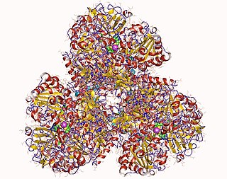
Formate dehydrogenase-N (EC 1.1.5.6, Fdh-N, FdnGHI, nitrate-inducible formate dehydrogenase, formate dehydrogenase N, FDH-N, nitrate inducible Fdn, nitrate inducible formate dehydrogenase) is an enzyme with systematic name formate:quinone oxidoreductase. This enzyme catalyses the following chemical reaction

Dimethyl sulfide:cytochrome c2 reductase (EC 1.8.2.4) is an enzyme with systematic name dimethyl sulfide:cytochrome-c2 oxidoreductase. It is also known by the name dimethylsulfide dehydrogenase (Ddh). This enzyme catalyses the following chemical reaction

In enzymology, a formylmethanofuran dehydrogenase (EC 1.2.99.5) is an enzyme that catalyzes the chemical reaction:

References

  1. Hille, Russ; Hall, James; Basu, Partha (2014). "The Mononuclear Molybdenum Enzymes". Chemical Reviews. 114 (7): 3963–4038. doi:10.1021/cr400443z. PMC   4080432 . PMID   24467397.
  2. Amao, Yutaka (2018). "Formate dehydrogenase for CO2 utilization and its application". Journal of CO2 Utilization. 26: 623–641. doi: 10.1016/j.jcou.2018.06.022 . S2CID   189383769.
  3. Popov VO, Lamzin VS (1994). "NAD(+)-dependent formate dehydrogenase". Biochem. J. 301 (3): 625–43. doi:10.1042/bj3010625. PMC   1137035 . PMID   8053888.
  4. Jormakka M, Byrne B, Iwata S (2003). "Formate dehydrogenase--a versatile enzyme in changing environments". Curr. Opin. Struct. Biol. 13 (4): 418–23. doi:10.1016/S0959-440X(03)00098-8. PMID   12948771.
  5. Graham A, Boxer DH (1981). "The organization of formate dehydrogenase in the cytoplasmic membrane of Escherichia coli". Biochem. J. 195 (3): 627–37. doi:10.1042/bj1950627. PMC   1162934 . PMID   7032506.
  6. Ruiz-Herrera J, DeMoss JA (1969). "Nitrate reductase complex of Escherichia coli K-12: participation of specific formate dehydrogenase and cytochrome b1 components in nitrate reduction". J. Bacteriol. 99 (3): 720–9. doi:10.1128/JB.99.3.720-729.1969. PMC   250087 . PMID   4905536.
  7. Stripp, Sven T.; Duffus, Benjamin R.; Fourmond, Vincent; Léger, Christophe; Leimkühler, Silke; Hirota, Shun; Hu, Yilin; Jasniewski, Andrew; Ogata, Hideaki; Ribbe, Markus W. (2022). "Second and Outer Coordination Sphere Effects in Nitrogenase, Hydrogenase, Formate Dehydrogenase, and CO Dehydrogenase". Chemical Reviews. 122 (14): 11900–11973. doi:10.1021/acs.chemrev.1c00914. PMC   9549741 . PMID   35849738.
  8. Fogeron, Thibault; Li, Yun; Fontecave, Marc (2022). "Formate Dehydrogenase Mimics as Catalysts for Carbon Dioxide Reduction". Molecules. 27 (18): 5989. doi: 10.3390/molecules27185989 . PMC   9506188 . PMID   36144724.
  9. Stiefel, Edward (2002-03-31). "Faculty Opinions recommendation of Molecular basis of proton motive force generation: structure of formate dehydrogenase-N". doi: 10.3410/f.1004770.61154 .{{cite journal}}: Cite journal requires |journal= (help)