Nucleic acid metabolism refers to the set of chemical reactions involved in the synthesis and degradation of nucleic acids (DNA and RNA). Nucleic acids are polymers (biopolymers) composed of monomers called nucleotides.
Nucleotide synthesis is an anabolic process that typically involves the chemical reaction of a phosphate group, a pentose sugar, and a nitrogenous base. In contrast, the degradation of nucleic acids is a catabolic process in which nucleotides or nucleobases are broken down, and their components can be salvaged to form new nucleotides.
Both synthesis and degradation reactions require multiple enzymes to facilitate these processes. Defects or deficiencies in these enzymes can lead to a variety of metabolic disorders.[1]
Composition of nucleotides, which make up nucleic acids.
Synthesis of nucleotides
Nucleotides are the monomers that polymerize to form nucleic acids. Each nucleotide consists of a sugar, a phosphate group, and a nitrogenous base. The nitrogenous bases found in nucleic acids belong to one of two categories: purines or pyrimidines.
In complex multicellular animals, both purines and pyrimidines are primarily synthesized in the liver, but they follow distinct biosynthetic pathways. However, all nucleotide synthesis requires phosphoribosyl pyrophosphate (PRPP), which donates the ribose and phosphate needed to form a nucleotide.
IMP serves as a precursor for both adenosine monophosphate (AMP) and guanosine monophosphate (GMP). AMP is synthesized from IMP using guanosine triphosphate (GTP) and aspartate, with aspartate being converted into fumarate. In contrast, the synthesis of GMP requires an intermediate step: IMP is first oxidized by NAD⁺ to form xanthosine monophosphate (XMP), which is subsequently converted into GMP via the hydrolysis of one ATP molecule and the conversion of glutamine to glutamate.[1]
Both AMP and GMP can be phosphorylated by kinases to form adenosine triphosphate (ATP) and guanosine triphosphate (GTP), respectively. ATP stimulates the production of GTP, while GTP stimulates the production of ATP. This cross-regulation maintains a balanced ratio of ATP and GTP, preventing an excess of either nucleotide, which could increase the risk of DNA replication errors and purine misincorporation.[1]
Lesch–Nyhan syndrome is caused by a deficiency of hypoxanthine-guanine phosphoribosyltransferase (HGPRT), an enzyme that catalyzes the salvage of guanine to GMP. This X-linked congenital disorder leads to the overproduction of uric acid and is associated with neurological symptoms, including intellectual disability, spasticity, and compulsive self-mutilation.[1][2][3]
Pyrimidine synthesis
Uridine-triphosphate (UTP), at left, reacts with glutamine and other molecules to form cytidine-triphosphate (CTP), on the right.
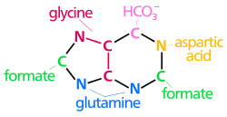
The synthesis of pyrimidine nucleotides begins with the formation of uridine monophosphate (UMP). This process requires aspartate, glutamine, bicarbonate, and two molecules of ATP to provide energy. Additionally, phosphoribosyl pyrophosphate (PRPP) provides the ribose-phosphate backbone. Unlike purine synthesis, in which the nitrogenous base is built upon PRPP, pyrimidine synthesis forms the base first and attaches it to PRPP later in the process.
Once UMP is synthesized, it undergoes phosphorylation using ATP to form uridine-triphosphate (UTP). UTP can then be converted into cytidine-triphosphate (CTP) in a reaction catalyzed by CTP synthetase, which utilizes glutamine as an amine donor.
The synthesis of thymidine nucleotides requires the reduction of UMP to deoxyuridine monophosphate (dUMP) via ribonucleotide reductase (see next section). dUMP is then methylated by thymidylate synthase to produce thymidine monophosphate (TMP). [1][5]
The regulation of pyrimidine synthesis is tightly controlled. ATP, a purine nucleotide, activates pyrimidine synthesis, while CTP, a pyrimidine nucleotide, acts as an inhibitor. This regulatory feedback ensures balanced purine and pyrimidine levels, which is essential for DNA and RNA synthesis.[1][6]
Deficiencies in enzymes involved in pyrimidine synthesis can lead to metabolic disorders such as orotic aciduria. This genetic disorder is characterized by excessive excretion of orotic acid in urine due to defects in the enzyme UMP synthase, which is responsible for the conversion of orotic acid into UMP.[1][7]
Converting nucleotides to deoxynucleotides
Nucleotides are initially synthesized with ribose as the sugar component, a characteristic feature of RNA. However, DNA requires deoxyribose, which lacks the 2'-hydroxyl (-OH) group on the ribose. The removal of this -OH group is catalyzed by ribonucleotide reductase, an enzyme that converts nucleoside diphosphates (NDPs) into their deoxy forms, deoxynucleoside diphosphates (dNDPs). The nucleotides must be in the diphosphate form for this reaction to occur.[1]
General outline of nucleic acid degradation for purines.
The breakdown of DNA and RNA occurs continuously within the cell. Purine and pyrimidine nucleosides can either be degraded into waste products for excretion or salvaged for reuse as nucleotide components.[5]
Purine degradation primarily occurs in the liver in humans and requires a series of enzymes to break down purines into uric acid. First, nucleotides lose their phosphate groups through the action of 5'-nucleotidase. The purine nucleoside adenosine is then deaminated by adenosine deaminase and hydrolyzed by a nucleosidase to form hypoxanthine. Hypoxanthine is subsequently oxidized to xanthine and then to uric acid via the enzyme xanthine oxidase.
The other purine nucleoside, guanosine, is cleaved to form guanine. Guanine is then deaminated by guanine deaminase to produce xanthine, which is further converted to uric acid. In both degradation pathways, oxygen serves as the final electron acceptor. The excretion of uric acid varies among different animals.[5]
Once nucleotides are synthesized, they can exchange phosphate groups to form nucleoside mono-, di-, and triphosphates. The conversion of a nucleoside diphosphate (NDP) to a nucleoside triphosphate (NTP) is catalyzed by nucleoside diphosphate kinase, which utilizes ATP as the phosphate donor. Similarly, nucleoside monophosphate kinase facilitates the phosphorylation of nucleoside monophosphates to their diphosphate forms.
Additionally, adenylate kinase plays a crucial role in regulating cellular energy balance by catalyzing the interconversion of two molecules of ADP into ATP and AMP (2 ADP ⇔ ATP + AMP).[1][8]
↑ "Lesch-Nyhan". Lesch-Nyhan.org. Retrieved 31 October 2014.
↑ Alqahtani, Saad Saeed; Koltai, Tomas; Ibrahim, Muntaser E.; Bashir, Adil H. H.; Alhoufie, Sari T. S.; Ahmed, Samrein B. M.; Molfetta, Daria Di; Carvalho, Tiago M. A.; Cardone, Rosa Angela; Reshkin, Stephan Joel; Hifny, Abdelhameed; Ahmed, Mohamed E.; Alfarouk, Khalid Omer (6 July 2022). "Role of pH in Regulating Cancer Pyrimidine Synthesis". Journal of Xenobiotics. 12 (3): 158–180. doi:10.3390/jox12030014. PMC9326563. PMID35893264.
This page is based on this Wikipedia article Text is available under the CC BY-SA 4.0 license; additional terms may apply. Images, videos and audio are available under their respective licenses.