Influenzavirus C

Last updated

Orthomyxoviridae
Virus classification
Group:Group V ((−)ssRNA)
Family: Orthomyxoviridae
Genus:Influenzavirus C
Genera

Influenza virus C is a genus in the virus family Orthomyxoviridae , which includes the viruses that cause influenza.

Virus Type of non-cellular infectious agent

A virus is a small infectious agent that replicates only inside the living cells of an organism. Viruses can infect all types of life forms, from animals and plants to microorganisms, including bacteria and archaea.

<i>Orthomyxoviridae</i> family of viruses that cause influenza

The Orthomyxoviruses are a family of RNA viruses that includes seven genera: Influenza virus A, Influenza virus B, Influenza virus C, Influenza virus D, Isavirus, Thogotovirus, and Quaranjavirus. The first four genera contain viruses that cause influenza in vertebrates, including birds, humans, and other mammals. Isaviruses infect salmon; the thogotoviruses are arboviruses, infecting vertebrates and invertebrates, such as ticks and mosquitoes.

Influenza infectious disease

Influenza, commonly known as the flu, is an infectious disease caused by an influenza virus. Symptoms can be mild to severe. The most common symptoms include: high fever, runny nose, sore throat, muscle pains, headache, coughing, sneezing, and feeling tired. These symptoms typically begin two days after exposure to the virus and most last less than a week. The cough, however, may last for more than two weeks. In children, there may be diarrhea and vomiting, but these are not common in adults. Diarrhea and vomiting occur more commonly in gastroenteritis, which is an unrelated disease and sometimes inaccurately referred to as "stomach flu" or the "24-hour flu". Complications of influenza may include viral pneumonia, secondary bacterial pneumonia, sinus infections, and worsening of previous health problems such as asthma or heart failure.

Contents

The species in this genus is called Influenza C virus. Influenza C viruses are known to infect humans and pigs. [1]

Human Species of hominid

Humans are the only extant members of the subtribe Hominina. Together with chimpanzees, gorillas, and orangutans, they are part of the family Hominidae. A terrestrial animal, humans are characterized by their erect posture and bipedal locomotion; high manual dexterity and heavy tool use compared to other animals; open-ended and complex language use compared to other animal communications; larger, more complex brains than other animals; and highly advanced and organized societies.

Pig genus of even-toed ungulates

A pig is any of the animals in the genus Sus, within the even-toed ungulate family Suidae. Pigs include the domestic pig and its ancestor, the common Eurasian wild boar, along with other species. Related creatures outside the genus include the peccary, the babirusa, and the warthog. Pigs, like all suids, are native to the Eurasian and African continents. Juvenile pigs are known as piglets. Pigs are highly social and intelligent animals.

Flu due to the Type C species is rare compared to Types A or B, but can be severe and can cause local epidemics. Type C has 7 RNA segments and encodes 9 proteins, while Types A and B have 8 RNA segments and encode at least 10 proteins.

Epidemic rapid spread of infectious disease to a large number of people in a given population within a short period of time

An epidemic is the rapid spread of infectious disease to a large number of people in a given population within a short period of time, usually two weeks or less. For example, in meningococcal infections, an attack rate in excess of 15 cases per 100,000 people for two consecutive weeks is considered an epidemic.

Influenza C virus

Influenza viruses are members of the family Orthomyxoviridae . [2] Influenza viruses A, B, C, and D represent the four antigenic types of influenza viruses. [3] Of the four antigenic types, influenza virus A is the most severe, influenza virus B is less severe but can still cause outbreaks, and influenza virus C is usually only associated with minor symptoms. [4]

Antigen molecule capable of inducing an immune response (to produce an antibody) in the host organism

In immunology, antigens (Ag) are structures specifically bound by antibodies (Ab) or a cell surface version of Ab ~ B cell antigen receptor (BCR). The terms antigen originally described a structural molecule that binds specifically to an antibody only in the form of native antigen. It was expanded later to refer to any molecule or a linear molecular fragment after processing the native antigen that can be recognized by T-cell receptor (TCR). BCR and TCR are both highly variable antigen receptors diversified by somatic V(D)J recombination. Both T cells and B cells are cellular components of adaptive immunity. The Ag abbreviation stands for an antibody generator.

<i>Influenza A virus</i> species of virus

Influenza A virus causes influenza in birds and some mammals, and is the only species of the Alphainfluenzavirus genus of the Orthomyxoviridae family of viruses. Strains of all subtypes of influenza A virus have been isolated from wild birds, although disease is uncommon. Some isolates of influenza A virus cause severe disease both in domestic poultry and, rarely, in humans. Occasionally, viruses are transmitted from wild aquatic birds to domestic poultry, and this may cause an outbreak or give rise to human influenza pandemics.

Influenza virus A can infect a variety of animals as well as humans, and its natural host or reservoir is birds, whereas influenza viruses B, C, and D do not have animal reservoirs. [4] [5] [6] Influenza virus C is not as easily isolated so less information is known of this type, but studies show that it occurs worldwide. [7] Influenza virus C currently has 6 lineages, which were estimated to have emerged around 1896 AD. [6]

This virus may be spread from person to person through respiratory droplets or by fomites (non-living material) due to its ability to survive on surfaces for short durations. [4] Influenza viruses have a relatively short incubation period (lapse of time from exposure to pathogen to the appearance of symptoms) of 18–72 hours and infect the epithelial cells of the respiratory tract. [4] Influenza virus C tends to cause mild upper respiratory infections. [8] Cold-like symptoms are associated with the virus including fever (38-40ᵒC), dry cough, rhinorrhea (nasal discharge), headache, muscle pain, and achiness. [4] [9] The virus may lead to more severe infections such as bronchitis and pneumonia. [8]

Fomite non living object capable of carrying infectious agent

A fomes or fomite is any inanimate object, that when contaminated with infectious agents, such as bacteria or viruses, can transfer disease to a new host.

Incubation period time between an infection and the onset of disease symptoms

Incubation period is the time elapsed between exposure to a pathogenic organism, a chemical, or radiation, and when symptoms and signs are first apparent. In a typical infectious disease, incubation period signifies the period taken by the multiplying organism to reach a threshold necessary to produce symptoms in the host.

Epithelium type of animal tissue and human

Epithelium is one of the four basic types of animal tissue, along with connective tissue, muscle tissue and nervous tissue. Epithelial tissues line the outer surfaces of organs and blood vessels throughout the body, as well as the inner surfaces of cavities in many internal organs. An example is the epidermis, the outermost layer of the skin.

After an individual becomes infected, the immune system develops antibodies against that infectious agent. This is the body’s main source of protection. [4] Most children between five and ten years old have already produced antibodies for influenza virus C. [9] As with all influenza viruses, type C affects individuals of all ages, but is most severe in young children, the elderly and individuals with underlying health problems. [4] [10] Young children have less prior exposure and have not developed the antibodies and the elderly have less effective immune systems. [4] Influenza virus infections have one of the highest preventable mortalities in many countries of the world. [10]

Structure and Variation

Influenza viruses, like all viruses in the family Orthomyxoviridae, are enveloped RNA viruses with single stranded genomes. [2] The antigens, matrix protein (M1) and nucleoprotein (NP), are used to determine if an influenza virus is type A, B, C, or D. [4] The M1 protein is required for virus assembly and NP functions in transcription and replication. [11] [12] These viruses also contain proteins on the surface of the cell membrane called glycoproteins. Type A and B have two glycoproteins: hemagglutinin (HA) and neuraminidase (NA). Types C and D have only one glycoprotein: hemagglutinin-esterase fusion (HEF). [4] [13] [6] These glycoproteins allow for attachment and fusion of viral and cellular membranes. Fusion of these membranes allows the viral proteins and genome to be released into the host cell, which then causes the infection. [14] Types C and D are the only influenza viruses to express the enzyme esterase. This enzyme is similar to the enzyme neuraminidase produced by Types A and B in that they both function in destroying the host cell receptors. [8] Glycoproteins may undergo mutations (antigenic drift) or reassortment in which a new HA or NA is produced (antigenic shift). Influenza virus C is only capable of antigenic drift whereas Type A undergoes antigenic shift, as well. When either of these processes occur, the antibodies formed by the immune system no longer protect against these altered glycoproteins. Because of this, viruses continually cause infections. [4]

Identification

Influenza virus C is different from Types A and B in its growth requirements. Because of this, it is not isolated and identified as frequently. Diagnosis is by virus isolation, serology, and other tests. [9] Hemagglutination inhibition (HI) is one method of serology that detects antibodies for diagnostic purposes. [7] Western blot (immunoblot assay) and enzyme-linked immunosorbent assay (ELISA) are two other methods used to detect proteins (or antigens) in serum. In each of these techniques, the antibodies for the protein of interest are added and the presence of the specific protein is indicated by a color change. [15] ELISA was shown to have higher sensitivity to the HEF than the HI test. [5] Because only Influenza viruses C and D produce esterase, In Situ Esterase Assays provide a quick and inexpensive method of detecting just Types C and D. [8] If more individuals were tested for Influenza virus C as well as the other three types, infections not previously associated with Type C may be recognized. [8]

Vaccination

Because influenza virus A has an animal reservoir that contains all the known subtypes and can undergo antigenic shift, this type of influenza virus is capable of producing pandemics. [5] Influenza viruses A and B also cause seasonal epidemics every year due to their ability to antigenic shift. [3] Influenza virus C does not have this capability and it is not thought to be a significant concern for human health. [5] Therefore, there are no vaccinations against influenza virus C. [4]

Related Research Articles

Antiviral drugs are a class of medication used specifically for treating viral infections rather than bacterial ones. Most antivirals are used for specific viral infections, while a broad-spectrum antiviral is effective against a wide range of viruses. Unlike most antibiotics, antiviral drugs do not destroy their target pathogen; instead they inhibit their development.

Hemagglutinin (influenza) Hemagglutinin of influenza virus

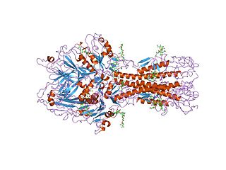
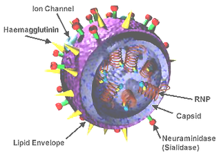
Influenza hemagglutinin (HA) or haemagglutinin[p] is a homotrimeric glycoprotein found on the surface of influenza viruses and integral to its infectivity.

Antigenic drift is a mechanism for variation in viruses that involves the accumulation of mutations within the genes that code for antibody-binding sites. This results in a new strain of virus particles which cannot be inhibited as effectively by the antibodies that were originally targeted against previous strains, making it easier for the virus to spread throughout a partially immune population. Antigenic drift occurs in both influenza A and influenza B viruses.

Rabies virus species of virus

Rabies lyssavirus, formerly Rabies virus, is a neurotropic virus that causes rabies in humans and animals. Rabies transmission can occur through the saliva of animals and less commonly through contact with human saliva. Rabies lyssavirus, like many rhabdoviruses, has an extremely wide host range. In the wild it has been found infecting many mammalian species, while in the laboratory it has been found that birds can be infected, as well as cell cultures from mammals, birds, reptiles and insects.

Hemagglutinin esterase

Hemagglutinin esterase (HEs) is a glycoprotein that certain enveloped viruses possess and use as invading mechanism. HEs helps in the attachment and destruction of certain sialic acid receptors that are found on the host cell surface. Viruses that possess HEs include influenza C, Toro-viruses, and coronaviruses. HEs is a dimer transmembrane protein consisting of two monomers, each monomer is made of three domains. The three domains are: membrane fusion, esterase, and receptor binding domains.

<i>Alphavirus</i> genus of viruses

In biology and immunology, an Alphavirus belongs to group IV of the Baltimore classification of the Togaviridae family of viruses, according to the system of classification based on viral genome composition introduced by David Baltimore in 1971. Alphaviruses, like all other group IV viruses, have a positive sense, single-stranded RNA genome. There are thirty alphaviruses able to infect various vertebrates such as humans, rodents, fish, birds, and larger mammals such as horses as well as invertebrates. Transmission between species and individuals occurs mainly via mosquitoes, making the alphaviruses a member of the collection of arboviruses – or arthropod-borne viruses. Alphavirus particles are enveloped, have a 70 nm diameter, tend to be spherical, and have a 40 nm isometric nucleocapsid.

<i>Thogotovirus</i> genus of viruses

Thogotovirus is a genus of enveloped RNA viruses, one of seven genera in the virus family Orthomyxoviridae. Their single-stranded, negative-sense RNA genome has six or seven segments. Thogotoviruses are distinguished from most other orthomyxoviruses by being arboviruses – viruses that are transmitted by arthropods, in this case usually ticks. Thogotoviruses can replicate in both tick cells and vertebrate cells; one subtype has also been isolated from mosquitoes. A consequence of being transmitted by blood-sucking vectors is that the virus must spread systemically in the vertebrate host – unlike influenza viruses, which are transmitted by respiratory droplets and are usually confined to the respiratory system.

H5N1 genetic structure

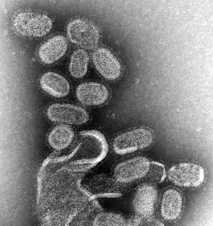
H5N1 genetic structure is the molecular structure of the H5N1 virus's RNA.

Antigenic variation refers to the mechanism by which an infectious agent such as a protozoan, bacterium or virus alters its surface proteins in order to avoid a host immune response. It is related to phase variation. Immune evasion is particularly important for organisms that target long-lived hosts, repeatedly infect a single host and are easily transmittable. Antigenic variation not only enables immune evasion by the pathogen, but also allows the microbes to cause re-infection, as their antigens are no longer recognized by the host's immune system. When an organism is exposed to a particular antigen an immune response is stimulated and antibodies are generated to target that specific antigen. The immune system will then "remember" that particular antigen, and defenses aimed at that antigen become part of the immune system’s acquired immune response. If the same pathogen tries to re-infect the same host the antibodies will act rapidly to target the pathogen for destruction. However, if the pathogen can alter its surface antigens, it can evade the host's acquired immune system. This will allow the pathogen to re-infect the host while the immune system generates new antibodies to target the newly identified antigen. Antigenic variation can occur by altering a variety of surface molecules including proteins and carbohydrates. There are many molecular mechanisms behind antigenic variation, including gene conversion, site-specific DNA inversions, hypermutation, as well as recombination of sequence cassettes. In all cases, antigenic variation and phase variation result in a heterogenic phenotype of a clonal population. Individual cells either express the phase-variable protein(s) or express one of multiple antigenic forms of the protein. This form of regulation has been identified mainly, but not exclusively, for a wide variety of surface structures in pathogens and is implicated as a virulence strategy.

Sendai virus (SeV), previously also known as murine parainfluenza virus type 1 or hemagglutinating virus of Japan (HVJ), is a negative sense, single-stranded RNA virus of the family Paramyxoviridae, a group of viruses featuring, notably, the genera Morbillivirus and Rubulavirus. SeV is a member of genus Respirovirus, members of which primarily infect mammals.

Env is a viral gene that encodes the protein forming the viral envelope. The expression of the env gene enables retroviruses to target and attach to specific cell types, and to infiltrate the target cell membrane.

Introduction to viruses A non-technical introduction to the subject.

A virus is a biological agent that reproduces inside the cells of living hosts. When infected by a virus, a host cell is forced to produce thousands of identical copies of the original virus at an extraordinary rate. Unlike most living things, viruses do not have cells that divide; new viruses are assembled in the infected host cell. But unlike still simpler infectious agents, viruses contain genes, which gives them the ability to mutate and evolve. Over 5,000 species of viruses have been discovered.

Avian sarcoma leukosis virus (ASLV) is an endogenous retrovirus that infects and can lead to cancer in chickens; experimentally it can infect other species of birds and mammals. ASLV replicates in chicken embryo fibroblasts, the cells that contribute to the formation of connective tissues. Different forms of the disease exist, including lymphoblastic, erythroblastic, and osteopetrotic.

Viral neuraminidase

Viral neuraminidase is a type of neuraminidase found on the surface of influenza viruses that enables the virus to be released from the host cell. Neuraminidases are enzymes that cleave sialic acid groups from glycoproteins and are required for influenza virus replication.

Virus quantification involves counting the number of viruses in a specific volume to determine the virus concentration. It is utilized in both research and development (R&D) in commercial and academic laboratories as well as production situations where the quantity of virus at various steps is an important variable. For example, the production of viral vaccines, recombinant proteins using viral vectors and viral antigens all require virus quantification to continually adapt and monitor the process in order to optimize production yields and respond to ever changing demands and applications. Examples of specific instances where known viruses need to be quantified include clone screening, multiplicity of infection (MOI) optimization and adaptation of methods to cell culture. This page discusses various techniques currently used to quantify viruses in liquid samples. These methods are separated into two categories, traditional vs. modern methods. Traditional methods are industry-standard methods that have been used for decades but are generally slow and labor-intensive. Modern methods are relatively new commercially available products and kits that greatly reduce quantification time. This is not meant to be an exhaustive review of all potential methods, but rather a representative cross-section of traditional methods and new, commercially available methods. While other published methods may exist for virus quantification, non-commercial methods are not discussed here.

Haemagglutinin-esterase fusion glycoprotein

In molecular biology, haemagglutinin-esterase fusion glycoprotein (HEF) is a multi-functional protein embedded in the viral envelope of several viruses, including influenza C virus, influenza D virus, coronaviruses, and toroviruses. HEF is required for infectivity, and functions to recognise the host cell surface receptor, to fuse the viral and host cell membranes, and to destroy the receptor upon host cell infection. The haemagglutinin region of HEF is responsible for receptor recognition and membrane fusion, and bears a strong resemblance to the sialic acid-binding haemagglutinin found in influenza A and B viruses, except that it binds 9-O-acetylsialic acid. The esterase region of HEF is responsible for the destruction of the receptor, an action that is carried out by neuraminidase in influenza A and B viruses. The esterase domain is similar in structure to Streptomyces scabies esterase, and to acetylhydrolase, thioesterase I and rhamnogalacturonan acetylesterase.

<i>Influenza D virus </i>

Influenza D virus is a species in the virus genus Influenzavirus D in the family Orthomyxoviridae, that causes influenza.

References

  1. Guo Y.; Jin F.; Wang P.; Wang M.; Zhu J.M. (1983). "Isolation of Influenza C Virus from Pigs and Experimental Infection of Pigs with Influenza C Virus". Journal of General Virology. 64: 177–82. doi:10.1099/0022-1317-64-1-177. PMID   6296296.
  2. 1 2 Pattison; McMullin; Bradbury; Alexander (2008). Poultry Diseases (6th ed.). Elsevier. p. 317. ISBN   978-0-7020-28625.
  3. 1 2 “Seasonal Influenza (Flu)” Centers for Disease Control and Prevention. March 22, 2012. https://www.cdc.gov/flu/about/viruses/types.htm
  4. 1 2 3 4 5 6 7 8 9 10 11 12 Margaret Hunt. “Microbiology and Immunology On-line” University of South Carolina School of Medicine. 2009. http://pathmicro.med.sc.edu/mhunt/flu.htm
  5. 1 2 3 4 World Health Organization (2006). "Review of latest available evidence on potential transmission of avian influenza (H5H1) through water and sewage and ways to reduce the risks to human health" (PDF).
  6. 1 2 3 Shuo Su; Xinliang Fu; Gairu Li; Fiona Kerlin; Michael Veit (25 August 2017). "Novel Influenza D virus: Epidemiology, pathology, evolution and biological characteristics". Virulence. 8 (8): 1580–1591. doi:10.1080/21505594.2017.1365216. PMC   5810478 . PMID   28812422.
  7. 1 2 Manuguerra JC, Hannoun C, Sáenz Mdel C, Villar E, Cabezas JA (1994). "Sero-epidemiological survey of influenza C virus infection in Spain". Eur. J. Epidemiol. 10 (1): 91–94. PMID   7957798.
  8. 1 2 3 4 5 Wagaman, Spence & O'Callaghan 1989
  9. 1 2 3 Matsuzaki Y, Katsushima N, Nagai Y, Shoji M, Itagaki T, Sakamoto M, Kitaoka S, Mizuta K, Nishimura H (2006). "Clinical features of influenza C virus infection in children". J. Infect. Dis. 193 (9): 1229–35. doi:10.1086/502973. PMID   16586359.
  10. 1 2 Ballada D, Biasio LR, Cascio G, D'Alessandro D, Donatelli I, Fara GM, Pozzi T, Profeta ML, Squarcione S, Riccò D (1994). "Attitudes and behavior of health care personnel regarding influenza vaccination". Eur. J. Epidemiol. 10 (1): 63–68. PMID   7957793.
  11. Ali A, Avalos RT, Ponimaskin E, Nayak DP (2000). "Influenza virus assembly: effect of influenza virus glycoproteins on the membrane association of M1 protein". J. Virol. 74 (18): 8709–19. PMC   116382 . PMID   10954572.
  12. Portela A, Digard P (2002). "The influenza virus nucleoprotein: a multifunctional RNA-binding protein pivotal to virus replication". J. Gen. Virol. 83 (Pt 4): 723–34. doi:10.1099/0022-1317-83-4-723. PMID   11907320.
  13. Gao Q, Brydon EW, Palese P (2008). "A seven-segmented influenza A virus expressing the influenza C virus glycoprotein HEF". J. Virol. 82 (13): 6419–26. doi:10.1128/JVI.00514-08. PMC   2447078 . PMID   18448539.
  14. Weissenhorn W, Dessen A, Calder LJ, Harrison SC, Skehel JJ, Wiley DC (1999). "Structural basis for membrane fusion by enveloped viruses". Mol. Membr. Biol. 16 (1): 3–9. PMID   10332732.
  15. Nelson, DL; Cox, MM (2013). Principles of Biochemistry (6th ed.). p. 179. ISBN   978-1-4292-3414-6.

Further reading